241
Iyeyasu JN et al. Langerhans cell histiocytosis in an elderly patient
Radiol Bras. 2012 Jul/Ago;45(4):241–243
Langerhans cell histiocytosis diagnosed in an elderly patient
*
Histiocitose de células de Langerhans diagnosticada em um paciente de idade avançada
Josie Naomi Iyeyasu1, Ana Carolina Marcos Vaz2, Fabiano Reis3, João Altemani4, Luciano de Souza Queiroz5, Keila Monteiro de Carvalho6
Langerhans cell histiocytosis is a rare disease characterized by proliferation of Langerhans cells. We report a case of Langerhans cell histiocytosis in a 63-year-old patient, who presented an expansile periorbital lesion as the first symptom, in whom computed tomography revealed characteristic lung commitment of the disease. The case management, as well as radiological findings and outcomes are described.
Keywords: Histiocytosis; Langerhans cell; Orbit.
A histiocitose de células de Langerhans é uma doença rara caracterizada proliferação de células de Langerhans. Neste artigo descrevemos um caso de histiocitose de células de Langerhans em um paciente de 63 anos, com uma lesão expansiva periorbital como primeiro sintoma e cuja tomografia computadorizada revelou acometimento pulmonar ca-racterístico da doença. A condução do caso, os achados radiológicos e os resultados são apresentados.
Unitermos: Histiocitose; Células de Langerhans; Órbita. Abstract
Resumo
* Study developed at Hospital de Clínicas – Universidade Es-tadual de Campinas (HC-Unicamp), Campinas, SP, Brazil.
1. Master, MD, Ophthalmologist at Hospital de Clínicas – Universidade Estadual de Campinas (HC-Unicamp), Campinas, SP, Brazil.
2. MD, Resident, Department of Radiology of Faculdade de Ciências Médicas – Universidade Estadual de Campinas (FCM-Unicamp), Campinas, SP, Brazil.
3. PhD, Teacher at Department of Radiology, Faculdade de Ciências Médicas – Universidade Estadual de Campinas (FCM-Unicamp), Campinas, SP, Brazil.
4. PhD, Physician Assistant at Department of Radiology, Fa-culdade de Ciências Médicas – Universidade Estadual de Cam-pinas (FCM-Unicamp), CamCam-pinas, SP, Brazil.
5. PhD, Teacher at Department of Pathological Anatomy, Fa-culdade de Ciências Médicas – Universidade Estadual de Cam-pinas (FCM-Unicamp), CamCam-pinas, SP, Brazil.
6. Private Docent, Teacher at Department of Ophthalmo-Otorhinolaryngology, Faculdade de Ciências Médicas – Universi-dade Estadual de Campinas (FCM-Unicamp), Campinas, SP, Brazil.
Mailing Address: Dr. Fabiano Reis. Hospital de Clínicas da Universidade Estadual de Campinas (HC-Unicamp). Rua Vital Brasil, 251, Cidade Universitária Zeferino Vaz, Caixa Postal 6142, Campinas, SP, Brazil, 13083-888. E-mail: fabianoreis2@ gmail.com
Received March 21, 2012. Accepted after revision June 15, 2012.
Iyeyasu JN, Vaz ACM, Reis F, Altemani J, Queiroz LS, Carvalho KM. Langerhans cell histiocytosis diagnosed in an elderly patient. Radiol Bras. 2012 Jul/Ago;45(4):241–243.
0100-3984 © Colégio Brasileiro de Radiologia e Diagnóstico por Imagem CASE REPORT
Immuno-histochemical analysis was positive for CD1a, CD 68, HAM56 and S-100 in the neoplastic cells, negative for HMB45, AE1, AE3, CD20, CD3, and posi-tive for KI67 in 5–10% of the nuclei, cor-roborating the diagnostic hypothesis of Langerhans cell histiocytosis (Figure 3).
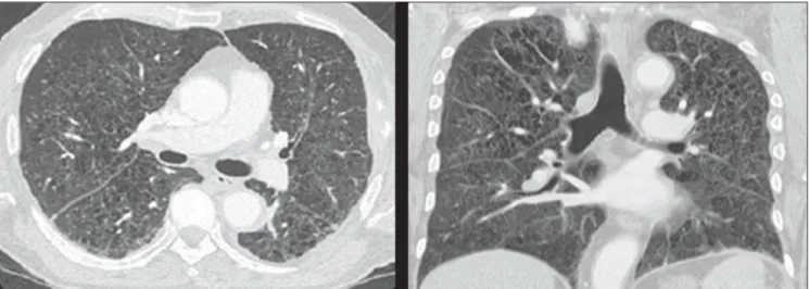
Then, the patient underwent further in-vestigation to determine the presence of ad-ditional foci of histiocytosis which demon-strated a lesion in the fifth left coastal arch and the pulmonary involvement typically observed in cases of histiocytosis (Figure 4). The patient underwent hematological follow-up and was submitted to two cycles of chemotherapy with vimblastine, etopo-side and prednisone, and progressed to death in February 2012 because of compli-cations resulting from his lung disease.
DISCUSSION
Langerhans cell histiocytosis is a rare disease (incidence of 2–5 cases per mil-lion)(1) characterized by proliferation of Langerhans cells(2), most commonly affect-ing male children in the age range between one and three years(1).
The etiology of this disease still remains unknown. It is believed that the disease is consequence of an immunological deregu-ing his imagderegu-ing studies and the whole case
progression. At the end, the authors also present a brief review of the literature on the disease.
CASE REPORT
The present report describes the case of a male, 63-year-old patient complaining of proptosis of his right eye for six months. Previous personal history: smoking and alcohol use. The screening for infecto-con-tagious diseases was negative.
Physical examination did not present any other finding, except the right-sided proptosis itself. Skull computed tomogra-phy (Figure 1) revealed a lesion affecting the lateral wall of the orbit (with bone de-struction) and the right intraorbital region, externally extending towards the perizygo-matic region. Right-sided ocular globe proptosis and invasion of the correspond-ing lateral extrinsic orbital muscle were observed. Then, the patient was submitted to complete exeresis of the lesion.
Histopathological study revealed prolif-eration of abundant macrophages (histio-cytes) with epithelioid cells and multi-nucleated gigantocytes with some figures of mitosis compatible with Langerhans cell histiocytosis (Figure 2).
INTRODUCTION
present-242
Iyeyasu JN et al. Langerhans cell histiocytosis in an elderly patient
Radiol Bras. 2012 Jul/Ago;45(4):241–243 Figure 1. Contrast-enhanced axial computed tomography of skull. Intraorbital expansile lesion extending toward the intra- and extraconal spaces, extracranial soft tissues and bone erosion of the lateral wall (zygomatic bone and major sphenoid wing) of the right orbit.
Figure 2. Hematoxylin eosin staining demonstrates fagocytosis of hemosid-erin by Langerhans cells recognizable by their irregular nuclei with a round or elongated contour, frequently with cleavage and indentation, loose and well-distributed cromatin.
Figure 3. Immuno-histochemical study demonstrates positivity for CD1a with a membranous pattern, which is a sine qua non condition for Langerhans cell diagnosis.
lation resulting in excessive production of cytokines and prostaglandins, which cause injury to several organs(1).
The disease may affect either single or multiple organs. The clinical presentation depends on the dysfunction degree and on the involved organs(1) and the main com-plications include hypophyseal dysfunc-tion, particularly diabetes insipidus(3), and neurodegenerative diseases(4).
The most common ophthalmological manifestation is the presence of a solitary bone lesion in the orbit(5), which, likewise the presence of lesions in the mastoid and temporal bones(6), is considered as a risk
factor for the onset of complications in the central nervous system (CNS)(7).
Langerhans cell histiocytosis, particu-larly in its disseminated presentation, may be associated with other conditions such as leukemia, Hodgkin’s and non Hodgkin’s lymphoma, history of neonatal infection, exposure to solvents, thyroid diseases, in-fectious diseases (adenovirus, parvovirus, Epstein-Barr virus, cytomegalovirus, HIV and HTLV) and smoking(1), like it may have occurred in the present case.
The imaging method of choice to assess the CNS is magnetic resonance imaging for its highest sensitivity in the detection of
243
Iyeyasu JN et al. Langerhans cell histiocytosis in an elderly patient
Radiol Bras. 2012 Jul/Ago;45(4):241–243 CD1a (representing diagnostic certainty)(1), as in the present case.
Local intervention is the treatment for Langerhans cell histiocytosis(6) in cases of a single lesion. Systemic treatment is re-served for cases of incomplete response to treatment, lesion reactivation or onset of other lesions(7), and chemotherapy is the gold standard(2), as observed in the present case.
The prognosis depends of the extent of the disease: patients with focal disease typi-cally have a good prognosis, while patients with disseminated disease and those with involvement of the central nervous system present a worse prognosis(6).
Thus, the authors conclude that Langer-hans cell histiocytosis is a rare and severe disease which may affect several organs.
Therefore, such disease must be included in the list of differential diagnosis for pa-tients with proptosis, since orbital involve-ment indicates a probable CNS compro-mise, i.e. worse prognosis. The authors consider the present case as being relevant, since it illustrates well the unfavorable natural progression of the disease, even with appropriate treatment, thus serving to emphasize the importance of an early diag-nosis. Additionally, the present case was rare, since the disease onset occurred in an elderly patient, while the onset in the child-hood is most frequent. Also, it should be observed that involvement of the orbit, like in the present case, is seldom found.
REFERENCES
1. Duda-Szyma½ska J, Wierzchniewska-ºawska A. Langerhans cell histiocytosis in a 3-year-old girl:
Figure 4. Chest computed tomography of the patient demonstrating multiple cysts diffusely scattered throughout the lung parenchyma, with irregular walls and variable dimensions (bizarre cysts) in association with diffuse micronodules, a typical pattern of pulmonary involvement by Langerhans cell histiocytosis.
a case report and literature review. Pol J Pathol. 2009;60:134–7.
2. Ng Wing Tin S, Martin-Duverneuil N, Idbaih A, et al. Efficacy of vinblastine in central nervous sys-tem Langerhans cell histiocytosis: a nationwide ret-rospective study. Orphanet J Rare Dis. 2011;6:83.
3. Grois N, Tsunematsu Y, Barkovich AJ, et al. Cen-tral nervous system disease in Langerhans cell his-tiocytosis. Br J Cancer Suppl. 1994;23:S24–S28. 4. Imashuku S. High dose immunoglobulin (IVIG) may reduce the incidence of Langerhans cell his-tiocytosis (LCH)-associated central nervous sys-tem involvement. CNS Neurol Disord Drug Tar-gets. 2009;8:380–6.
5. Margo CE, Goldman DR. Langerhans cell histio-cytosis. Surv Ophthalmol. 2008;53:332–58.
6. Allen CE, McClain KL. Langerhans cell histiocy-tosis: a review of past, current and future therapies. Drugs Today (Barc). 2007;43:627–43. 7. Harris GJ. Langerhans cell histiocytosis of the