Simultaneous meningioma and brain metastasis from
renal cell carcinoma – a rare presentation. Case report
Meningioma e metástase cerebral de carcinoma de células renais simultâneos –
uma apresentação rara. Relato de caso
Aline Lariessy Campos Paiva
I, João Luiz Vitorino Araujo
II, Vinicius Ricieri Ferraz
I, José Carlos Esteves Veiga
IIIFaculdade de Ciências Médicas da Santa Casa de São Paulo (FCMSCSP), São Paulo (SP), Brazil
ABSTRACT
CONTEXT: Brain metastases are the most common tumors of the central nervous system. Because of their high frequency, they may be associated with rare situations. Among these are tumor-to-tumor metastasis and an even a rarer situation called simultaneous brain tumors, which are more related to primary tumors of the reproductive and endocrine systems.
CASE REPORT: A 56-year-old male patient with a history of renal cell carcinoma (which had previously been resected) presented with a ventricular lesion (suggestive of metastatic origin) and simultaneous olfactory groove lesion (probably a meningioma). First, only the ventricular lesion was dealt with, but after a year, the meningothelial lesion increased and an occipital lesion appeared. Therefore, both of these were resected in a single operation. All the procedures were performed by the same neurosurgeon. The patient evolved without neurological deicits during the postoperative period. After these two interventions, the patient remained well and was referred for adjuvant treatment.
CONCLUSIONS: This study provides the irst description of an association between these two tumors. Brain metastases may be associated with several lesions, and rare presentations such as simultaneity with meningioma should alert neurosurgeons to provide the best oncological treatment.
RESUMO
CONTEXTO: As metástases cerebrais são os tumores mais comuns do sistema nervoso central e, devido à sua elevada frequência, podem estar associadas a situações raras. Entre estas estão as “tumor to tumor metastasis” e uma situação ainda mais rara chamada de tumores cerebrais simultâneos, mais relacionados a tumores primários dos sistemas endocrinológico e reprodutivo.
RELATO DE CASO: Um homem de 56 anos com histórico de câncer de células renais (extirpado previa-mente) apresentou-se com lesão ventricular (sugestiva de origem metastática) e simultaneamente com uma lesão em topograia de goteira olfatória (provavelmente meningioma). Primeiramente, apenas a lesão ventricular foi abordada, porém após um ano, a lesão meningotelial aumentou e uma lesão occipital apare-ceu e então ambas foram ressecadas em uma única cirurgia. Todos os procedimentos foram realizados pelo mesmo neurocirurgião. O paciente evoluiu sem déicits neurológicos no período pós-operatório. Após essas duas intervenções, o paciente permaneceu bem, sendo encaminhado para tratamento adjuvante.
CONCLUSÕES: O presente trabalho é a primeira descrição da associação encontrada entre esses dois tumores. As metástases cerebrais podem associar-se a várias lesões, e manifestações raras, tais como apre-sentação simultânea com meningioma, devem alertar o neurocirurgião a fornecer o melhor tratamento oncológico.
IMD. Neurosurgery Resident, Faculdade de Ciências Médicas da Santa Casa de São Paulo (FCMSCSP), São Paulo (SP), Brazil.
IIPhD. Assistant Neurosurgeon, Faculdade de Ciências Médicas da Santa Casa de São Paulo (FCMSCSP), and Neurosurgeon, Instituto do Câncer Arnaldo Vieira de Carvalho (ICAVC), Oncocenter and Hospital Nove de Julho, São Paulo (SP), Brazil.
IIIPhD. Full Professor and Head, Discipline of Neurosurgery, Faculdade de Ciências Médicas da Santa Casa de São Paulo (FCMSCSP), São Paulo (SP), Brazil.
KEY WORDS: Neoplasm metastasis. Brain neoplasms. Carcinoma, renal cell. Meningioma.
Cerebral ventricle neoplasms.
PALAVRAS-CHAVE: Metástase neoplásica. Neoplasias encefálicas. Carcinoma de células renais. Meningioma.
INTRODUCTION
Brain metastases constitute a common complication of advanced primary tumors. herefore, they are an important issue that guides the approach taken towards patients with a diagnosis of cancer.1
he incidence of brain metastases is about 9 to 17%, based on various studies.1 However, the exact incidence is thought to be
higher, possibly because there are many asymptomatic patients. In several studies, only surgical metastatic disease is included in the statistical analysis.1
Brain metastases are observed in 2 to 17% of patients with metastatic renal cell carcinoma (mRCC).2,3 hese patients usually
require a neurosurgical approach and adjuvant therapies, espe-cially radiotherapy. However, despite optimal treatment, patients presenting with brain metastasis have a very poor prognosis and probably also have other compromised organs. Another factor associated with increased mortality is that mRCC does not have a good response to radiation.2-4
here are two entities that are rarely related to brain metastases but which, when they occur, it is important to be aware of. he irst of these is tumor-to-tumor metastasis5 (collision tumor is used as
a synonym by some authors), which was irst described in 1902. his is a well-documented phenomenon in which a host tumor that is usually more indolent serves as the source for growth of a more aggressive neoplasm such as a meningioma, thus leading to growth of a high-grade glioma or metastatic lesion.6-8
he second of these is an even rarer phenomenon that has been named synchronous or simultaneous tumors, and which forms the topic of the present report. hese occur when two his-tological tumors compromise the central nervous system (CNS) at the same time but there is no histopathological evidence that one tumor served as the source of growth for the other, as occurs in the tumor-to-tumor entity.9,10
his report aimed to present a unique case of simultaneous benign meningioma and brain metastasis from renal cell carcinoma in a male adult. We were unable to ind any similar cases reported in the literature, through reviewing the MEDLINE database.
CASE REPORT
A 56-year-old male patient came to our neuro-oncology ser-vice in 2013, with a history of mild frontal headache, but with-out neurological symptoms. He had a history of renal cell car-cinoma in his right kidney and had undergone nephrectomy in 2011. In the same year, he underwent follow-up examina-tions but without evidence of brain metastatic disease. He had no other comorbidities.
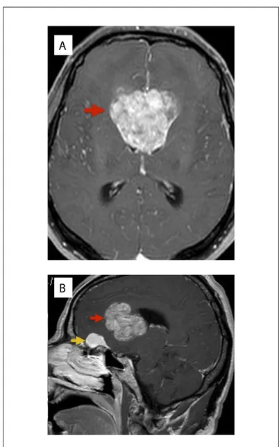
he headache became progressively worse and was associated with nausea, photophobia and phonophobia. In 2015, on control brain magnetic resonance imaging (MRI), the presence of an intraventric-ular tumor was noticed (Figure 1), along with another lesion in the
olfactory groove (on MRI, it was suggestive of a meningioma). A neu-rosurgical approach was used to treat the ventricular lesion, consisting of transcallosal tumor resection, which was performed in May 2015. he procedure was free from complications, gross total removal was achieved and the patient reported improvement of the headache. He was referred for neuro-oncology outpatient follow-up and for radio-therapy evaluation. Only the larger of the two lesions was resected on this occasion because two diferent approaches performed at the same time might have increased the morbidity and, moreover, the patient did not have any neurological deicits at this time.
Five months ater the irst procedure, the patient complained of visual impairment (which upon physical examination was found to
Figure 1. Magnetic resonance imaging of a patient with simultaneous brain tumors: A - axial image showing ventricular tumor; B – sagittal image showing two lesions: olfactory groove meningioma and ventricular tumor.
A
he patient underwent whole-brain radiotherapy at this time, without any surgical indication.
he olfactory groove lesion increased ater radiation therapy had been completed (Figure 3) and the patient reported that his headache had returned. At this time, neurosurgical resection of the two lesions (the olfactory groove meningioma and the occipital lesion) in a single procedure was proposed. Given the possibility of tumor-to-tumor metastasis, it was very important to determine whether the meningioma had served as a basis for the metastatic lesion, in order to better deine the complementary treatment.
he surgery was performed without complications and the his-topathological and immunohistochemical analyses conirmed that the olfactory groove lesion was a grade I meningioma (Figure 4), without evidence of tumors of another origin difering from the meningothelial lineage. he only radionecrosis was observed at his-topathological analysis of the occipital lesion. Simpson II resection was performed (Figure 5). hese two surgical procedures (one to treat the intraventricular metastasis and the other to deal with the olfactory groove meningioma and the occipital lesion) were per-formed by the same oncological neurosurgeon (JLVA).
he patient reported improvement of his headache and was discharged for outpatient follow-up with the neurosurgical and neuro-oncology team. Complete screening with the aim of reveal-ing any other metastatic lesion that might have been present was performed, consisting of computed tomography (CT) scans on the chest, abdomen and pelvis. here was no evidence of any local or metastatic recurrence.
DISCUSSION
Brain metastases constitute one of the most common neurologi-cal complications in oncologineurologi-cal patients with advanced disease.1
In some cases, they may be the initial manifestation, which then Figure 2. Axial contrasted T1-weighted magnetic resonance
imaging exam showing a right occipital lesion after radiation.
Figure 3. Axial contrasted T1-weighted magnetic resonance imaging exam showing olfactory groove meningioma
be due to let hemianopia) and frontotemporal headache. Because of this, MRI was performed again and this revealed another lesion, in the right occipital lobe (Figure 2), probably of metastatic origin.
leads to diagnosing the primary tumor.11 he signs and
symp-toms are nonspeciic and may vary according to the site and size. In rare cases, they may manifest as lesions in the scalp and skull.12,13 he incidence rate is about 9 to 17%, based on various
studies, although the exact incidence is thought to be higher.1,3
he blood-brain barrier (BBB) and absence of a lymphatic sys-tem are factors that make dissemination of cancerous cells more diicult. hus, patients with brain metastases generally also have extracranial lesions. his shows that when tumor cells invade brain structures, the disease is more advanced and has a worse progno-sis.2,3 Conditions that alter immunological defenses, such as human
immunodeiciency virus infection, may be associated with brain metastasis. here are some hypotheses stating that such conditions could favor appearance of some tumors.14
Regarding metastatic renal cell carcinoma, brain lesions gener-ally do not occur at the same time as the primary tumor. Some stud-ies have shown that the incidence is highest around 10 to 13 years ater the initial nephrectomy.1 he treatment may be diicult because
central nervous system lesions are usually resistant to chemotherapy and radiotherapy.15 Nonetheless, these lesions may respond to
immu-notherapy using alpha-interferon16 or interleukin (IL)-2. Ater
treat-ment of brain metastases, the median survival is about 4-5 months,3
but aggressive surgical resection signiicantly increases this period. here are some hypotheses explaining why brain metastasis may appear ater a long period, in the absence of other metastatic lesions16
in renal cell carcinoma cases. One hypothesis is that this might be
because, in the initial stage, the brain metastasis is microscopic and does not cause any neurological symptoms. Another hypothesis is that adjuvant therapy for renal cell carcinoma decreases host immu-nopotency and thus leads to faster development of brain lesions.16
In the present report, the patient evaluated initially did not present central nervous system impairment: it was only ater some years that it was found that he had brain metastasis.
Ater disruption of the BBB, migration of inlammatory cells, including tumor-associated macrophages (TAMs)17,18 may
contrib-ute towards persistence of increased vascular permeability. TAMs are recruited to tumors through speciic chemokine/ chemokine receptor interactions. When neoplastic cells invade the central nervous system and a metastasis develops, the lesion is seen to be well vascularized and is susceptible to spontaneous intracranial hemorrhage,4 which
may include intraventricular bleeding. Metastatic renal cell carcinoma has a unique ainity to the ventricular system, in close association with the choroid plexus, probably due to a chemokine cascade.11
Multiple primary intracranial tumors of diferent histological types are rare, except for cases observed ater radiotherapy or in situation of phacomatosis19 such as Von Recklinghausen syndrome.
However, multiple brain tumors in the absence of these conditions constitute an even rarer phenomenon.10
When a lesion serves as the source for growth of another neopla-sia, this is considered to constitute an entity named tumor-to-tumor metastasis (or collision tumor). A more indolent tumor is generally the substratum for an aggressive lesion.5 In the present case,
how-ever, the histopathological analysis (Figure 5) did not reveal that one tumor had served for growth of another but, rather, that two difer-ent tumors had simultaneous occurrence at diferdifer-ent sites.
Because brain metastases have higher incidence than primary central nervous system tumors, they present greater involvement in cases of multiple brain tumors, such as collision tumors or simul-taneous tumors. Systemic cancers rarely metastasize into preex-isting intracranial neoplasms; meningiomas are the major recipi-ent of these metastases.8
Neuroimaging is unable to predict which entity was present. Only when accurate histopathological and immunohistochemical analysis is performed is it possible to conirm whether one tumor has served as source of growth for another or whether the observed tumor represents two diferent lesions occurring simultaneously. Brain metastases can sometimes behave on CT and MRI as images of typical meningiomas and thus confuse the diagnosis.20 In the
present case, the appearance of the metastasis was not confused with a meningothelial origin (Figure 1).
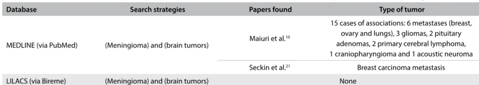
Simultaneous occurrence of an intracranial meningioma and brain metastases in the same patient at the same time is a rather unusual event. hus, some thought is needed regarding the patho-genic relationship, pathological diagnosis, surgical indications10
and imaging patterns. We conducted a search in the MEDLINE Figure 5. Brain computed tomography showing Simpson II
Database Search strategies Papers found Type of tumor
MEDLINE (via PubMed) (Meningioma) and (brain tumors) Maiuri et al.
10
15 cases of associations: 6 metastases (breast, ovary and lungs), 3 gliomas, 2 pituitary adenomas, 2 primary cerebral lymphoma, 1 craniopharyngioma and 1 acoustic neuroma
Seckin et al.21 Breast carcinoma metastasis
LILACS (via Bireme) (Meningioma) and (brain tumors) None
Table 1. Metastatic brain tumors reported in the literature (PubMed database) as simultaneous presentation with meningiomas
database (using the terms: simultaneous/synchronous, menin-gioma and metastasis) and only found two papers (Table 1).10,21
Neither of them reported on simultaneous renal cell carcinoma. here are few reports in the literature describing this condition and the largest review on these simultaneous lesions only brought together iteen cases. Six of them were metastatic lesions, but none of them was from renal cell carcinoma.9
In the case reported here, the patient had a known diagnosis of renal cell carcinoma. Brain MRI showed a ventricular lesion sug-gestive of metastatic origin. Because the simultaneous olfactory groove meningioma was small at this time, it was preferred to only operate the larger lesion, in order to reduce morbidity that would occur if two diferent approaches were used. However, ater some months, the olfactory groove lesion was found to have increased and a new occipital and symptomatic lesion had appeared.
Neoplasms from the female endocrine and reproductive sys-tem are generally more related to meningiomas10 and, because of
this, are usually present in women. In the present report, however, an even rarer situation was discussed: a male patient with menin-gioma and brain metastasis, for whom the primary form was renal cell carcinoma. We did not ind any reports of this association in the literature review that we conducted.
CONCLUSIONS
Brain tumors may present through diferent patterns and, even if they are benign lesions, as meningiomas generally are, they may be associated with rare situations. An occurrence of two brain tumors is one of these situations, and this constitutes a challenge. Simultaneous lesions are an even rarer phenomenon. Metastases are more oten reported as part of this entity, although in most cases endocrine and reproductive system tumors have a closer and larger relationship with meningioma growth and therefore are seen more frequently in females. Renal cell carcinoma had not reported until now as part of this association.
REFERENCES
1. Nayak L, Lee EQ, Wen PY. Epidemiology of brain metastases. Curr Oncol
Rep. 2012;14(1):48-54.
2. Sheehan JP, Sun MH, Kondziolka D, Flickinger J, Lunsford LD. Radiosurgery
in patients with renal cell carcinoma metastasis to the brain: long-term
outcomes and prognostic factors inluencing survival and local tumor
control. J Neurosurg. 2003;98(2):342-9.
3. Vogl UM, Bojic M, Lamm W, et al. Extracerebral metastases determine the
outcome of patients with brain metastases from renal cell carcinoma.
BMC Cancer. 2010;10:480.
4. Shuch B, La Rochelle JC, Klatte T, et al. Brain metastasis from renal cell
carcinoma: presentation, recurrence, and survival. Cancer. 2008;113(7):1641-8.
5. Erdogan H, Aydin MV, Tasdemiroglu E. Tumor-to-tumor metastasis of
the central nervous system. Turk Neurosurg. 2014;24(2):151-62.
6. Berent W. Seltene Metastasenbildung. Zentralbl Allg Pathol. 1902;13:406-10.
7. Carr K, He L, Weaver K, Highield Nickols H. Renal cell carcinoma
metastatic to meningioma: tumor-to-tumor metastasis. Clin
Neuropathol. 2014;33(2):152-6.
8. Chahlavi A, Staugaitis SM, Yahya R, Vogelbaum MA. Intracranial collision
tumor mimicking an octreotide-SPECT positive and FDG-PET negative
meningioma. J Clin Neurosci. 2005;12(6):720-3.
9. Lenarz M, Durisin M, Becker H, Brandis A, Lenarz T. A case of multiple
primary tumors of the anterior skull base. Skull Base. 2007;17(2):153-6.
10. Maiuri F, Cappabianca P, Iaconetta G, Esposito F, Messina A.
Simultaneous presentation of meningiomas with other intracranial
tumours. Br J Neurosurg. 2005;19(4):368-75.
11. Shapira Y, Hadelsberg UP, Kanner AA, Ram Z, Roth J. The ventricular
system and choroid plexus as a primary site for renal cell carcinoma
metastasis. Acta Neurochir (Wien). 2014;156(8):1469-74.
12. Ferraz VR, Vitorino-Araújo JL, Sementilli L, Neto JF, Veiga JC. Lesion in
Scalp and Skull as the First Manifestation of Hepatocellular Carcinoma.
Case Rep Neurol Med. 2016;2016:2897048.
13. Vitorino-Araujo JL, Veiga JC, Barboza VR, et al. Scalp, skull and brain
metastasis of squamous cell carcinoma of the cervix--a rare entity. Br
J Neurosurg. 2013;27(4):519-20.
14. Badke GL, de Aguiar GB, Silva JM, et al. Cerebral Metastasis from
Breast Cancer in a Male Patient with HIV. Case Rep Neurol Med.
2015;2015:482839.
15. Wyler L, Napoli CU, Ingold B, et al. Brain metastasis in renal cancer
patients: metastatic pattern, tumour-associated macrophages
and chemok ine/chemoreceptor expression. Br J Cancer.
2014;110(3):686-94.
16. Sadatomo T, Yuki K, Migita K, et al. Solitary brain metastasis from renal
cell carcinoma 15 years after nephrectomy: case report. Neurol Med
17. Doolittle ND, Peereboom DM, Christoforidis GA, et al. Delivery of
chemotherapy and antibodies across the blood-brain barrier and the
role of chemoprotection, in primary and metastatic brain tumours:
report of the Eleventh Annual Blood-Brain Barrier Consortium meeting.
J Neurooncol. 2007;81(1):81-91.
18. Jin G, Hao S, Xie J, Mi R, Liu F. Collision tumors of the sella: coexistence
of pituitary adenoma and craniopharyngioma in the sellar region.
World J Surg Oncol. 2013;11:178.
19. Iacoangeli M, Di Rinzo A, Colasanti R, et al. Rare synchronous association
of vestibular schwannoma and indolent insular oligodendroglioma in
a patient without neuroibromatosis: controversial issue of timing for
surgical treatment of asymptomatic low-grade gliomas. Onco Targets
Ther. 2012;5:357-61.
20. Tagle P, Villanueva P, Torrealba G, Huete I. Intracranial metastasis or
meningioma? An uncommon clinical diagnostic dilemma. Surg Neurol.
2002;58(3-4):241-5.
21. Seckin H, Yigitkanli K, Ilhan O, Han U, Bavbek M. Breast carcinoma
metastasis and meningioma. A case report. Surg Neurol.
2006;66(3):324-7; discussion 327.
Conlict of interest: None
Sources of funding: None
Date of irst submission: October 10, 2016
Last received: October 25, 2016
Accepted: October 28, 2016
Address for correspondence:
Aline Lariessy Campos Paiva
Neurosurgery Resident, Faculdade de Ciências Médicas da Santa Casa de
São Paulo (FCMSCSP)
Rua Dr. Cesário Mota Júnior, 112
São Paulo (SP) — Brasil
CEP 01221-020
Tel./Fax. (+55 11) 2176-7000