ORIGINAL |
ORIGINAL
RGO, Rev Gaúch Odontol, Porto Alegre, v.65, n.1, p. 83-86, jan./mar., 2017 http://dx.doi.org/10.1590/1981-86372017000100003243
ABSTRACT
The simple bone cyst,or traumatic bone cyst, solitary or idiopathic bone cavity is an intraosseous pseudocyst discovered through a panoramic radiographic examination. Commonly, the SBC is presented as an asymptomatic lesion, radiolucent, unilocular, without cortical expansion, with margins adorning vital tooth roots of the affected region. The literature has recommended that clinical and radiographic aspects of simple bone cyst are quite convincing as to diagnosis, so follow-up through clinical and radiographic examination has been suggested. This case illustrates an exuberant clinical case of simple bone cyst, which was presented as multilocular and expansive lesion in a 7-year-old patient. Surgical exploration was performed because her parents were anxious about the presumptive diagnosis of other more aggressive lesions. The diagnosis of SBC was conirmed. The case was monitored and, after three years, total bone remodeling of the affected area was observed. In conclusion, asymptomatic radiolucent lesions that suggest a strong presumptive diagnosis of a simple bone cyst can be clinically and radiographically monitored. However, clinical variations, as in the present clinical case, require conirmation of the diagnosis, which is performed through exploratory surgery. The establishment of the diagnosis affords the safest approach for both the professional and the patient.
Indexing terms:Bone cyst. Child. Differential diagnosis.
RESUMO
O cisto ósseo simples, ou cisto ósseo traumático, solitário ou cavidade óssea idiopática é um pseudocisto intra-ósseo descoberto através de um exame radiográico panorâmico. Comumente, o cisto ósseo simples se apresenta como uma lesão assintomática, radiolúcida, unilocular, sem expansão de corticais, com margens festonando as raízes dentárias vitais da região afetada. A literatura tem recomendado que os aspectos clínicos e radiográicos do cisto ósseo simples são bastante convincentes do seu diagnóstico, portanto a proservação através de exame clínico e radiográico tem sido sugerida. O presente caso ilustra um caso clínico de um grande cisto ósseo simples, o qual se apresentava multilocular e expansivo, numa criança de 7 anos. Diante da ansiedade dos pais e diagnóstico presuntivo de outras lesões mais agressivas, foi realizada a exploração cirúrgica, a qual conirmou o diagnóstico de cisto ósseo simples. O caso foi acompanhado e, após três anos, foi observada a total remodelação óssea da área afetada. Como conclusão, as lesões radiolúcidas assintomáticas que sugerirem um diagnóstico presuntivo muito indicativo de cisto ósseo simples, podem ser acompanhadas clínica e radiograicamente. Entretanto, as variações de apresentação clínica, como no presente caso clínico, requerem uma conirmação do diagnóstico, a qual é realizada através da exploração cirúrgica. O estabelecimento do diagnóstico torna a condução do caso mais segura para ambos o proissional e o paciente.
Termos de indexação: Cisto ósseo solitário. Criança. Diagnóstico diferencial.
Simple bone cyst in a 7-year-old child
Cisto ósseo simples em criança de 7 anos
Heitor ALBERGONI DA SILVEIRA1
Camila LOPES CARDOSO1
Mariane PEXE1
Rafael ZETEHAKU ARAUJO1
Anthony BENITES CONDEZO1
Marcos MARTINS CURI2
1 Universidade do Sagrado Coração, Departamento de Cirurgia. Rua Irmã Arminda,10-50, Jardim Brasil, 17011-160, Bauru, SP, Brasil. Correspondência para / Correspondence to: CL CARDOSO. E-mail: <[email protected]>.
2 Hospital Santa Catarina, Departamento de Oncologia. São Paulo, SP, Brasil.
with the margins adorning the roots of the teeth in the affected region1-4. Clinically it is asymptomatic and its inal
diagnosis is obtained during exploratory surgery, when the surgeon conirms that the cavity is empty1-3. In terms of
how this entity should be handled, there is little consensus in the literature, whether it should be by monitoring or through exploratory surgery1-5. The aim of the present
case is to illustrate an atypical case of SBC which, given its large size and moderate bone expansion, was treated via exploratory surgery.
INTRODUCTION
A simple bone cyst (SBC), also referred to as traumatic or solitary bone cyst, or idiopathic bone cavity, is considered an intraosseous pseudocyst devoid of epithelial lining, containing serosanguineous luids or none at all1-4.
The SBC is seen more in the second decade of life, the mandible being the region most frequently affected1.
84 RGO, Rev Gaúch Odontol, Porto Alegre, v.65, n.1, p. 83-86, jan./mar., 2017
HA SILVEIRA et al.
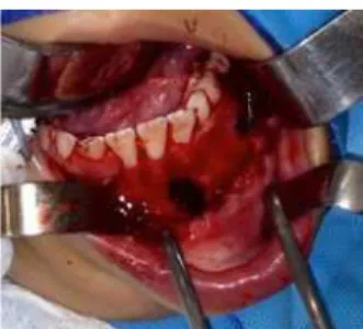
Figure 4. Image of exploratory surgery with the aim of diagnosis. The presence of an empty cavity was noted.
Figure 5. Panoramic x-ray 3 years later, showing the repair of the area affected by the lesion and the permanence of the teeth that were involved in the alteration.
DISCUSSION
SBC, described for the irst time in 1929 by Lucas et al.6, is a pathology that usually aflicts long bones such
as the femur1,3, however, in the maxillary bones there is a
rate of incidence of 1.25%4. In the oral cavity, the literature
has shown the mandible to be the bone most affected. As for the age group affected by this entity, the vast majority of studies have reported an age range between 15 and 207-9. In the quest to ind other studies reporting SBC
in children, as in the present case, only one report was found for the same age group, associated with a previous trauma10. Other SBC studies in children relate to children
10 years of age, or older7,9.
The majority of SBC cases are discovered by means of panoramic x-rays, as the lesion is asymptomatic1-5. As
yet, there are no completely plausible theories to explain its etiopathogenesis. The literature has most commonly accepted the hypothesis that the trauma in the altered region results in the formation of a hematoma which, unable to repair itself, instead liqueies11. Although this
theory has been fairly well disseminated, the patient in this report did not present any history of trauma in the region of the alteration, similar to other reports12. Other reported
theories relating to SBC development include cystic degeneration of ibro-osseous lesions, vascular anomalies and altered bone metabolism11-12.
CASE REPORT
Caucasian, female patient aged 7, was referred to our team with a panoramic x-ray that revealed an asymptomatic, unilocular, radiolucent lesion in the anterior region and body of the left mandible (igure 1). According to her reported medical history, there was no associated causal factor, nor history of trauma in the region. Cone-beam computed tomography showed a large, multilocular, hypodense area in the affected region. The axial reconstructions revealed moderate cortical expansion (igures 2 and 3). The main diagnostic hypotheses were keratocystic odontogenic tumor (KOT) and SBC. Given the extent of the alteration and the anxiety of the parents, exploratory surgery of the altered area was scheduled in order to establish a deinitive diagnosis. During the intraoperative phase, an empty cavity was found conirming the diagnosis of SBC (igure 4). After exploratory surgery, over the course of 3 years, the affected region was completely repaired, revealing the formation of a lamina dura and no alteration to the permanent erupted teeth, thus corroborating the literature with regard to prognosis (igure 5).
Figure 1. Initial appearance. A) Panoramic x-ray showing a well-circumscribed, uni-locular, radiolucent region, from the region of the incisors to the left-side molars. The teeth presented no tooth resorption. B) Intraoral appearance, demonstrating normal mucosa lining the affected region, without bone expansion.
Figure 2. Axial reconstructions of the cone-beam computed tomography demon-strating a moderately expansive, multilocular, hypodense area when ob-served closer to the lower edge of the mandible.
RGO, Rev Gaúch Odontol, Porto Alegre, v.65, n.1, p. 83-86, jan./mar., 2017 85 Simple bone cyst in a 7-year-old child
4. Andrade LE, Cerqueira LS, Rebouças DS, Marchionni AMT. Simple boné cyst: a case report. Rev Cir Traumatol Buco-Maxilo-Fac. 2016;16(2):36-39.
5. Damante JH, Da S Guerra EN, Ferreira Jr O. Spontaneous resolution of simple bone cysts. Dentomaxillofac Radiol. 2002 May;31(3):182-6. doi: 10.1038/sj/dmfr/4600696
6. Lucas CD. Do all cysts of the jaw originate from the dental system? J Am Dental Assoc. 1929;16:647. doi: 10.14219/jada. archive.1929.0086
REFERENCES
1. Barnes L, Eveson JW, Reichart P, Sidransky D (Eds.). World health organization classiication of tumours. Pathology and genetics of head and neck tumours. p.327. Lyon: WHO Press; 2005.
2. Paiva LCA, Menezes FS, Porto GG, Cerqueira PRF. Simple bone cyst: case report. Rev Cir Traumatol Buco-Maxilo-Fac. 2011;11(2):15-22.
3. Teixeira RG, Bueno CES, Höling RTB, Bussadori SK. Cisto ósseo simples. RGO, Rev Gaúcha Odontol. 2003;51(4):243-48.
In terms of the clinical and radiographic aspects, SBC shares a differential diagnosis mainly with the keratocystic odontogenic tumor (KOT) as both lesions have a benign character represented by growth, evolution and absence of symptoms. Another characteristic of SBC is the pattern of growth in the anterior-posterior direction, without expansion of the buccal and lingual cortices, similar to KOT. According to the authors, the absence of growth is observed in 92% of cases of SBC8. Moreover, the
ornate appearance, contouring the vital teeth in the region of the alteration, leads to two diagnostic hypotheses. In the present case, given the multilocular nature and cortical expansion of the alteration, the following diagnostic hypotheses were raised: central giant-cell lesions, KOT, multicystic ameloblastoma or intraosseous vascular malformations, in addition to SBC. This was yet another motive for us to report on this clinical case. Few studies have described the variations of SBC’s clinical presentation10,13.
When retrospective SBC studies or case studies are analyzed, it has been seen that the prognosis is very favorable12. Authors have advocated exploratory surgery as
the approach for concluding the diagnosis and managing the case through monitoring. In one study, conducted in 2000, 10 cases of SBC were clinically and radiographically monitored over a period of 2 to 7 years. As far as the evolution of the lesions is concerned, 2 cases exhibited an increase in size over time, 1 case remained static, 6 cases presented a remodeling and just one was fully resolved. Based on the study, the authors suggested that a follow-up of asymptomatic and very likely cases of SBC should be carried out irst after 2 months, and thereafter at 3, 6 and 12 months5. Other authors have also emphasized
the success of the non-intervention appraoch14.Meanwhile
some authors consider the clinical and radiographic aspects paramount in establishing an SBC diagnosis15.
Despite the conservative approach found in the literature, some larger and expansive, clinical cases of SBC,
as in the present study, need conirmation of the diagnosis through exploratory surgery10,13. The need for this is
justiied by the clinical similarity to KOT, which presents with the behavior of a locally aggressive lesion with a high potential for recurrence. Moreover, in the present case, the child’s parents were particularly anxious and distressed by their ignorance of the bone alteration. In addition to KOT, other lesions could come into the differential diagnosis of SBC, such as vascular malformations and central giant-cell lesions, principally.
Exploratory surgery to conirm the diagnosis of SBC has shown itself to be a good prognosis over time4, as in the present report. It must be stressed that the intervention proposed here was conservative, with no complications and the permanent tooth germs were preserved, as observed in the 3-year control, which monitors the image of the complete bone remodeling of the area initially affected.
CONCLUSION
To conclude, asymptomatic, radiolucent lesions that suggest a presumptive diagnosis indicative of SBC, may be clinically and radiographically monitored and the follow-up should be carried out in a short interval of time in the irst few months and, if it is found to be static, increase the control interval, as suggested by Damante et al., 20005. Nevertheless, atypical cases such as the present clinical case require conirmation of the diagnosis, which is carried out via exploratory surgery. The establishment of the diagnosis makes the handling of the case safer for both professional and patient.
Collaborators
86 RGO, Rev Gaúch Odontol, Porto Alegre, v.65, n.1, p. 83-86, jan./mar., 2017
HA SILVEIRA et al.
7. Discacciati ED, de Faria VM, Garcia NG, Sakai VT, Pereira AA, Hanemann JA. Idiopathic bone cavity: case series involving children and adolescents. J Investig Clin Dent. 2012;3(2):103-8. doi: 10.1111/j.2041-1626.2011.0087.x
8. Martins-Filho PR, Santos Tde S, Araújo VL, Santos JS, Andrade ES, Silva LC. Traumatic bone cyst of the mandible: a review of 26 cases. Braz J Otorhinolaryngol. 2012;78(2):16-21. doi: 10.1590/ S1808-86942012000200004
9. An SY, Lee JS, Benavides E, Aminlari A, McDonald NJ, Edwards PC, et al. Multiple simple bone cysts of the jaws: review of the literature and report of three cases. Oral Surg Oral Med Oral Pathol Oral Radiol. 2014;117(6):e458-69. doi: 10.1016/j. oooo.2014.03.004
10. Shigematsu H, Fujita K, Watanabe K. Atypical simple bone cyst of the mandible. A case report. Int J Oral Maxillofac Surg. 1994;23(5):298-9.
11. Harnet JC, Lombardi T, Klewansky P, Rieger J, Tempe MH, Clavert JM. Solitary bone cyst of the jaws: a review of the etiopathogenic hypotheses. J Oral Maxillofac Surg. 2008;66(11):2345-8. doi: 10.1016/j.joms.2007.08.035
12. Madiraju G, Yallamraju S, Rajendran V, SrinivasaRao K. Solitary bone cyst of the mandible: a case report and brief review of literature. BMJ Case Rep. 2014;30: doi:10.1136/bcr-2013-200945.
13. Tong AC, Ng IO, Yan BS. Variations in clinical presentations of the simple bone cyst: report of cases. J Oral Maxillofac Surg. 2003;61(12):1487-91. doi: 10.1016/j.joms.2003.05.003
14. Blum T. An additional report on traumatic bone cysts; also a discussion of Dr. John G. Whinery’s paper, “Progressive Bone Cavities of the Mandible”. Oral Surg Oral Med Oral Pathol. 1955 Sep;8(9):917-39.
15. Szerlip L. Traumatic bone cysts. Resolution without surgery. Oral Surg Oral Med Oral Pathol. 1966 Feb;21(2):201-4.