314 Radiol Bras. 2017 Set/Out;50(5):314–322
Congenital Zika syndrome and neuroimaging indings: what
do we know so far?
Síndrome congênita pelo vírus Zika e achados de neuroimagem: o que sabemos até o momento?
Bruno Niemeyer de Freitas Ribeiro1, Bernardo Carvalho Muniz2, Emerson Leandro Gasparetto3, Nina Ventura4, Edson Marchiori5
Niemeyer B, Muniz BC, Gasparetto EL, Ventura N, Marchiori E. Congenital Zika syndrome and neuroimaging indings: what do we know so far? Radiol Bras. 2017 Set/Out;50(5):314–322.
Abstract
Resumo
Although infection with the Zika virus was irst recognized in 1942, it received little attention until 2007, when a true pandemic spread throughout Africa, Asia, and the Americas. Since then, numerous forms of central nervous system involvement have been described, mainly malformations related to congenital infection. Although the neuroimaging indings in congenital Zika syndrome are not pathognomonic, many are quite suggestive of the diagnosis, and radiologists should be prepared to interpret such indings accordingly. The objective of this article is to review the computed tomography and magnetic resonance imaging indings in con -genital Zika syndrome.
Keywords: Magnetic resonance imaging; Computed tomography; Zika virus; Congenital Zika syndrome; Congenital infection.
A infecção pelo vírus Zika, apesar de conhecida desde 1942, apresentou destaque somente a partir de 2007, quando uma verda
-deira pandemia se espalhou pela África, Ásia e Américas. Durante este período, numerosas formas de acometimento do sistema nervoso central têm sido descritas, principalmente as malformações relacionadas a infecção congênita. Apesar de os achados de neuroimagem na síndrome congênita pelo vírus Zika não serem patognomônicos, muitos são bastante sugestivos, devendo o ra
-diologista estar preparado para saber interpretar e sugerir o diagnóstico. O objetivo deste artigo é revisar os achados de tomograia computadorizada e ressonância magnética da síndrome congênita pelo vírus Zika.
Unitermos: Ressonância magnética; Tomograia computadorizada; Vírus Zika; Síndrome congênita pelo vírus Zika; Infecção congê -nita.
Study conducted in the Radiology Department of the Instituto Estadual do Cére-bro Paulo Niemeyer, Rio de Janeiro, RJ, Brazil.
1. Masters Student, MD, Neuroradiologist at the Instituto Estadual do Cérebro Paulo Niemeyer, Rio de Janeiro, RJ, Brazil.
2. Full Member of the Colégio Brasileiro de Radiologia e Diagnóstico por Imagem (CBR), MD, Neuroradiologist at the Instituto Estadual do Cérebro Paulo Niemeyer, Rio de Janeiro, RJ, Brazil.
3. PhD, MD, Neuroradiologist, Head of the Instituto Estadual do Cérebro Paulo Niemeyer, Rio de Janeiro, RJ, Brazil.
4. PhD, MD, Neuroradiologist at the Instituto Estadual do Cérebro Paulo Nie-meyer, Rio de Janeiro, RJ, Brazil.
5. Full Professor at the Universidade Federal do Rio de Janeiro (UFRJ), Rio de Janeiro, RJ, Brazil.
Mailing address: Dr. Bruno Niemeyer de Freitas Ribeiro. Instituto Estadual do Cé-rebro Paulo Niemeyer – Departamento de Radiologia. Rua do Rezende, 156, Centro. Rio de Janeiro, RJ, Brazil, 22231-092. E-mail: [email protected].
Received May 9, 2017. Accepted after revision July 28, 2017.
humans was not reported until 1952(1–8). Typically, it
oc-curs in tropical and subtropical areas of the world, mainly in Africa and Asia, the two major lineages (Asian and
Afri-can) originating from a common ancestor(1–8). Like other
arboviruses, ZIKV presents many barriers to the accumu-lation of mutations, as a consequence of double replica-tion in mammalian and invertebrate hosts, and the rate
of ixation of its mutations is therefore relatively slow(5).
Beginning in 2007, ZIKV, which had until then been conined to a narrow equatorial zone in Africa and Asia, began to become more widespread, affecting Micronesia; in the 2013–2014 period, there was a ZIKV epidemic in French Polynesia and New Caledonia. Since then, there has been a progressive expansion of the virus, cases of ZIKV infection having been reported in South America,
Central America, and the Caribbean Islands(1,4), the
dis-placement of people and the presence of vectors being important factors for its dissemination.
The irst autochthonous ZIKV transmission in Brazil
occurred in May 2015(4,8), with probable dissemination
from the Paciic region, given that phylogenetic studies have shown high similarity (99.7% for nucleotides and 99.9% for amino acids) with the virus circulating among
islands in the Paciic Ocean(2). Brazil was the Latin
American country most affected by ZIKV, approximately
INTRODUCTION
Flaviviruses are among the most important emerging viruses known to man, being transmitted by mosquitoes and ticks. The Zika virus (ZIKV) pandemic is the most recent of the arthropod-borne viral diseases, following on the heels of dengue, West Nile virus, and chikungunya, for which outbreaks were recorded in 1990, 1999, and
2013, respectively(1). ZIKV is an arbovirus of the family
1,500,000 cases being reported in 2015 and 2016(3). Global warming and climate change, together with the El Niño phenomenon, might have contributed to accelerat-ing the spread of the virus and its vector, additional fac-tors including the low socioeconomic status and lack of awareness of the population, as evidenced by the fact that the epidemic was more intense in impoverished areas of the northern and northeastern regions of the country,
espe-cially in the states of Pernambuco, Bahia, and Paraíba(3).
During epidemics, various forms of central nervous system (CNS) involvement associated with ZIKV infec-tion have been reported, such as meningoencephalitis, Guillain-Barré syndrome, and acute disseminated
en-cephalomyelitis(2,3,5,7,9). Simultaneously, numerous cases
of CNS malformations potentially related to congenital ZIKV infection, characterizing congenital Zika syndrome, have gained prominence in the scientiic community, hav-ing been widely documented by computed tomography (CT) and magnetic resonance imaging (MRI).
Although the neuroimaging indings in congenital Zika syndrome are not pathognomonic, many are quite suggestive of the diagnosis, and radiologists should be prepared to recognize those indings, interpret them, and propose the diagnosis. The objective of this article is to review the CT and MRI indings in congenital Zika syn-drome. To that end, we searched the PubMed database using the following search terms: “Zika virus”; “congeni-tal Zika virus infection”; “zika neuroimaging”; “zika mag-netic resonance imaging”; “zika computed tomography”; and “zika ultrasound”. We analyzed articles indexed up through May 2017.
ZIKV TRANSMISSION
The transmission of ZIKV occurs mainly by the bite of mosquitoes of the genus Aedes, which are common in the tropics and are also recognized vectors of dengue fever, yellow fever, and chikungunya. Although the main vector of ZIKV transmission is A. aegypti, other Aedes spe-cies, such as A. albopictus, A. africanus, A. luteocephalus,
A. vittatus, A. furcifer, A. hensilli and A. apicoargenteus,
can transmit the virus, as can mosquitoes of the genera
Anopheles, Eretmapodites, Culex, and Mansonia(1,3,5,8).
Aedes aegypti is widely distributed in the Americas, usually lives in close proximity to people and their homes, lays its eggs in still waters, as well as in water that collects in buckets, lowerpots, empty pipes, and other containers,
and bites mainly during the daytime(3,4,7,8).
The infection cycle begins when Aedes species ingest ZIKV-infected blood, which initiates a process of viral rep-lication in the epithelial cells of the midgut and migration of the virus to the salivary glands of the mosquito; after 5–10 days, the mosquito is able to transmit the virus to
healthy individuals(3,4). Other forms of transmission, such
as blood transfusion, sexual (oral, anal, or vaginal) trans-mission, vertical (transplacental or perinatal) transtrans-mission,
and transmission via urine, have been described(1,2,4,8).
Because most patients infected with ZIKV are asymptom-atic, there is an extremely high risk of blood donors
act-ing as a source of transmission in endemic areas(8). Other
suspected routes of ZIKA transmission include monkey
bite, organ transplantation, and hemodialysis(3). In kidney
transplantation, the risk of ZIKV infection should be con-sidered if the donor is a resident of or is returning from an endemic area, given ZIKV can be detected in the urine
of people infected for more than 30 days(3). Despite the
fact that the viral particle has been isolated in breast milk, there is as yet no evidence of transmission through breast-feeding, although that route of transmission has been de-scribed for other laviviruses(3,5,10).
CLINICAL ASPECTS OF ZIKV INFECTION
In 75–80% of cases, ZIKV infection is
asymptom-atic(3). In symptomatic cases, after an incubation period
that typically lasts for 3–12 days, the most common mani-festation is a self-limiting proile—low-grade fever (37.8– 38.5°C); headache; muscle aches; pain in the small joints of the hands and feet; non-purulent conjunctivitis; ocular pain; prostration; and pruritic maculopapular rash—simi-lar to that of dengue, albeit milder and typically evolving
to regression of the symptoms in 2–7 days(1,3,4).
Hema-tological and biochemical parameters are usually nor-mal(1,3). After the irst infection, the person develops im-munity and will not develop the disease if again exposed to the virus(2–5).
Although severe and fatal forms of ZIKV are quite rare, cases of Guillain-Barré syndrome, meningoencepha-litis, and acute disseminated encephalomyelitis have been
reported(1,2,8,9,11), such cases having been attributed to
the high degree of tropism of ZIKV for the CNS(8).
DIAGNOSIS
Fauci et al.(12) reported that the diagnosis of ZIKV
infection can be made mainly on the basis of the clini-cal indings. However, the authors stated that this is only applicable in areas in which there is a current ZIKV epi-demic and other diseases with a similar clinical presenta-tion, such as dengue and chikungunya, are not endemic.
The diagnosis of ZIKV can be conirmed through am-pliication of the viral genome by reverse transcriptase-polymerase chain reaction (RT-PCR) in samples of blood, saliva, urine, cerebrospinal luid and amniotic luid, al-though the RT-PCR procedure is expensive and subject to contamination. In addition, due to the kinetics of ZIKV viremia, the timing of the investigation depends on the
material collected(1,3,8): in blood and saliva, RT-PCR can
tests for the detection of immunoglobulin M antibodies against ZIKV are usually conducted 4–5 days after the on-set of symptoms, and those antibodies can remain detect-able for an additional 2–3 months, as has been reported for other laviviruses(2–4,8)
.
TREATMENT
Because ZIKV infection is usually self-limiting, the treatment consists of increased luid intake and rest. Pharmacological treatment should be restricted to symp-tom relief(3).
There are as yet no vaccines or antiviral therapies for ZIKV infection. Therefore, preventive measures, focusing on vector eradication—such as reducing the number of water reservoirs available for oviposition and using insec-ticides—and on mosquito bite avoidance—such as the use of repellents—are fundamental as the best form of
controlling the disease(3,8). It is also recommended that
to that travelers returning from endemic areas to nonen-demic areas be advised to use repellents for a minimum of 14 days, in order to prevent local mosquitoes from getting
the virus(3). Patients with clinical signs of the disease and
individuals returning from endemic areas should also be advised to use condoms or abstain from sex, especially if
their partner is pregnant(2,3). Some health authorities also
recommend that women avoid becoming pregnant dur-ing ZIKV epidemics and that those who are pregnant or intending to become pregnant avoid traveling to endemic
areas(3). Pregnant women who are symptomatic should
be tested for ZIKV by RT-PCR, as should those in whom there is evidence of microcephaly on ultrasound. In the latter cases, follow-up ultrasound examinations should be
performed every 3–4 weeks(2).
Recently, the vector control group of the World Health Organization discussed the use of genetically modiied mosquitoes for the control of A. aegypti. By competing with wild A. aegypti males, the male OX513A mosquito
was successful in controlling dengue in Brazil(2).
CAUSALITY OF MALFORMATIONS AND CONGENITAL ZIKV INFECTION
The cautious approach to implicating ZIKV as a cause of birth defects is not surprising, given that the last epi-demic of birth defects caused by an infectious pathogen
(the rubella virus) occurred more than 50 years ago(13).
In addition, no laviviruses have been deinitively demon-strated to be a cause of birth defects in humans and there have been no reports of adverse pregnancy events during
previous ZIKV outbreaks(13,14).
Two methods have been used in order to identify po-tential teratogens: the identiication of the combination of a rare exposure and presentation of a rare defect, as was the case for rubella virus, which was identiied by an ophthalmologist after noticing a characteristic form of cataract in children born to mothers with rubella infection
during pregnancy(13,15); and the identiication of a causal
relationship, which can be achieved through the use of epidemiological studies, as in the case of valproic acid, which was identiied as a teratogen after a case-control study demonstrated a 20-fold increase in the incidence of spina biida when valproic acid was administered during
the irst trimester of pregnancy(13,16). In 1994, Thomas
Shepard, a pioneer in the ield of teratology, proposed seven criteria, including those previously cited, to conirm
tera-togenicity(17). According to the Shepard system,
causal-ity is established through a rare exposure-rare defect ap-proach—when criteria 1, 3, and 4 (as deined below) are met—or through an epidemiological approach—when
criteria 1, 2, and 3 (as deined below) are met(13).
In the Shepard system, criterion 1 is exposure to a given agent at a critical time during the course of the pregnancy. Microcephaly and other brain abnormalities that have been observed in many infants are consistent with infection occurring in the irst trimester or at the be-ginning of the second trimester of pregnancy. An analysis of the time to biochemical conirmation of ZIKV trans-mission in certain Brazilian states and of the increased incidence of microcephaly identiied the irst trimester of
pregnancy as the critical period of ZIKV infection(18).
Criterion 2 in the Shepard system is the existence of at least two high-quality epidemiological studies support-ing the association. Data from Brazil and French Poly-nesia demonstrated temporal and geographic associations between ZIKV infection and the subsequent appearance
of infants with congenital microcephaly(18–30). In one of
those studies, conducted during the outbreak in Brazil, 88 pregnant women with a skin rash were tested for ZIKV RNA. Of the 72 who tested positive, 42 underwent ultra-sound. Among those 42 women, fetal abnormalities were observed in 12 (29%). The 16 women who tested negative did not present abnormalities on the ultrasound
examina-tion(21). Although those studies provided important
evi-dence of a causal relationship, they presented limitations,
as cited by their authors(18,19,21), including a lack of
con-trol for confounding factors and a relatively small number of cases. Therefore, criterion 2 is not strictly met.
Shepard system criterion 3 is a speciic defect or syndrome occurring after exposure to a given teratogen. In fact, many fetuses and infants with congenital ZIKV infection have presented a characteristic appearance, in-cluding severe microcephaly, intracranial calciications and other brain abnormalities, sometimes accompanied by ocular alterations, cutis verticis gyrata (excessive scalp skin), arthrogryposis, and talipes equinovarus (clubfoot), leading some authors to coin the term “congenital Zika
syndrome” to describe the presentation(22,23). In addition,
cutis verticis gyrata is not a typical inding in other forms of microcephaly.
of a rare defect, the rationale of which is that it is unlikely that two rare events will occur in conjunction, especially with the appropriate chronology to suggest causation. Mi-crocephaly is a rare defect estimated to occur in 6 out
of 10,000 live births in the United States(13). Although
exposure to ZIKV was not uncommon among women re-siding in Brazil during the outbreak, there were reports of adverse events in fetuses among female travelers who were in endemic areas for only a short time, constituting
a rare exposure(24,25). In one report, a European woman
presented fever and a skin rash at the end of week 13 of gestation, while in the state of Rio Grande do Norte, in northeastern Brazil. Prior to that, the woman had under-gone a number of ultrasound examinations, none of which had shown any abnormalities. However, ultrasound per-formed at 29 weeks of gestation revealed multiple cerebral malformations. The fetus was aborted, and an autopsy was conducted. All organs were examined, as were the placenta and umbilical cord. The indings included mi-crocephaly, pachygyria/lissencephaly, and hydrocephalus, together with multifocal dystrophic calciications in the cortex and subcortical white matter, as well as signs of lo-cal inlammation and placental lo-calciications. There were no relevant pathological abnormalities in the remaining organs, and RT-PCR of the brain tissue was positive only for ZIKV, no other laviviruses being identiied. The ZIKV strain identiied bore a high (99.7%) resemblance to the Asian lineage identiied in the outbreak that occurred in
French Polynesia in 2013(25).
Shepard system criterion 5 is the inding of terato-genicity in animal models. Animal studies of ZIKV have demonstrated not only its neurotropism but also its tera-togenic potential(26–29).
Criterion 6 in the Shepard system is that the associa-tion makes biological sense. Such sense has clearly been demonstrated, given that other viral agents have similar effects, as well as the fact that studies have shown that ZIKV has a high degree of tropism for and a deleterious effect on fetal brain tissue(24,25).
The criterion 7 in the Shepard system is used only for exposure to medications and chemicals. Therefore, it does not apply to exposure to infectious agents.
In view of the above, according to the Shepard sys-tem, there is strong evidence that ZIKV is a teratogen.
MANIFESTATIONS OF CONGENITAL ZIKV INFECTION
The effects of intrauterine ZIKV infection are more severe when they occur in the irst or second trimesters of pregnancy, especially in the irst trimester, ranging from fetal death to various congenital abnormalities, which in-clude cutis verticis gyrata (of the scalp, neck, or both), low birth weight, polyhydramnios, anasarca, arthrogrypo-sis, and hearing loss, as well as ocular and CNS
malfor-mations with craniofacial manifestations(3,25,30–33).
Of all the congenital manifestations of ZIKV infec-tion, the one that has had the greatest impact is micro-cephaly, which has been well documented, in Brazil and French Polynesia, and has been associated with the Asian lineage of the virus. During the ZIKV epidemic in Brazil (from March 2015 to February 2016), there was a 20-fold increase in the incidence of microcephaly in comparison with previous years(3).
In the literature, there is no consensus regarding the deinition of microcephaly. Microcephaly is a brain growth disorder in which the head circumference is smaller than that expected for age, gender, and race; the cut-off value can be the 3rd or 5th percentile or 2–3 standard
devia-tions below the mean(31–33). It can be caused by numerous
conditions, including genetic disorders and infectious dis-eases (rubella, cytomegalovirus infection, toxoplasmosis, herpes virus infection, HIV infection), as well as exposure to alcohol, drugs, or other toxic substances present in the environment. Clinically, patients with microcephaly pres-ent severe neurological impairmpres-ent, common symptoms
being hypertonia, spasticity, and seizures(13,15–17,33,34)
. The pathogenesis of ZIKV microcephaly is not fully understood, although it is believed to begin with placen-tal infection. In the placenplacen-tal tissue of pregnant women
infected with ZIKV, Noronha et al.(29) detected viral
pro-teins in Hofbauer cells and some histiocytes in the in-tervillous spaces, suggesting that ZIKV damages the
pla-cental barrier, inducing chronic placentitis(29), as has also
been documented in rats(28). As gestation progresses, the
virus disseminates to the fetal brain, where it preferen-tially infects neuronal progenitor cells, decreasing their viability and impeding their growth, thus inhibiting cell proliferation, cell differentiation, and neuronal apoptosis, with consequent thinning of the cortex and macroscopic
signs of microcephaly(3,30). It has also been suggested that
the placental inlammatory process can act in synergy with cerebral ZIKV infection in the genesis of brain mal-formations(32).
Despite the neurotropism of ZIKV, there have been no reports of microcephaly in infected children soon after birth. That is probably attributable to the low infective
potential of ZIKV in developed neural cells(3,32,33).
NEUROIMAGING FINDINGS IN CONGENITAL ZIKA SYNDROME
In CT and MRI studies of congenital Zika syndrome, the indings most commonly reported are a reduction in head circumference and a dramatic reduction in brain volume, both of which are indicative of microcephaly and are more common when the infection occurs in the irst
trimester of pregnancy, with a risk of 1–13%(34–41).
Micro-cephaly can be asymmetric. The condition is mild in 25%
of cases and moderate-to-severe in 75%(35).
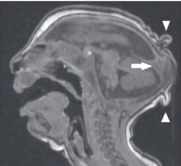
in microcephaly of other causes, is characterized by a collapsed aspect of the skull cap, with overlapping su-tures and prominent occipital bones, creating a “shelf” appearance, commonly accompanied by excessive, folded skin on the scalp (Figure 1). That presentation could be due, in part, to the continued growth of the skull and skin while the size of the brain regresses, or even, at some point, collapse of the skull, which previously had larger
dimensions due to cerebral ventriculomegaly(34–40). One
unusual inding that occurs in some newborns and sug-gests the latter hypothesis is herniation of the orbital fat to the skull, which could be secondary to the abrupt de-formation of the skull, rather than direct infection of the
eye, as occurs in other congenital infections(34).
Abnormalities of cortical development are frequent indings, occurring in 94–100% of cases, commonly pre-senting as agyria-pachygyria (Figure 2), and probably varying according to the stage of cortical development in
which the infection occurs(34–43). The involvement is
usu-ally diffuse, predominantly occurring in the frontal, insu-lar, and parietal lobes, with varying degrees of severity; it is frequently accompanied by wide sylvian and interhemi-spheric issures, as well as by an increase in the extra-axial cerebrospinal luid (CSF) space, the last being associated with a loss of brain volume and impaired CSF reabsorp-tion(34–43). Gray matter heterotopia is rare(38).
Calciications are common in congenital Zika syn-drome, occurring in 88–100% of patients; unlike classic infections (toxoplasmosis, rubella, cytomegalovirus, her-pes simplex, HIV, and syphilis), in which calciications are periventricular and cortical, ZIKV infection tends to
involve the cortical-subcortical junction (Figure 3); one possible explanation is that ZIKV infection has a vascular component, given that other processes that mainly affect
this region are associated with vascular alterations(34–43).
As illustrated in Figure 4, there are other sites that
pres-ent calciications in ZIKV infection(34–40), which are, in
descending order of frequency, the following: the basal nuclei and thalamus (in 29–65% of cases); the periven-tricular region (in 14–65%); the cortical region (in 14– 24%); and the infratentorial region (in 4–18%). It is note-worthy that periventricular and cortical involvement are more common in newborns in whom there is a signiicant loss of brain parenchymal volume; therefore, the precise location of calciications in such newborns is dificult to determine(34). According to Oliveira-Szejnfeld et al., in-fratentorial calciications are present in more severe man-ifestations, being accompanied by brainstem
malforma-tion, aqueduct stenosis, and secondary hydrocephalus(34).
There are inherent differences between CT and MRI, the former presenting greater sensitivity in the detection of calciications even when compared with susceptibility-weighted MRI sequences, whereas MRI presents a greater capacity for characterizing cortical abnormalities and the
development of the corpus callosum(34–40).
Enlargement of the lateral ventricles, as depicted in Figure 5, is common, occurring in 94–100% of cases; in Figure 1. A 3-month-old patient. Sagittal T1-weighted MRI slice, without con
-trast, showing craniofacial disproportion with a microcephalic aspect, together with occipital prominence and cutis verticis gyrata (arrowheads). Note also the conluence of the enlarged dural venous sinuses and the heterogeneous mate -rial (arrow).
most cases, it is moderate-to-pronounced and symmetric; it is accompanied by septations in 10–29% of cases, and occipital horns are commonly seen, sometimes making it dificult to distinguish periventricular cysts(34–43)
. It should also be borne in mind that the presence of ven-triculomegaly is directly related to a reduction in brain volume and can cause the head circumference to remain
normal or even increase(34–40,43).
Hypoplasia, dysgenesis, and agenesis of the corpus callosum are often seen in congenital Zika syndrome (Figure 6), reportedly occurring in 75–94% of cases and presenting a direct correlation with parenchymal
dam-age(34–43). Other associated abnormalities are
hippocam-pal malformation and thickening of the fornix(34–40).
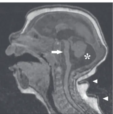
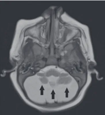
Brainstem abnormalities, which have been reported in 21–70% of patients with congenital ZIKV infection, are characterized by a tapered, atrophic encephalic trunk with preferential involvement of the pons (Figure 7), are com-monly associated with more severe conditions and can be related to synergism between the reduction in the number of descending ibers and the direct effects of the virus(34–43). Other abnormalities of the posterior fossa include cerebel-lar hypoplasia, which is usually diffuse and symmetric (Figure 8), occurring in 27–82% of cases, and an enlarged Figure 4. A: A 5-month-old patient.
Axial CT scan, without contrast, show -ing foci of calciication in the basal nuclei (arrows) and left thalamus (ar -rowhead). B: A 5-month-old patient. Axial CT scan, without contrast, show -ing calciications in the dorsolateral regions of the mesencephalic tegmen -tum (arrows), as well as at the cortical-subcortical junction in the temporal
lobes (arrowheads). A B
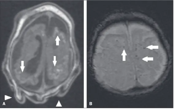
Figure 3. A: A 2-month-old patient. Ax -ial T1-weighted MRI slice, without con -trast, showing multiple punctate foci of hyperintensity at the cortical-sub -cortical junction of the frontal and pa -rietal lobes, indicative of calciications (arrows). Note also the cutis verticis gyrata (arrowheads). B: A 6-month-old patient. Axial susceptibility-weighted MRI sequence showing foci of hy -pointensity at the cortical-subcortical junction, affecting both hemispheres of the brain and more conspicuous in the frontal regions, because of the
Figure 5. A: A 7-month-old patient. Magnetic resonance imaging, T2, axial section showing diffuse enlargement of the lateral ventricles (asterisks) and simpliication of the gyral pattern (ar -rowheads). B: An 8-month-old patient. Axial T2-weighted MRI slice showing asymmetric dilation of the posterior portions of the lateral ventricles (as -terisks), constituting a colpocephaly coniguration.
A B
cisterna magna, the frequency of which increases in pro-portion to the severity of the infection, although there is no direct correlation between enlarged cisterna magna and
the occurrence of cerebellar hypoplasia(34–43).
Alteration of the white matter MRI signal secondary to a delay in myelination is seen in 88–100% of cases of congenital ZIKV infection. Conluence of the enlarged dural venous sinuses with heterogeneous material (Fig-ure 1), which can correspond to a thrombus or a hemato-crit effect due to dehydration with hemoconcentration, is seen in 28–53% of cases(34,35,38,41,42).
Currently, there are three severity spectra in
con-genital Zika syndrome(41): with microcephaly at birth,
Figure 6. A 3-month-old patient. Sagittal T1-weighted MRI slice, without con -trast, showing diffuse tapering of the corpus callosum (arrows). Note also the craniofacial disproportion with a microcephalic aspect.
presenting all of the abnormalities described in the lit-erature and with a symmetric appearance; with postnatal microcephaly, the presentation of which is comparable to that of microcephaly at birth, minus the calciications outside the cortical-subcortical junction and the agyria; and without microcephaly, which presents with calciica-tions restricted to the cortical-subcortical junction, areas of pachygyria, a delay in myelination, and slight ventricu-lar enventricu-largement, with an asymmetric appearance. In
ad-dition, as proposed by Aragão et al.(41), the presence of
polymicrogyria mainly in the frontal lobes is seen only in patients without microcephaly or with postnatal micro-cephaly.
Recently, spinal and nerve root changes have been described in congenital Zika syndrome, the severity of those changes presenting an apparent correlation with ar-throgryposis, deined as contracture of two or more joints
from birth(42). Visual inspection of T2-weighted MRI
se-quences in the sagittal and axial planes has revealed a signiicant reduction in the thickness of the entire spinal cord, accompanied by a signiicant reduction in the thick-ness of the anterior roots of the medullary cone, in pa-tients with congenital Zika syndrome and arthrogryposis. However, patients who do not present such joint changes have been found to show a reduction in spinal thickness only in the dorsal region and only discrete tapering of the
anterior roots of the medullary cone (Figure 9)(42). Clear
involvement of the descending anterior medullary tracts, with apparent preservation of ascending posterior tracts,
has also been seen, as reported by Mlakar et al.(25).
Aragão et al.(42) showed that arthrogryposis correlates
with greater severity of brain damage, with a greater num-ber of cerebral calciications and a greater chance of in-fratentorial calciications, as well as a greater chance of hypoplasia of the brainstem and cerebellum. In addition, all of the cases in which there was arthrogryposis have presented with pachygyria and an absence of polymicro-gyria, which could indicate that congenital Zika syndrome with arthrogryposis occurs in the earlier stages of fetal development, because pachygyria results from failure of
neuronal migration and of cortical organization between weeks 12 and 16 of gestation, whereas polymicrogyria
oc-curs around week 20(42).
CONCLUSION
Fetal infection with ZIKV causes severe CNS de-velopmental abnormalities. Although the neuroimaging indings are not pathognomonic, they can be suggestive of congenital Zika syndrome when the clinical and bio-chemical data are consistent with the diagnosis.
The main indings in congenital Zika syndrome are craniofacial disproportion with a microcephalic aspect, accompanied by calciications (predominantly at the cortical-subcortical junction), malformations of cortical development, ventriculomegaly, and abnormalities in the formation of the corpus callosum. However, attention should be paid to the spectrum of potential presentations of congenital Zika syndrome, and the possibility of ZIKV involvement should not be ruled out when microcepha-ly is not present or when the neuroimaging indings are more subtle.
REFERENCES
1. Yadav S, Rawal G, Baxi M. Zika virus: an emergence of a new arbo
-virus. J Clin Diagn Res. 2016;10:DM01–3.
2. Abushouk AI, Negida A, Ahmed H. An updated review of Zika virus. J Clin Virol. 2016;84:53–8.
3. Atif M, Azeem M, Sarwar MR, et al. Zika virus disease: a current
review of the literature. Infection. 2016;44:695–705.
4. Aziz H, Zia A, Anwer A, et al. Zika virus: global health challenge,
threat and current situation. J Med Virol. 2017;89:943–51.
Figure 8. A 2-month-old patient. Axial T2-weighted MRI slice showing diffuse, symmetric cerebellar hypoplasia (arrows), with prominence of the CSF spaces in the posterior fossa.
5. Blázquez AB, Saiz JC. Neurological manifestations of Zika virus in -fection. World J Virol. 2016;5:135–43.
6. Dick GW, Kitchen SF, Haddow AJ. Zika virus. I. Isolations and
se-rological speciicity. Trans R Soc Trop Med Hyg. 1952;46:509–20.
7. Pinheiro TJ, Guimarães LF, Silva MTT, et al. Neurological mani-festations of Chikungunya and Zika infections. Arq Neuropsiquiatr. 2016;74:937–43.
8. Younger DS. Epidemiology of Zika virus. Neurol Clin. 2016;34: 1049–56.
9. Niemeyer B, Niemeyer R, Borges R, et al. Acute disseminated en-cephalomyelitis following Zika virus infection. Eur Neurol. 2017; 77:45–6.
10. Hinckley AF, O’Leary DR, Hayes EB. Transmission of West Nile
virus through human breast milk seems to be rare. Pediatrics. 2007; 119:666–71.
11. Fontes CAP, Santos AASMD, Marchiori E. Magnetic resonance
imaging indings in Guillain-Barré syndrome caused by Zika virus
infection. Neuroradiology. 2016;58:837–8.
12. Fauci AS, Morens DM. Zika virus in the Americas—yet another ar
-bovirus threat. N Engl J Med. 2016;374:601–4.
13. Rasmussen SA, Jamieson DJ, Honein MA, et al. Zika virus and
birth defects—reviewing the evidence for causality. N Engl J Med.
2016;374:1981–7.
14. Duffy MR, Chen TH, Hancock WT, et al. Zika virus outbreak on Yap Island, Federated States of Micronesia. N Engl J Med. 2009;360;2536–43.
15. Webster WS. Teratogen update: congenital rubella. Teratology. 1998;58:13–23.
16. Lammer EJ, Sever LE, Oakley GP Jr. Teratogen update: valproic
acid. Teratology. 1987;35:465–73.
17. Shepard TH. “Proof” of human teratogenicity. Teratology. 1994;50: 97–8.
18. Oliveira WK, Cortez-Escalante J, Oliveira WTGH, et al. Increase in
reported prevalence of microcephaly in infants born to women
liv-ing in areas with conirmed Zika virus transmission durliv-ing the irst trimester of pregnancy – Brazil, 2015. MMWR Morb Mortal Wkly
Rep. 2016;65:242–7.
19. Cauchemez S, Besnard M, Bompard P, et al. Association between
Zika virus and microcephaly in French Polynesia, 2013-15: a retro-spective study. Lancet. 2016;387:2125–32.
20. Teixeira MG, Costa MCN, Oliveira WK, et al. The epidemic of Zika virus-related microcephaly in Brazil: detection, control, etiology,
and future scenarios. Am J Public Health. 2016;106:601–5. 21. Brasil P, Pereira JP Jr, Moreira ME, et al. Zika virus infection in
pregnant women in Rio de Janeiro. N Engl J Med. 2016;375:2321– 34.
22. Chan JF, Choi GK, Yip CC, et al. Zika fever and congenital Zika
syndrome: an unexpected emerging arboviral disease. J Infect.
2016;72:507–24.
23. Costa F, Sarno M, Khouri R, et al. Emergence of congenital Zika syndrome: viewpoint from the front lines. Ann Intern Med. 2016; 164:689–91.
24. Driggers RW, Ho CY, Korhonen EM, et al. Zika virus infection with prolonged maternal viremia and fetal brain abnormalities. N Engl J Med. 2016;374:2142–51.
25. Mlakar J, Korva M, Tul N, et al. Zika virus associated with micro-cephaly. N Engl J Med. 2016;374:951–8.
26. Bell TM, Field EJ, Narang HK. Zika virus infection of the central nervous system of mice. Arch Gesamte Virusforsch. 1971;35:183– 93.
27. Dick GW. Zika virus. II. Pathogenicity and physical properties. Trans R Soc Trop Med Hyg. 1952;46:521–34.
28. Miner JJ, Cao B, Govero J, et al. Zika virus infection during preg-nancy in mice causes placental damage and fetal demise. Cell. 2016;165:1081–91.
29. Noronha L, Zanluca C, Azevedo ML, et al. Zika virus damages the
human placental barrier and presents marked fetal neurotropism.
Mem Inst Oswaldo Cruz. 2016;111:287–93.
30. Klase ZA, Khakhina S, Schneider AB, et al. Zika fetal neuro-pathogenesis: etiology of a viral syndrome. PLoS Negl Trop Dis. 2016;10:e0004877.
31. Wang J, Ling Feng. Zika virus infection and microcephaly: evidence for a causal link. Int J Environ Res Public Health. 2016;13. pii 1031.
32. Hajra A, Bandyopadhyay D, Heise L, et al. Zika and pregnan-cy: a comprehensive review. Am J Reprod Immunol. 2017;77. doi:10.1111/aji.12607. Epub 2016 Nov 25.
33. Ticconi C, Pietropolli A, Rezza G. Zika virus infection and preg -nancy: what we do and do not know. Pathog Glob Health. 2016; 110:262–8.
34. Oliveira-Szejnfeld PS, Levine D, Melo ASO, et al. Congenital brain abnormalities and Zika virus: what the radiologist can expect to see
prenatally and postnatally. Radiology. 2016;281:203–18.
35. Aragão MFV, van der Linden V, Brainer-Lima AM, et al. Clinical
features and neuroimaging (CT and MRI) indings in presumed
Zika virus related congenital infection and microcephaly: retrospec-tive case series study. BMJ. 2016;353:i1901.
36. Hazin NA, Poretti A, Martelli CMT, et al. Computed tomographic indings in microcephaly associated with Zika virus. N Engl J Med.
2016;374:2193–5.
37. Mehrjardi MZ, Keshavarz E, Poretti A, et al. Neuroimaging indings
of Zika virus infection: a review article. Jpn J Radiol. 2016;34:765– 70.
38. Mehrjardi MZ, Poretti A, Huisman TAGM, et al. Neuroimaging
indings of congenital Zika virus infection: a pictorial essay. Jpn J
Radiol. 2017;35:89–94.
39. Moore CA, Staples JE, Dobyns WB, et al. Characterizing the pat
-tern of anomalies in congenital Zika syndrome for pediatric clinics. JAMA Pediatr. 2017;171:288–95.
40. Werner H, Sodré D, Hygino C, et al. First trimester intrauterine Zika virus infection and brain pathology: prenatal and postnatal
neuroimaging indings. Prenat Diagn. 2016;36:785–9.
41. Aragão MFVV, Holanda AC, Brainer-Lima AM, et al. Nonmicroce-phalic infants with congenital Zika syndrome suspected only after neuroimaging evaluation compared with those with microcephaly at birth and postnatally: how large is the Zika virus “iceberg”. AJNR Am J Neuroradiol. 2017;38:1427–34.
42. Aragão MFVV, Brainer-Lima AM, Holanda AC, et al. Spectrum of spinal cord, spinal root, and brain MRI abnormalities in congenital Zika syndrome with and without arthrogryposis. AJNR Am J Neuro-radiol. 2017;38:1045–53.
43. van der Linden V, Pessoa A, Dobyns W, et al. Description of 13
infants born during October 2015-January 2016 with congenital Zika virus infection without microcephaly at birth – Brazil. MMWR