André Gonçalves Carvalho Preto
Licenciatura em Bioquímica na Faculdade de Ciências
da Universidade de Lisboa
The Role of DsrC in Dissimilatory
Sulfite Reduction
Dissertação para obtenção do Grau de Mestre em
Bioquímica para a Saúde
Orientador:
Dra. Inês A. C. Pereira, ITQB
Co-orientador:
Dra. Sofia Venceslau, ITQB
Júri
Presidente: Dr. Pedro Matias, Investigador Principal do ITQB UNL
Arguente: Dr. João Bogalho Vicente, Investigador Auxiliar do ITQB UNL
Vogais: Dra. Margarida Archer, Investigadora Principal do ITQB UNL e Dr. Inês Cardoso Pereira, Investigadora Principal do ITQB UNL
Instituto de Tecnologia Química e Biológica António Xavier
Copyright
I
Copyright
O Instituto de Tecnologia Química e Biológica António Xavier e a Universidade Nova de Lisboa têm o
direito, perpétuo e sem limites geográficos, de arquivar e publicar esta dissertação através de exemplares
impressos reproduzidos em papel ou de forma digital, ou por qualquer outro meio conhecido ou que venha
a ser inventado, e de a divulgar através de repositórios científicos e de admitir a sua cópia e distribuição
com objetivos educacionais ou de investigação, não comerciais, desde que seja dado credito ao autor e
Acknowledgments
III
Acknowledgments
First of all I would like to take this opportunity to thank my supervisor Dra. Inês Cardoso Pereira for
accepting me in this project and letting me work and learn in the Bacterial Energy Metabolism Lab. Her
guidance, knowledge and motivation helped me a lot during this past year.
I also want to thank my co-supervisor Dr. Sofia Venceslau for teaching me all the experimental procedures
presented here and the patience, attention and time she spent with me. Furthermore the freedom she
offered me to plan my experiences gave me enthusiasm and confidence in my work.
I want to thank my colleagues Américo Duarte, Mónica Martins, Cláudia Mourato, Sónia Zacarias, André
Santos and Irene Racagni who provided such a good environment at the work place and gave me so many
good advices.
As it should be I have to thank all my friends that accompanied me through my learning process since
school days till college. They are the ones that helped through the whole process and made this such a
wonderful and enjoyable journey. I also have to acknowledge my colleges and friends from my master’s,
especially Francisca Arez, Valeria Girlus and Alina Kulakova for all those hilarious coffee breaks that turned
ITQB such a good place to work in.
Finally I thank the most important people in my life, my parents and brother, for all their support, love and
care. Their sacrifices both financially and emotionally throughout the years is what made this all possible
Abstract
V
Abstract
Studies involving the human gastrointestinal microbiota and its metabolites have revealed the presence of
sulfate-reducing bacteria (SRB) within the gut. These microorganisms have been implicated in inflammatory
bowel diseases due to the toxic effects of sulfide, produced during dissimilatory sulfate reduction, leading
to cell inflammation. The reduction of sulfite to sulfide is carried out by the dissimilatory sulfite reductase,
DsrAB, and also involves the DsrC protein, which is a major protein in the cell and contains two conserved
redox-active cysteines in a flexible C-terminal arm. The disulfide bond formed between these two conserved
cysteines during sulfite reduction is believed to be reduced by several proteins that are related to the
catalytic subunits of the heterodisulfide reductases (Hdr) of methanogens, namely HdrB and HdrD.
This work aimed to study the impact of DsrC variant strains from Desulfovibrio vulgaris Hildenborough in
cell growth while providing ethanol or pyruvate as an electron donor and search for new physiological
partners of DsrC during dissimilatory sulfate reduction in lactate-sulfate or ethanol-sulfate conditions. This
work shows that a single point mutation in one of the strictly conserved cysteines (Cys93) of DsrC results
in a severe decrease in cell growth indicating that DsrC, and in particular Cys93, is of major importance in
the energy metabolism of these organisms. Pull down assays of DsrC coupled to mass spectrometry data
and Western blot analysis showed that under ethanol-sulfate conditions DsrC interacts with the
FloxABCD/HdrABC complex, ferredoxin and alcohol dehydrogenase, suggesting that these proteins are
organized in a supramolecular structure. This result is in agreement with DsrC variant strains being affected
under ethanol-sulfate condition and it experimentally validates a previous proposal that implicated DsrC in
the ethanol oxidation pathway via the Flox/Hdr complex. Here, through DsrC, it is shown a link between the
carbon metabolism and the sulfate reduction metabolism that has never been identified before.
Resumo
VII
Resumo
As doenças inflamatórias intestinais (DII) são doenças crónicas que provocam a inflamação do tecido
intestinal. Vários estudos realizados à flora intestinal em pacientes com DII revelaram o envolvimento de
bactérias redutoras de sulfato (BRS) no desenvolvimento destas doenças devido aos efeitos tóxicos
provocados pelo produto final do seu metabolismo, o sulfureto, que é produzido durante a redução
dissimilativa de sulfato. A redução de sulfito a sufureto, nestes microorganismos é levada a cabo pela
redutase dissimilativa de sulfito, DsrAB, e envolve uma outra proteína designada de DsrC, que possui um
papel crucial dentro da célula. Esta proteína possui duas cisteínas altamente conservadas no braço fléxivel
do C-terminal que durante a produção de sulfureto forma uma ligação disulfureto entre as cisteínas. Nesta
forma, a DsrC encontra-se no estado oxidado e acredita-se que para voltar à sua forma reduzida, várias
proteínas relacionadas com as subunidades catalíticas das reductases de heterodisulfureto de organismos
metanogénicos (HdrB e HdrD) actuam como possivéis parceiros fisiológicos.
Este trabalho focou-se no estudo do impacto da DsrC no crescimento de Desulfovibrio vulgaris
Hildenborough durante a redução dissimilativa de sulfato ou em condições fermentativas, fornecendo
respectivamente etanol ou piruvato como dadores de electrões. Outro foco foi encontrar os possíveis
parceiros fisiológicos da DsrC em células crescidas em meio lactato-sulfato e etanol-sulfato.
Este trabalho mostra que a mutação provocada numas das cisteínas altamente conservadas (Cys93)
resulta num decréscimo acentuado no crescimento das células, o que indica que a DsrC, e em particular
a Cys93, desempenha um papel crucial no metabolismo energético da célula. Os ensaios de pull down da
DsrC acoplados à análise por espectrometria de massa e Western blot mostraram que a DsrC interage
com o complexo FloxABCD/HdrABC e com as proteínas ferredoxina e alcoól desidrogenase na condição
etanol-sulfato. Este resultado valida uma proposta já efectuada anteriormente onde a DsrC tinha sido
implicada na oxidação de etanol via complexo Flox-Hdr.
Table of contents
IX
Table of Contents
Abbreviations List XI Figures and Tables List XIII
1. Introduction 1
1.1. Sulfate-Reducing Bacteria in the Human Gut 3
1.1.1. Gut Microbiota 3
1.1.2. Inflammatory Bowel Diseases 4
1.1.3. SRB Pathogenic Role in IBD 5
1.2. Sulfate-Reducing Bacteria(SRB) 7
1.2.1. Sulfur Transformations 8
1.2.2. Distribution and Diversity 9
1.2.3. Physiology and Metabolism 9
1.2.4. Desulfovibrio vulgaris Hildenborough 11
1.3. The Dissimilatory Sulfate Reduction Pathway 11
1.3.1. The DsrC Protein 13
2. Objectives 19
3. Materials and Methods 23
3.1. Growth Studies of DsrC Variant Strains 25
3.1.1.Strains 25
3.1.2. Media 26
3.1.3. Growth curves 26
3.1.4. Ethanol Quantification 27
3.2. DsrC Physiological Partners Studies 28
3.2.1. Media and Cell Growth 28
3.2.2. DsrC Pull Down Assays 28
Table of contents
X
3.2.4. Electrophoretic Techniques 29
3.2.5. Mass Spectrometry Protein Identification 30
3.2.6. Western Blot Analysis 30
4. Results 33
4.1. Growth Curves of DsrC Variant Strains 35
4.2. DsrC Physiological Partners 38
4.2.1. Mass Spectrometry 40
4.2.2. Western Blot Analysis 41
5. Discussion 45
References 53
Abbreviations List
XI
Abbreviations List
Abs – Absorbance
Adh – Alcohol dehydrogenase AL-DH – Aldehyde dehydrogenase AMP – Adenosine monophosphate AprBA – APS reductase
APC – Antigen presenting cells APS – Adenosine 5’-phosphosulfate ATP – Adenosine triphosphate
BCIP – 5-bromo-4-chloro-3-indolyl phosphate CD – Crohn’s disease
CoM-S-S-CoB – Heterodisulfide of CoM-SH and CoB-SH Dsr – Dissimilatory sulfite reductase
DvH – Desulfovibrio vulgaris Hildenborough E0’ – Standard electron potential
FAD – Flavin adenine dinucleotide Fdx – Ferredoxin
Flox – Flavin oxidoreductase Hdr – Heterodisulfide reductase IDB – Inflammatory Bowel Diseases IFN-γ– Interferon- γ
IgA – Immunoglobulin A IL – Interleukin
IMAC – Immobilized ion metal affinity chromatography Km – Kanamycin
Ldh – Lactate dehydrogenase
MOY – MO basal medium with yeast extract MQ – Menaquinone
MQH2– Menaquinol
MvhDGA – Methyl viologen-reducing hydrogenase NAD+– Nicotinamide adenine dinucleotide
NADH – Nicotinamide adenine dinucleotide reduced form NAD(P)H –Nicotinamide adenine dinucleotide phosphate NBT – Nitro-blue tetrazolium chloride
Figures and Tables List
XII
Pi – Inorganic phosphate PPi – Inorganic pyrophosphate PVDF – Polyvinylidene difluoride rRNA – ribosomal RNA
Sat – ATP sulfurylase or Sulfate adenylyltranferase SCFA – Short chain fatty acids
SDS-PAGE – Sodium dodecyl sulfate-polyacrylamide gel electrophoresis SRB – Sulfate-reducing bacteria
SRO – Sulfate-reducing organisms
Qmo – Quinone-interacting membrane-bound oxidoreductase TBS – Tris-buffered saline
TGF-β– Transforming growing factor-β Th – Helper-T-cells
TLR – Toll-like receptors TNF-α – Tumor necrosis factor-α
Tris – Tris(hydroxymethyl)aminomethane Treg – Regulatory T cells
Figures and Tables List
XIII
Figures and Tables List
Figures
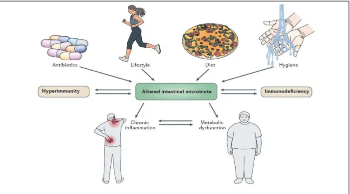
Figure 1.1– Factors responsible for changing the gut microbiota composition and the effects of dysbiosis
on hosts’ health.
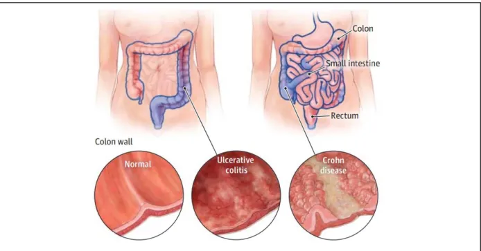
Figure 1.2 – Localization and extension of chronic inflammation induced by ulcerative colitis and Crohn’s disease.
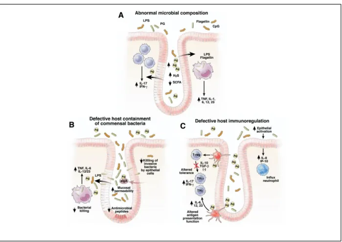
Figure 1.3 – Potential mechanism by which SRB induce chronic inflammation in IBD.
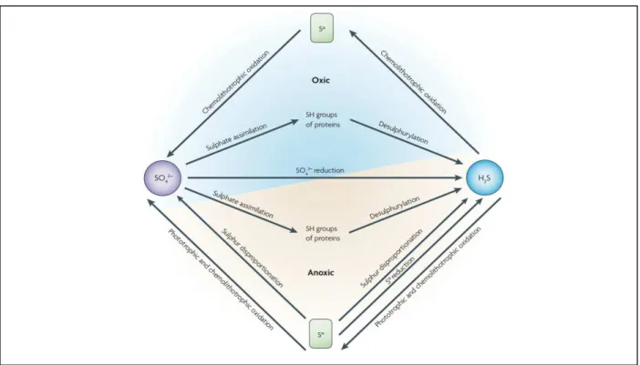
Figure 1.4 –Sulfur transformations.
Figure 1.5 – Microbial degradation of organic matter in anoxic environments in the presence of sulfate reducers.
Figure 1.6 –Schematic representation of the proposed sulfate reduction mechanism.
Figure 1.7 –Three dimensional structure of DsrC.
Figure 1.8 –Structure of DsrAB in complex with DsrC.
Figure 1.9 –Proposed mechanism of sulfite reduction by DsrAB and DsrC.
Figure 1.10 –Proposed model and possible physiological partners of DsrC in sulfite reduction of SRB.
Figure 3.1 –Schematic representation of the different DvH strains used in this work.
Figure 3.2 –Schematic representation of the experimental procedure used to monitor all growth curves.
Figure 3.3 –Schematic representation of the experimental procedure used to grow WT and IPFG07 DvH cells for pull down assays.
Figure 4.1 –Growth curves of DvH WT and DsrC variant strains.
Figure 4.2 – Ethanol (mM) accumulated during growth of DvH WT and DsrC variant strains during pyruvate fermentation.
Figure 4.3 – Tricine-SDS-PAGE 10 % acrylamide protein separation of the fractions obtained from the DsrC pull down assays.
Figure 4.4 – Western Blot analysis of protein fractions obtained from DsrC pull down assays of the soluble fraction of cells grown in ES4 medium.
Figure 4.5 –Relative band intensity of the proteins bands analyzed by Western blot.
Figure 5.1 – Mechanism for the function of the HdrABC-FloxABCD complex.
Figure 5.2 – DsrC as a link between dissimilatory sulfite reduction and ethanol oxidation pathways.
Tables
Table 3.1 –List of conditions used for Western blot analysis.
Table 4.1 –Specific growth rate (μg), doubling time (Td), and maximal OD (600 nm) for DvH WT and DsrC variant strains in different conditions.
Figures and Tables List
XIV
Introduction
3
1. Introduction
1.1. Sulfate-Reducing Bacteria in the Human Gut
1.1.1 Gut Microbiota
An ecological community of commensal, symbiotic and pathogenic microorganisms, inhabits the body
surfaces of all vertebrates, including man. In the gut these microorganisms are very condensed and are
involved in various roles such as extracting nutrients and energy from food, immune defense and in
extending the metabolic repertoire of the host (1, 2, 3).
The diversity and composition of the body microbiota depends on topographical and temporal variations
since the microbial community varies within each body part and are influenced by particular growth or
mat-uration phases of the host. In humans, the microbiota composition depends not only on those but also on
other factors such as diet, life-style, hygiene, antibiotic use and host’s genetic disposition (Figure 1.1) (1, 4). Thus, the composition of the gut microbiota varies between individuals. All adult humans have a core
microbiota which is constituted by bacteria that belong to just a few phyla. In adults, Bacteroidetes and
Firmicutes usually dominate, whereas Actinobacteria, Proteobacteria, and Verrucomicrobia are frequent
but generally minor constituents (2).
The colonization of the human gut begins after birth and by adulthood the gut presents about 1014 bacterial
cells which is ten times the number of cells in the human body (4). The combined genomes of these
microorganisms, called microbiome, provide a large variety of biochemical and metabolic activities that
complement the host physiology. For example, they help the metabolism of otherwise indigestible
polysaccharides and produce essential vitamins. They are also required for the development and
differentiation of the human gut and confer protection against many pathogens having a crucial role in
immune defense (4). Through this symbiotic relationship, the microbiota helps the development of the
immune system and the latter shapes the microbe fauna in the gut. Those microbes influence the immune
system through many signaling pathways that involve various classes of molecules and the resulting
immune-mediated signaling processes act not only in the gut but in other organs like the brain, liver and
muscle (3). The disruption of the homeostasis between the gut microbiota and the host (dysbiosis) can
cause chronic inflammation and metabolic dysfunction, which ultimately can be associated with health
Introduction
4
Figure 1.1 – Factors responsible for changing the gut microbiota composition and the effects of dysbiosis on hosts’ health. The composition of the microbe community on the host’s gut can be shaped by many factors like lifestyle, diet, hygiene or frequent use of antibiotics. In addition, the host’s genetic disposition like hyperimmunity, characterized by the overexpression of pro-inflammatory mediators, or immunodeficiency, owing to mutations in regulatory immune proteins can influence that community. In turn, dysbiosis affects the levels of immune mediators and induces both
chronic inflammation and metabolic dysfunction. Taken from (4).
1.1.2. Inflammatory Bowel Diseases
Inflammatory bowel diseases (IBD) are chronic inflammatory disorders that occur within the gastrointestinal
tract and arise from the disruption of the immune tolerance to the gut microbiota especially in genetically
predisposed hosts. The disruption is caused by a decrease in the prevalence of the major members of the
human commensal microbiota (e.g. Clostridium, Bacteroides and Bifidobacteria) and an increase in
detrimental bacteria like sulfate-reducing bacteria (SRB) (5). The detected dysbiosis is associated with the
reduction of the levels of mucosal defensins, Immunoglobulin A (IgA) and malfunction of phagocytosis
which weakens innate immunity and bacterial killing. Additionally, there is an overaggressive adaptive
immune response to the altered gut microbiota due to increased cell reaction, dysfunctional regulatory
T-cells (Treg) and antigen presenting T-cells (APC) (5).
Ulcerative colitis (UC) and Crohn’s disease (CD) are considered two of the main disorders among IBD. However, they have distinct pathogenesis, inflammatory profiles and symptomatology. Both involve severe
diarrhea, pain, fatigue and weight loss but inflammation linked to CD can extend deeply into submucosal
regions and occurs anywhere along the gut whereas inflammation related to UC affects only the superficial
Introduction
5
Figure 1.2 – Localization and extension of chronic inflammation induced by ulcerative colitis and Crohn’s disease. Ulcerative colitis inflammation usually begins in the rectum and may extend continuously to involve the entire colon but only affects the inner layer of the bowel wall. Crohn’s disease may affect any part of the gastrointestinal tract and the inflammation affects all layers of the bowel wall. Taken from (7).
The lymphocytes recruited and cytokines produced in those two diseases are also different. CD is
associ-ated with type 1 helper-T-cell (Th1) and type 17 helper-T-cell (Th17) overaggressive immune responses
that increase production of interleukin (IL)-12, IL-23, IL-27, interferon-γ (IFN- γ) and tumor necrosis
factor-α (TNF- α). UC seems to be associated with type 2 helper-T cell (Th2) immune responses leading to higher levels of IL-5 and transforming growth factor-β (TGF-β) (8).
1.1.3. SRB Pathogenic Role in IBD
Although not being part of the dominant members of the human gut, SRB seem to be always present in the
intestinal mucosa (9, 10, 11) even though in fecal samples they are only detected in approximately 50% of
healthy individuals (12). These bacteria can oxidize a wide variety of organic compounds within the large
intestine like ethanol, succinate, pyruvate and lactate and are also able to use other electron donors to
reduce sulfate to hydrogen sulfide (H2S) such as volatile fatty acids (acetate, butyrate, propionate), amino
acids (glutamate, alanine, serine) and indolic and phenolic compounds. Furthermore, most SRB are
capa-ble of metabolizing hydrogen resulting from bacterial fermentation (12, 13).
Distinct species of SRB belonging to the genera Desulfovibrio, Desulfobacter, Desulfobulbus and
Desulfotomaculum have been identified in the human gut but the most predominant is the Desulfovibrio
Introduction
6
directly through the diet or from co-colonizing bacteria. For instance, D. piger uses sulfate directly from
ingested food or indirectly from Bacteroidetes via sulfatases that cleave sulfated glycoproteins or even from
mucopolysaccharides (long chain sugar molecules usually found in mucous surfaces of the human body)
and can use hydrogen derived from fermentation (14).
Numerous studies have implicated SRB in the development of inflammatory bowel diseases especially UC
and CD (5, 9, 12, 15). The hydrogen sulfide produced by these bacteria may have a pro-inflammatory action
that leads genetically-susceptible individuals to develop IBD (5, 6, 11, 16). Its potential mechanism of
path-ogenesis is resumed in Figure 1.3.
Figure 1.3 – Potential mechanism by which SRB induce chronic inflammation in IBD. (A) Intestinal dysbiosis in inflammatory bowel diseases is caused by decrease of putative beneficial bacteria and increase in detrimental bacteria
like SRB. This imbalance causes increased levels of H2S due to the consumption of short chain fatty acids (SCFA) like
butyrate thus contributing to the down regulation of epithelial tight junction’s proteins and up regulation of pore forming
proteins that enhances mucosal permeability (B) Defective secretion of defensins or secretory IgA (also known as IgA)
leads to mucosal bacterial overgrowth and increased epithelia permeability to antigens. Defective killing of phagocy-tosed bacteria raises intracellular bacteria concentration and consequentially increases the stimulation of bacterial toll
like receptors (TLR) ligands and antigens that activate pathogenic innate and T-cell immune responses. (C) The
Introduction
7
The increased growth of SRB and decreased growth of Clostridium in patients with IBD can explain thereduced intraluminal levels of SFCA like butyrate. Similarly, the overgrowth of SRB can enhance the
pro-duction of hydrogen sulfide blocking the use of butyrate by colonocytes (17). This contributes to the down
regulation of epithelial tight junction proteins and to the upregulation of pore forming proteins thus increasing
the epithelial permeability to bacteria and also a large variety of antigens (Figure 1.3 A) (18, 19). This
epithelial barrier dysfunction is worsened by impaired defensin and IgA production. Defensins are
antimi-crobial peptides that are used to assist in killing of phagocytosed bacteria and IgA being the most secreted
antibody within mucosal surfaces is responsible to entrap antigens and to down regulate epitope expression
on bacterial cell surfaces and, therefore, regulate microbial intestinal colonization. Furthermore, IgA
pre-vents attachment of pathogens, evasion of epithelial cells and removes bacteria breaching the epithelial
barrier by translocating them back to the lumen and by promoting their clearance by dendritic cells,
neutro-phils and phagocytes (Figure 1.3 B) (20, 21). Killing of invasive bacteria that reach the lamina propria, a
thin mucous layer that lies beneath gut epithelium, through the “leaky” epithelium is reduced by defective phagocytosis by macrophages. This ineffective clearance leads to the overwhelming exposure of bacterial
TLR ligands and antigens that activate pathogenic innate and T-cell immune responses responsible for
increasing the secretion of pro-inflammatory cytokines like TNF- α, IL-6, IL-12 and IL-23 (Figure 1.3 A and 1.3 B). The disrupted mechanism of tolerance in epithelial cells and APC amplifies innate immunity by
increasing the recruitment of neutrophils. Additionally, defective Treg and APC causes activation of the
nuclear factor κB (NF-κB) signaling pathway, which is usually suppressed by TGF-β and IL-10 in healthy hosts, producing overaggressive T-cell responses (Th1 and Th17) and thus intensifying the production
of
chemokines and pro-inflammatory cytokines such as IFN- γ and IL-17 (Figure 1.3 C) (5).1.2. Sulfate-Reducing Bacteria (SRB)
SRB are anaerobic microorganisms that use sulfate as final electron acceptor for the degradation of organic
compounds, producing sulfide in the process. It has been estimated that microbial sulfate reduction is
responsible for the oxidation of 12-29% of the organic carbon flux to the sea floor, which indicates their critical role in both the sulfur and carbon cycle (22).
The study of the physiology and metabolism of SRB becomes very important not only for their implications
in health but also in the environment. SRB intervene in biocorrosion of ferrous metals, as well as corrosion
of concrete and stonework (23). Also, sulfide constitutes a serious problem to the petroleum industry since
sulfide produced from their metabolism causes a problem called “oil souring” that affects oil fields and
pipelines (23, 24, 25). The study of SRB becomes also essential for their biotechnological applications since
they are able to immobilize hazardous and toxic metals released by metallurgic and nuclear plants or oil
Introduction
8
1.2.1. Sulfur Transformations
Sulfur is amongst the most abundant elements on Earth. The largest reservoirs of sulfur are iron sulfides
(pyrite; FeS2) and gypsum (CaSO4) in rock and sediments, and sulfate (SO42-) in seawater (~28 mM). The
sulfur cycle is very complex since sulfur can have a wide range of oxidation states and can either be
trans-formed chemically or biologically. The most relevant oxidation states in nature are sulfate (SO42-; oxidation
state +6), elemental sulfur (S0; oxidation state0), and sulfide (S2-; oxidation state -2) (27).
Sulfur is taken as a nutrient by microorganisms, plants and animals that is then assimilated and
incorpo-rated in proteins and several other biological molecules, such as enzymes and vitamins. Decomposition of
dead organisms in the absence of oxygen causes the release of hydrogen sulfide by desulphurylation of
those molecules. Microorganisms play a very important role in all sulfur transformations (Figure 1.4).
Figure 1.4 – Sulfur transformations. Sulfate (SO42-) can either be reduced by means of assimilation to incorporate
sulfur in amino acids or can be reduced by SRB through the dissimilatory sulfate reduction pathway. The sulfide (H2S)
produced in the process can be oxidized again to sulfate by chemolithotrophic and phototrophic microorganisms. Other transformations are carried out by specialized bacteria, such as elemental sulfur reduction and sulfur disproportionation. Taken from (28).
As mentioned before SRB use sulfate as a final electron acceptor to produce sulfide, which then can be
oxidized to elemental sulfur and sulfate by chemolithotrophic bacteria under oxic conditions or by
chemolithotrophic and phototrophic organisms under anoxic conditions. Other sulfur transformation may
Introduction
9
Desulfovibrio sulfodismutans), which is a process that simultaneously produces sulfide and sulfate fromelemental sulfur (28).
1.2.2. Distribution and Diversity
SRB are ubiquitous within anoxic environments such as marine sediments, fresh waters, soil and mucous
surfaces of animals. They have successfully adapted to almost all ecosystems of the planet since there is
evidence of their existence in high temperature and pressure environments and in a wide range of pH
values (29). SRB have been identified in extremely low pH environments like acid-mine drainage sites
where the pH can be as low as 2 and have been also detected in very high pH environments such as soda
lakes where pH can be 10, establishing their survival capability under harsh conditions (28). Most SRB are
free living, but some live in community with other microorganisms in a symbiotic way providing each other
essential nutrients for their growth, such as with methanotrophic archaea and sulfate-oxidizing
Gammap-roteobacteria (30, 31).
More than 220 species of 60 genera of sulfate-reducing organisms (SRO) have been described until now
(23). Based on comparative analysis of 16S ribosomal RNA (rRNA) sequences, SRO can be distributed
into seven different phylogenetic lineages, five classes within the Bacteria and two classes within Archaea.
Among the bacteria domain most SRB belong to the Deltaproteobacteria class, followed by Clostridia,
Ni-trospirae, Themodesulfobacteria and Thermodesulfobiaceae classes. Within Archaea SRO belong only to
just a few genus of the Euryarchaeota and Crenarchaeota classes (23, 28). Almost 100 SRO already have
their complete genome sequenced, e.g., Desulfotalea psychrophila, Archaeoglobus fulgidus, Desulfovibrio
vulgaris Hildenborough (DvH) and Desulfovibrio desulfuricans G20. The genomes of SRO belonging to the
Archaea domain are usually much smaller compared to those of Bacteria. When the Desulfotalea
psy-chrophila (Bacteria) and Archaeoglobus fulgidus (Archaea) genomes were compared small similarities were
found, and these were mainly between genes for proteins involved in the dissimilatory sulfate reduction,
indicating that only a small fraction of genes are necessary for sulfate reduction (32).
1.2.3. Physiology and Metabolism
Sulfate reducers are able to perform dissimilatory sulfate reduction which is an energy conservation process
associated with the reduction of large amounts of sulfate necessary for their growth (23, 28, 33) and most
are able to incorporate sulfide into amino acids through dissimilatory sulfite reduction (34).
SRB can use an extensive variety of electron donors and some electron acceptors, and so are metabolically
versatile and can inhabit a large range of different environments. Most SRB are chemoheterotrophic
organisms since they need organic carbon for cell growth and generate energy through chemical reactions.
Introduction
10
carbon dioxide (CO2) and those who incompletely oxidize organic compounds to acetate (Figure 1.5) (28).
Regarding their electron donor metabolism, SRB can use many substrates for their growth such as
monocarboxylic and dicarboxylic acids (23), sugars (35), amino acids (36), SCFA (17) and aromatic
compounds (37).
Many organic macromolecules like proteins, polysaccharides and lipids cannot be directly used by SRB so
they depend on other microorganisms to degrade these polymeric substrates to products that are
substrates for SRB (28).
Figure 1.5 – Microbial degradation of organic matter in anoxic environments in the presence of sulfate reducers. Macromolecules like proteins, polysaccharides and lipids are hydrolyzed by hydrolytic bacteria to produce amino acids, sugars, SCFA, etc. These monomers are then fermented by fermentative bacteria into lactate, butyrate, propionate, hydrogen, acetate and other fermentative products which serve as substrates for SRB growth. Taken from (28).
Although the main electron acceptor of SRB is sulfate, other sulfur compounds such as sulfite (SO32-),
thiosulfate (S2O32-), elemental sulfur (S0), and sulfonates can support growth of some species. Furthermore,
a few species of SRB can use alternative electron acceptors like iron (FeIII), uranyl (UVI), selenite (SeVI),
Introduction
11
1.2.4.
Desulfovibrio vulgaris
Hildenborough
According to the Integrated Microbial Genomes website there are over 60 known species of the
Desulfovib-rio genus, from which 43 genomes are available. Within the Proteobacteria the DesulfovibDesulfovib-rio genus belongs
to the Deltaproteobacteria class, Desulfovibrionales order and Desulfovibrionaceae family.
The most extensive biochemical and physiological studies have been done with members of the genus
Desulfovibrio, which are the most easily and rapidly cultured sulfate reducers. The Desulfovibrio vulgaris
Hildenborough (DvH) strain was the first SRB to have its genome sequenced (38, 39). These cells have a
single polar flagellum and stain Gram negative meaning they have a cytoplasmic cell membrane (inner
membrane), a thin peptidoglycan layer and an outer cell membrane with lipopolysaccharides in its outer
leaflet and phospholipids in the inner leaflet. DvH uses hydrogen, alcohols or organic acids as electron
donors for sulfate reduction. It uses lactate preferentially as a carbon source although other compounds
can be used such as pyruvate, formate and certain primary alcohols like ethanol. The optimal growth
tem-perature ranges from 25 to 40 ºC while the optimal pH goes from 6.6 to 7.5 (38). There is also evidence
that these microorganisms are not strict anaerobes, as it was previously considered because they tolerate
low levels of oxygen even though it limits growth (40).
1.3. The Dissimilatory Sulfate Reduction Pathway
The dissimilatory sulfate reduction is a process carried out by SRB that involves the eight-electron reduction
of sulfate (SO42-) to hydrogen sulfide (H2S). This process occurs in the cytoplasm and requires the presence
of sulfate transporters and three soluble enzymes - adenosine triphosphate (ATP) sulfurylase (also known
as sulfate adenylyltranferase), adenosine 5’-phosphosulfate (APS) reductase and dissimilatory sulfite re-ductase (DsrAB/DsrC) (41). The dissimilatory reduction pathway is outlined in Figure 1.6.
First, SO42- is transported inside the cell by symport with sodium (Na+) or protons (H+) (42). Due to the
stability of SO42- and its low redox potential [E0’ (SO42- / SO32-) = - 526 mV], when it enters the cytoplasm,
SO42- has first to be activated by reaction with ATP to form APS, a reaction that is catalyzed by ATP
sulfu-rylase (Sat) (Eq. 1). This endergonic reaction is driven by the hydrolysis of inorganic pyrophosphate (PPi)
by a pyrophosphatase forming inorganic phosphate (Pi) (Eq. 2) (33, 42). Most of the times this reaction is
carried out by soluble pyrophosphatases but in some organisms there are membrane-associated
pyrophos-phatases that allow proton translocation thus allowing energy conservation from hydrolysis of inorganic
pyrophosphate (41).
SO42- + ATP + 2H+ APS + PPi ΔGo’ = + 46 kJ/mol (1)
Introduction
12
Therefore, to turn SO42- into APS the cell needs to spend two ATP equivalents molecules, ATP and PPi. In
the following step, the reduction of APS to sulfite (SO32-) [Eo’ (APS/SO32-) = - 60 mV] is performed by APS
reductase (AprBA) and involves 2 electrons (Eq. 3) (33, 42).
APS + 2e- + 2H+ HSO3- + AMP ΔGo’ = - 69 kJ/mol (3)
The physiological electron donor for AprBA is believed to be the quinone-interacting membrane bound
oxidoreductase complex (QmoABC) (43). This Qmo complex is essential for SO42-, but not SO32- reduction
as it was confirmed in a study involving a mutant of DvH lacking the qmoABC genes. This mutant is
inca-pable of growing on sulfate but can grow on sulfite or thiosulfate as electron donor (44). Finally, SO32- is
reduced by six electrons to H2S [Eo’ (SO32-/H2S) = - 116 mV] (Eq. 4) via the dissimilatory sulfite reductase,
DsrAB (33, 42) with the involvement of the small protein DsrC and the membrane complex DsrMKJOP (33).
HSO3- + 6e- + 6H+ HS- + 3H2O ΔGo’ = - 172 kJ/mol (4)
Figure 1.6 – Schematic representation of the proposed sulfate reduction mechanism. After entering the cell
sulfate(SO42-)is activated to APS by Sat. In the second step, APS receives 2 electron from the Qmo complex in a
reaction catalyzed by AprBA. Finally, sulfite (SO32-) formed in the last reaction is reduced by 6 electrons to form sulfide
Introduction
13
1.3.1. The DsrC protein
In SRB the dissimilatory sulfite reductase, DsrAB, is an essential enzyme that catalyzes the reduction of
sulfite to sulfide. This reaction also involves a small protein of 12-14kDa named DsrC, which comprises a
highly conserved C-terminal region containing two strictly conserved cysteine residues. One of these Cys
is the penultimate residue of the C-terminus (C104 in D. vulgaris which was named as CysA) and the other
one is located 10 residues upstream (C93 in D. vulgaris which was named as CysB) (51). DsrC was first
reported as being a subunit of DsrAB in a α2β2γ2 composition. However, the dsrC gene is not in the same
transcriptional unit as the dsrAB genes (46) and recently it was reported that the majority of DsrC in cell
extracts of D. vulgaris was not associated with DsrAB, suggesting that DsrC is an interacting partner of
DsrAB, and possibly of other proteins (47). DsrC is suggested to have an important role in cellular
metabolism since dsrC is a highly expressed gene in D. vulgaris at the same or higher levels when
compared to other proteins involved in sulfate reduction (48, 49, 50). Several 3D structures of DsrC have
been determined (including D. vulgaris) and all present a globular shape with the exception of the C-terminal
arm which adopts an extended and disordered configuration in solution but a retracted configuration in the
crystal structure (45). The globular part of DsrC presents a helix-turn-helix (HTH) structural motif which is
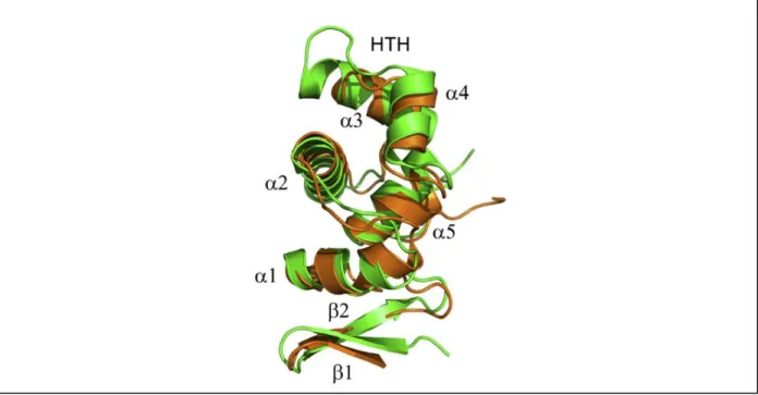
be responsible for protein binding to DNA and protein-protein interactions (Figure 1.7) (51).
Figure 1.7 – Three dimensional structure of DsrC. Overlapping representation of DsrC from the sulfur oxidizer
Allochromatium vinosum (green) and D. vulgaris (brown). DsrC presents a globular shape composed by five α–helixes,
Introduction
14
The reaction mechanism of DsrAB has been studied for many years because, in contrast to the assimilatory
sulfate reduction where sulfite is directly reduced to sulfide, in dissimilatory sulfite reduction DsrAB
produces in vitro a combination of products, mainly trithionate (S3O62-) and thiosulfate (S2O32-) (52). This
led to believe that the reduction of sulfite to sulfide was dependent on the formation of trithionate and
thiosulfate as intermediates. However, this suggestion was also disputed since these products of DsrAB
are highly dependent on the reaction conditions which don’t correspond to the ones presented in vivo (53). A major step to understand this mechanism was achieved when the crystal structure of this enzyme was
determined. It was shown that only one catalytic siroheme-[4Fe-4S] cofactor is present per αβ unit, bound to DsrB, whereas the equivalent cofactor bound to DsrA, sirohydrochlorin (siroheme without iron) seems to
have a structural role (45). Additionally through the crystal structure of DsrAB of D. vulgaris it was revealed
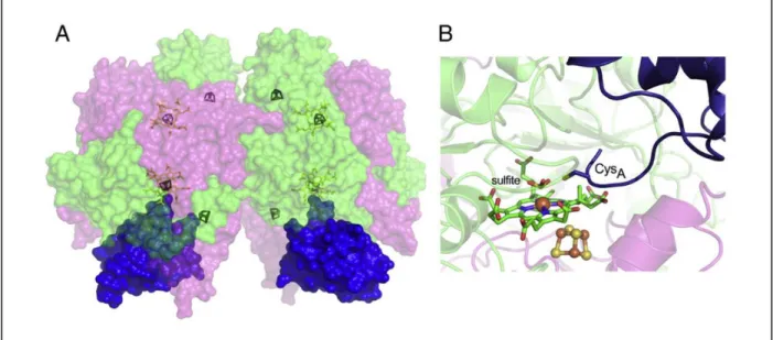
that this enzyme is present in a α2β2γ2 composition with DsrC (Figure 1.8 A). It was also shown that the
C-terminal of DsrC projects inside DsrAB bringing the CysA residue near the catalytic site where the sulfite
molecule is located (Figure 1.8 B) (45).
Figure 1.8 – Structure of DsrAB in complex with DsrC. (A) Molecular surface representation of DsrAB in a α2β2γ2
composition with DsrC. (B) Detailed image of the Desulfovibrio gigas catalytic site where it can be seen the C-terminal
arm reaching for the siroheme-[4Fe-4S] active site. DrsA – green, DsrB – pink, DsrC – blue. Taken from (51).
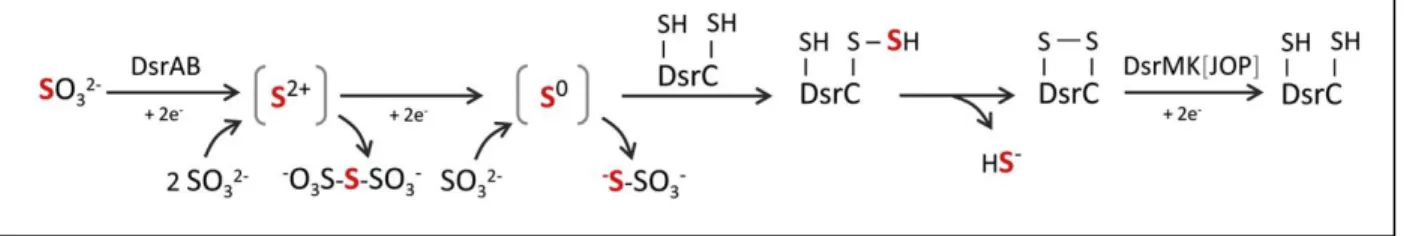
The crystal structure of these enzymes led to the proposal of a mechanism for sulfite reduction (Figure 1.9).
Sulfite is reduced by 4 electrons forming two intermediates in a SII and S0 valence state that stay bound to
the DsrAB catalytic site. The S0 intermediate then binds to DsrC CysA producing a persulfide as a key
intermediate. This persufide upon reacting with the other conserved cysteine (CysB) forms a disulfide bond
in DsrC (which is now in the oxidized form) and releases sulfide (45). This disulfide bond formed between
the two conserved cysteines of DsrC is believed to be reduced by several proteins present in these
Introduction
15
The putative physiological partners of DsrC belong to the CCG protein family, known to have a conservedcysteine-rich sequence (CXnCCGXmCXXC) (51). This motif binds a [4Fe-4S] 3+ cluster, which in Hdr is the
catalytic cofactor responsible for the reduction of the heterodisulfide, CoM-S-S-CoB formed in the last step
of methanogenesis (54). The heterodisulfide is not present in SRB, so this lead to the proposal that the
disulfide bond of DsrC after the release of sulfide during sulfite reduction, can be the equivalent of the
heterodisulfide in SRB. This oxidized form of DsrC can work as a substrate for proteins related to HdrB and
HdrD (51).
Figure 1.9 – Proposed mechanism of sulfite reduction by DsrAB and DsrC. Upon receiving 4 electrons sulfite is
reduced to a S0 valence state. This intermediate still bound to DsrAB catalytic site then reacts with CysA forming a
persulfide. Internal reaction of this persulfide with the other cysteine forms a disulfide bond and releases sulfide. The
SII and S0 intermediates can react with excess sulfide to form trithionate and thiosulfate, respectively. DsrMKJOP and
maybe other HdrB/HdrD related proteins are responsible to restore the oxidized DsrC into its reduced form. Taken from (51).
The possible disulfide reductases of DsrC are presented in Figure 1.10 and include the membrane
complexes DsrMKJOP, HmcABCDEF and TmcABCD that are involved in electron transfer with the
periplasm and/or menaquinone (MQ) pool, and also soluble proteins involved in oxidation of several
substrates, such as lactate (lactate dehydrogenase, Ldh), ethanol [alcohol dehydrogenase (Adh), flavin
oxidoreductase (FloxABCD) and HdrABC] and H2 [methyl viologen-reducing hydrogenase (MvhDGA) and
HdrABC) (51).
DsrMKJOP is a transmembrane complex composed by two modules, DsrJOP that is facing the periplasm
and DsrMK facing the cytoplasm (41). The DsrMK module of this complex has a CCG motif in the DsrK
subunit analogous to the one found in HrdD, thus suggesting that it can reduce the oxidized form of DsrC.
This can have an important role in the sulfite reduction pathway since the Dsr complex can possibly mediate
electron transfer between the menaquinol (MQH2) pool to the oxidized form of DsrC, contributing to energy
conservation. The DsrJOP module is most likely involved in electron flow between the periplasm and the
MQ pool, although the mechanism behind this process is not fully understood yet (55). Furthermore, there
is evidence that DsrK interacts with DsrC both in A. vinosum (56) and D. desulfuricans and in the majority
of SRO the genes that encode this protein complex are usually clustered with dsrAB and dsrC genes,
suggesting their physiological interaction (51).
HmcABCDEF and TmcABCD are both transmembrane complexes with a cytoplasmic subunit belonging to
Introduction
16
found in the HmcF subunit and in the Tmc complex it is present in the TmcB subunit (41). Since these
complexes have a similar architecture to DsrMKJOP and both have a subunit belonging to the CCG protein
family this suggests that they might be alternative electron donors to the oxidized form of DsrC (51).
Besides the HdrD-like proteins associated with membrane complexes, there are many other Hdr-related
proteins belonging to the CCG protein family that might act as electron donors to DsrC in SRB. Some of
these proteins are the soluble complexes FloxABCD/HdrABC and MvhDGA/HdrABC both containing the
HdrB subunit with the CCG motif. In SRB the hdrABC genes are found within two different sets of genes
clusters: hdrABC/floxABCD and hdrABC/mvhDGA (41). The floxABCD genes are found in many SRB and
code for a NAD(P)H (nicotinamide adenine dinucleotide phosphate) oxidoreductase that oxidizes NAD(P)H
formed during ethanol oxidation by an alcohol dehydrogenase to transfer electrons to HdrABC and
ferredoxin (Fdx) (57, 58). The HdrABC/MvhDGA is a complex characteristic of methanogens and present
in few SRO (41). It is involved in the oxidation of H2 by reduction of Fdx (54). These two complexes
(FloxABCD/HdrABC and MvhDGA/HdrABC) are involved in a flavin-based electron bifurcation mechanism
where the oxidation of ethanol and H2, respectively, is used to reduce both Fdx and a heterodisulfide, which
in SRO is proposed to be the oxidized form of DsrC (58). The link between FloxABCD and DsrC is further
supported by the fact that the genes that encode these proteins are found next to each other in some
organism (e.g. Desulfobacterium autotrophicum, Desulfosarcina sp. BuSS) (51).
Figure 1.10 – Proposed model and possible physiological partners of DsrC in sulfite reduction of SRB. During
sulfite reduction, after DsrC releases sulfide there’s the formation of a disulfide between CysA and CysB. This disulfide
Introduction
17
Other set of conserved proteins related to HdrB/HdrD are lactate dehydrogenases which are encoded bythree different gene loci. The first gene loci is involved in oxidation of organic acids and encodes, among
other proteins, the Ldh catalytic subunit (ldhA), a Ldh iron-sulfur subunit with two CCG domains (ldh1a) and
a HdrD related protein (ldh1b). The second gene locus has the genes lldEFG that encode a small HdrB
related protein called LldE that contains two CCG domains. The third gene locus is constituted by two genes
that encode the Ldh catalytic subunit (ldhA) and an HdrD-related protein encoded by the gene ldh3. These
three gene loci encode Ldhs with HdrB/HdrD like subunits suggesting that they might transfer electrons to
DsrC, coupling the oxidation of lactate to the reduction of sulfite (51).
Other Hdr-like proteins have also been identified such as the HdrD-related protein HdrF, which is a
multidomain protein composed by an iron-sulfur domain, a transmembrane domain and two CCG domains,
and HdrG which is a flavin adenine dinucleotide (FAD)-containing oxidoreductase containing a FAD-binding
domain, one or two FAD oxidase domains, an iron sulfur domain and two CCG domains. Since these
proteins have never been isolated their exact function is not yet known but it is suggested they are involved
in electron transfer from NAD(P)H, fatty acids and other metabolites to oxidized DsrC or the MQ pool (51).
The possible interaction between DsrC and the membrane complexes DsrMKJOP, HmcABCDEF and
TmcABCD might provide a connection between sulfite reduction and chemiosmotic energy conservation
associated with proton translocations across the membrane, whereas the interaction of DsrC with the
HdrABC soluble complexes may be associated with the energy coupling mechanism of electron bifurcation
Objectives
21
2. Objectives
It has been recently shown that DsrC is a central and important protein in the bioenergetic metabolism of
sulfate reducers, namely by having a strong impact on sulfite reduction by DsrAB. Thus, the main objective
of this project was to further study the role of this small protein in the sulfite reduction pathway and discover
other possible interaction partners of DsrC.
This work focused on two objectives:
The first one was to investigate how DsrC impacts growth under sulfate reducing and fermentative
conditions using Desulfovibrio vulgaris wild-type versus DsrC variant strains, using ethanol or pyruvate as
electron donors. To achieve this objective growth studies of those strains were performed, aiming to
elucidate the role of DsrC on the bioenergetic metabolism.
The second objective was to search for additional DsrC physiological partners during sulfate reduction while
providing different electron donors such as lactate and ethanol. To achieve this objective DsrC pull down
assays were performed in order to isolate DsrC in complex with other proteins. The co-eluted proteins were
analysed by mass spectrometry, Sodium dodecyl sulfate-polyacrylamide gel electrophoresis (SDS-PAGE)
Materials and Methods
25
3. Material and Methods
3.1. Growth Studies of DsrC Variant Strains
3.1.1. Strains
The strains used in this work were previously constructed in the lab (60) and are represented in Figure 3.1.
The wild-type DVH (WT DvH) contains only the chromosomal copy of the dsrC gene (cDsrC). IPFG06 strain
contains both the chromosomal and a plasmid copy of dsrC (pDsrC). IPFG07 strain contains only the
plasmid-copy of dsrC and IPFG09 strain contains only the plasmid copy of dsrC, containing the Cys93Ala
mutation in a strictly conserved cysteine, and a Cys26Ala mutation in a structural cysteine (not conserved
among DsrC).
Figure 3.1 – Schematic representation of the different DvH strains used in this work.
x
pDsrC
pDsrC
cDsrC
IPFG06
–
DvH + pMO DsrC
x
pDsrC
X
IPFG09
– DvH ∆
dsrC
+
pMODsrC C26A
C93A
IPFG07
– DvH ∆
dsrC
+
pMODsrC
cDsrC
Materials and Methods
26
3.1.2. Media
MOY medium used to grow all DvH strains contained 8 mM MgCl2, 20 mM NH4Cl, 0.6 mM CaCl2, 2 mM
K2HPO4-NaH2PO4, 0.6 % (v/v) Trace Elements, 0.06 mM FeCl, 0.12 mM EDTA, 30 mM Tris-HCl pH 7.4,
0.1 % (v/v) Thauers vitamins, 1 g/L yeast extract, 1.2 mM thioglycolate (used as a reducing agent), as
described by Zane and coworkers (42). Trace Elements solution contained 2.5 mM MnCl2.4H2O, 1.26 mM
CoCl2.6H2O, 1.47 mM ZnCl2, 0.21 mM Na2MoO4.2H2O, 0.024 mM Na2WO4.2H2O, 0.32 mM H3BO3,
0.035 mM Na2SeO3.5H2O, 0.012 mM CuCl2.2H2O and 0.38 mM NiCl2.6H2O. Thauers vitamin solution
con-tained 82 μM biotin, 45 μM folic acid, 468 μM pyridoxine hydrochloride, 148 μM thiamine hydrochloride, 133 μM riboflavin, 406 μM nicotinic acid, 210 μM DL-panthotenic acid, 365 μM p-aminobenzoic acid, 242
μM lipoic acid, 14 mM choline chloride, and 7.4 μM vitamin B12. The pH of the medium was adjusted with HCl to a final value of 7.2. All flasks and tubes were degassed with N2, sealed and autoclaved to assure
proper anaerobic growth. For different culture conditions MOY medium was supplied with different electron
acceptors and donors. Pre-inoculum medium was provided with sodium pyruvate as an electron donor and
sodium sulfate as an electron acceptor (60 mM pyruvate – 3 mM sulfate). MOY medium for ethanol-sulfate growth (respiration) was supplied with ethanol as an electron donor and sodium sulfate as an electron
acceptor (40 mM ethanol – 20 mM sulfate). MOY medium for pyruvate growth (fermentation) was supple-mented with sodium pyruvate as electron donor (60 mM pyruvate), and was provided with 3 mM sulfide
working as a reducing agent and sulfur source. In this work ethanol-sulfate and pyruvate growth medium is
going to be referred as MOY ES4 and MOY PYR, respectively.
3.1.3. Growth Curves
DvH WT and mutant strains were grown anaerobically at 37 oC. The experimental procedure to generate
the growth curves is outlined in Figure 3.2. Hungate tubes containing MOY medium (60 mM Pyruvate – 3 mM sulfate) were inoculated with 1 mL of stock cells to a final volume of 5 mL (20 % (v/v) inoculum). Cells
were grown overnight and then transferred into 100 mL flasks containing the same medium to a final volume
of 50 mL (10 % (v/v) inoculum). The optical density (OD) of these pre-culture flasks was monitored until an
OD600nm of 0.6. Upon reaching the desired OD 100 mL flasks containing either MOY ES4 (40 mM ethanol
– 20 mM sulfate) or MOY PYR (60 mM pyruvate) were inoculated with 4 % of these pre-cultured cells to a final volume of 50 mL. Antibiotics were added to MOY medium as follows: spectinomycin (100 µg/mL) was
added to IPFG06, IPFG07 and IPFG09 strains in all growth steps and geneticin (400 µg/mL) was
supplemented in pre-inoculum growths of IPFG07 and IPFG09. To the WT strain no antibiotics were added.
The OD of the cultures was monitored at various time points with a Shimadzu UV-1603 spectrophotometer
Materials and Methods
27
Figure 3.2 – Schematic representation of the experimental procedure used to monitor all growth curves.
3.1.4. Ethanol Quantification
Ethanol accumulation in the growth medium was determined with an enzymatic kit from NZYTech. This
method is based on the quantification of NADH formed from ethanol through the combined action of alcohol
dehydrogenase (Adh) and aldehyde dehydrogenase (AL-DH). First ethanol is oxidized by Adh with NAD+
to form acetaldehyde and NADH (Eq. 1). AL-DH then oxidizes acetaldehyde with NAD+ to produce acetate
and NADH (Eq. 2). Since 2 molecules of NADH are created per molecule of ethanol, the quantification of
NADH measured is twice the amount of ethanol in the sample.
Ethanol + NAD+ Acetaldehyde + NADH + H+ (1)
Acetaldehyde + NAD+ + H2O Acetate + NADH + H+ (2)
The samples were collected from the flasks with MOY PYR growing strains at different time points (after
inoculation, mid-exponential, late exponential, and stationary phase) and were centrifuged at 13000 rpm
during 15 minutes to separate the cells from the medium. The enzymatic assay was performed in sealed
cuvettes at room temperature and was processed in two steps. In the first step 50 µL of sample (or water
for blank) was mixed with 1 mL of H2O, 100 µL of potassium pyrophosphate buffer (1.5M, pH 9.0), 100 µL
of NAD+ and 10 µL of AL-DH (75 U/mL). Absorbance was then measured at 340 nm after 2 minutes (Abs1).
In the second step 10 µL of Adh (167 U/mL) was added to the previous mixture and absorbance was again
measured at the same wavelength after 5 minutes (Abs2).
Ethanol concentration was calculated by the absorbance difference for the blank and the samples (Abs2 – Abs1). The absorbance difference of the blank was subtracted from the absorbance difference of the
sam-ple thereby obtaining ΔAbsethanol. The concentration of ethanol, based on the ε of NADH at 340 nm, was
Stock cells
5 mL 50 mL
Pre inoculum
Pre inoculum
MOY ES4 or MOY PYR
10 % inoculum 4 % inoculum IPFG06WT
IPFG07 IPFG09 50 mL
Materials and Methods
28
calculated using the following expression: [Ethanol] (g/L) = 0.09287 ΔAbsethanol. All reported ethanol
con-centrations are the mean of three replicates.
3.2. Studies on DsrC Physiological Partners
3.2.1. Media and Cell Growth
To perform pull down assays using DsrC as a bait DvH WT and IPFG07 cells were grown either with
ethanol or lactate as electron donors by using MOY ES4 medium (40 mM ethanol – 20 mM sulfate) or MOY LS4 medium (30 mM lactate – 30 mM sulfate), respectively. Cells were grown anaerobically at 37 oC in 1 L
reagent bottles using a final volume of 800 mL. All pre-culture cells were grown according to the procedure
outlined in section 3.1.2. Reagent bottles were inoculated with 2 % (v/v) of fresh pre-cultured cells grown
in MOY medium (60 mM pyruvate – 3 mM sulfate) as defined in Figure 3.3. Spectinomycin antibiotic was added to all tubes, flasks and reagent bottles growing IPFG07 cells. Pre-cultures of IPFG07 were also
supplemented with geneticin. The OD of cultures was monitored at various time points with a Shimadzu
UV-1603 spectrophotometer at 600 nm until they reached late exponential phase. Afterwards, cells were
harvested by centrifugation at 13000 rpm for 15 minutes and the supernatant was discarded.
Figure 3.3 – Schematic representation of the experimental procedure used to grow WT and IPFG07 DvH cells for pull down assays.
3.2.2. DsrC Pull Down Assays
DsrC pull down assays were performed by affinity chromatography, specifically by immobilized metal ion
affinity chromatography (IMAC), taking advantage of the His tag on plasmid-expressed DsrC from the
IPFG07 strain. Harvested cells of DvH WT and IPFG07 grown either in MOY ES4 or MOY LS4 medium
were resuspended in buffer A (25 mM potassium phosphate pH 7.4, 100 mM NaCl and 10 % glycerol) and
disrupted in a French Press in the presence of DNase. The cell lysates were centrifuged for 20 minutes at
13000 rpm to remove cell debris (pellet) from the protein crude extract (supernatant) and this was then Stock cells
5 mL 50 mL
800 mL Pre inoculum Pre inoculum MOY ES4
or MOY LS4
10 % inoculum 2 % inoculum WT
IPFG07
Cell
Materials and Methods
29
ultra-centrifuged for 2 h at 42000 rpm to separate cell membranes (pellet) from the soluble fraction(supernatant). Before pull down assays, the total protein content of soluble fractions obtained from WT and
IPFG07 cells was quantified using the Bradford method to ensure that the same amount of protein would
be injected in the IMAC column (HiTrap Chelating HP column (GEHealthcare) charged with nickel). Using
a FPLC AKTATM system the soluble fractions were injected into the column already equilibrated with buffer
A. DsrC was then eluted with a mixture of buffer A and buffer B (25 mM potassium phosphate pH 7.4, 100
mM NaCl, 500 mM imidazole and 10 % glycerol) by using increasing concentrations of imidazole (30, 50,
70, 90, 110, 130, 150, 170, 200 and 500 mM). The chromatography was performed at 4 ºC. All fractions
eluted were recovered and concentrated by centrifugation at 4000 rpm with concentrators using 3000 MW
membranes (Millipore). Proteins eluted in each fraction were separated by SDS-PAGE (Sodium Dodecyl
Sulfate-Polyacrylamide Gel Electrophoresis) and were then analyzed by mass spectrometry and Western
blot.
3.2.3. Protein Quantification
Total protein concentration was determined by the Bradford method using a standard curve prepared with
bovine serum albumin (NZYTech) in the following concentrations: 0, 125, 250, 500, 750 and 1000 µg/mL.
The assay was prepared by mixing 20 µL of sample and 1 mL of Bradford reagent (Bio-rad). The blank was
prepared by replacing sample with buffer A. Absorbance was then measured at 595 nm using a Shimadzu
UV-1603 spectrophotometer.
3.2.4. Electrophoretic Techniques
Protein samples of each fraction obtained from the pull down assays were separated using
Tricine-SDS-PAGE 10 % acrylamide (v/v). Each gel contained a stacking layer for protein concentration and a resolving
layer for protein separation. To load the gels 20 µg of protein of each fraction was treated with the same
volume of loading buffer (6 M Urea, 5 % w/v SDS, 0.1 % w/v glycerol, 0.05 % w/v bromophenol blue) and
1 µL of β-Mercaptoethanol and were then boiled for 4 minutes. The gels were run at 100 V for 30 minutes and after that 130 V for 2 hours using two different buffers: anode buffer (100 mM Tris pH 8.9) and cathode
buffer (100 mM Tricine, 100 mM Tris and 0,1 % SDS pH 8.25). Lastly, the gels were stained using a
Coomassie Blue solution.
The same protocol was used to separate proteins for Western blot analysis but instead of
Tricine-SDS-PAGE 10 % acrylamide (v/v), Tricine-SDS-Tricine-SDS-PAGE 13 % acrylamide (v/v) or SDS-Tricine-SDS-PAGE 12 % acrylamide
Materials and Methods
30
3.2.5. Mass Spectrometry Protein Identification
Mass spectrometry data was obtained by the Mass Spectrometry Unit (UniMS) at ITQB/iBET, Oeiras,
Portugal. The protein bands were cut from the SDS-PAGE Coomassie Blue stained gels and then were
de-stained, reduced with dithiothreitol, alkylated with iodoacetamide and digested with trypsin (Promega, 6.7
ng/µL) overnight at 37 ᵒC. The peptides were desalted and concentrated using POROS R2 (Applied Biosystems) and eluted directly onto the MALDI plate using 0.6 µL of 5mg/mL
alpha-cyano-4-hydroxycinnamic acid (Sigma) in 50 % (v/v) acetonitrile and 5 % (v/v) formic acid. The data was acquired
in positive reflector MS and MS/MS modes using a 4800plus MALDI-TOF/TOF (AB Sciex) mass
spectrometer and the 4000 Series Explorer Software v.3.5.3 (Applied Biosystems). External calibration was
performed using Pepmix1 (Laser BioLabs). The twenty-fifth most intense precursor ions from the MS
spectra were selected for MS/MS analysis. The raw MS and MS/MS data was analyzed using Protein Pilot
Software v. 4.5 (ABSciex) with the Mascot search engine (MOWSE algorithm). The searches were
performed against the protein database NCBI (35149712 sequences; 12374887350 residues) with bacterial
taxonomy restrictions (23117303 sequences). The search parameters were as follows: monoisotopic
peptide mass values were considered, maximum precursor mass tolerance (MS) of 100 ppm and a
maximum fragment mass tolerance (MS/MS) of 0.3 Da. A maximum of two tryptic missed cleavages were
allowed. Carboxyamidomethylation of cysteines, oxidation of methionine, deamidation of asparagine and
glutamine, and N-Pyro Glu of the N-terminal Q were set as variable modifications. Protein identification was
only accepted when significant protein homology scores were obtained (p<0.05).
3.2.6. Western Blot Analysis
Protein samples were separated by SDS-PAGE [12 % acrylamide (v/v)] or Tricine-SDS-PAGE [13 %
acryla-mide (v/v)] and transferred to 0.45 μm polyvinylidene difluoride (PVDF) membranes (Roche) at 100 V and 400 mA in a Mini Trans-Blot® dry electrophoretic transfer cell (Bio-Rad) containing transfer buffer (48 mM
Tris, 39 mM glycine pH 9.2). The type of SDS-PAGE gel and transfer time used for each protein is presented
in Table 3.1. Protein volume to be loaded in the gel was calculated for each IPFG07 fraction and then the
same volume was used for the corresponding WT fractions in order to calibrate the assay. The PVDF
membranes were dried overnight, washed two times with TBS (Tris-Buffered Saline, 20 mM Tris-HCl pH
7.5, 150 mM NaCl) and then were treated with blocking buffer [20 mM Tris-HCl pH 7.5, 150 mM NaCl,
0.05 % Tween 20 (v/v), and 1 % nonfat milk (w/v)] and incubated with a primary antibody diluted in blocking
buffer for 1 hour at room temperature. The dilutions of primary antibodies used were as follows: anti-FloxA
at 1:3000, anti-HdrA at 1:1000, anti-Fdx at 1:1000, anti-Adh at 1:5000 and anti-Apr 1:3000 (see Table 3.1).
After primary antibody incubation, membranes were washed three times and then incubated with anti-rabbit
Materials and Methods
31
buffer for 45 minutes at room temperature. After three washing steps with TBS, protein detection wasper-formed using Alkaline Phosphatase Buffer (100 mM Tris-HCl pH 9.5, 100 mM NaCl, and 5 mM MgCl2) and
NBT (nitro-blue tetrazolium chloride)/BCIP (5-bromo-4-chloro-3-indolyl phosphate) substrates (Carl Roth®).
Table 3.1 – List of conditions used for Western blot analysis.
Primary/Secondary antibody
Gel used for protein separation Total Protein mass loaded (μg) Transference time (min) Primary/Secondary antibody dilution Anti-FloxA/Anti-rabbit IgG SDS-PAGE 12 % acrylamide
(v/v)
10 30 1:3000/1:15000
Anti-Adh/Anti-rabbit IgG
SDS-PAGE 12 % acrylamide
(v/v)
10 30 1:5000/1:15000
Anti-HdrA/Anti-rabbit IgG
SDS-PAGE 12 % acrylamide
(v/v)
10 30 1:1000/1:15000
Anti-Apr/Anti-rabbit IgG
SDS-PAGE 12 % acrylamide
(v/v)
10 30 1:3000/1:15000
Anti-Fdx/Anti-rabbit IgG
Tricine-SDS-PAGE 13 % acrylamide (v/v)