COMPUTED TOMOGRAPHY FINDINGS IN PATIENTS LESS THAN
20 YEARS OLD WITH LYMPHOMA*
Adriana Moreira Viana Borba1, Alexandra Maria Vieira Monteiro2, Cláudio Marcio Amaral de Oliveira Lima3, Érica Barreiros Ribeiro3, Stella Beatriz Gonçalves de Lucena4, Luis Flávio Skinner5
OBJECTIVE: To describe the general findings of lymphoma and their histological patterns in patients less than 20 years old. MATERIALS AND METHODS: Twenty-two cases (16 male and 6 female, mean age 11.5 years) from the digital archive of computed tomography at the Cancer Control Center of “Hospital Universitário Pedro Ernesto – Universidade do Estado do Rio de Janeiro”, Rio de Janeiro, RJ, Brazil, were retrospectively analyzed in the period between March 2003 and July 2005. Of these 22 cases, 12 were Hodgkin’s and 10 were non-Hodgkin’s. RESULTS: Overall, mediastinal lymphadenomegaly was the most frequent finding (59%), with predominance in the Hodgkin’s subgroup (75%), followed by hepatosplenomegaly (50%) and cervical and retroperitoneal lymphadenomegaly (27.3%). The Hodgkin’s subgroup presented a prevalence of lym-phadenopathy, in many lymph node chains, followed by hepatosplenomegaly (50%). One case was found with unilateral tonsillar mass, pulmonary ground-glass opacities, and renal nodules. In the non-Hodgkin’s subgroup, the disease was predominantly extranodal, characterized by hepatosplenomegaly (50%), thick-ening of the intestinal wall (40%), pleural effusion (30%), pulmonary nodule (20%), ascites (10%), pericar-dial effusion (10%) and mixed bone lesions (10%). CONCLUSION: Computed tomography is an extremely useful method for detection, staging and follow-up of lymphomas, with alert findings like mediastinal lym-phadenopathy, hepatosplenomegaly, unilateral tonsillar mass and thickening of intestinal wall.
Keywords: Lymphoma; Computed tomography.
Aspectos da tomografia computadorizada no linfoma em pacientes abaixo de 20 anos de idade.
OBJETIVO: Descrever os achados gerais do linfoma em pacientes abaixo de 20 anos de idade e por subtipo histológico. MATERIAIS E MÉTODOS: Estudo retrospectivo do arquivo digital de tomografia computado-rizada do Centro de Controle do Câncer do Hospital Universitário Pedro Ernesto da Universidade do Estado do Rio de Janeiro, no período de março de 2003 a julho de 2005. Dos 22 casos — 16 do sexo masculino e 6 do sexo feminino, com média de idade de 11,5 anos —, 12 eram do subtipo Hodgkin e 10 eram não-Hodgkin. RESULTADOS: Dos achados gerais, verificamos as linfonodomegalias mediastinais como o mais freqüente (59%), com predomínio no grupo Hodgkin (75%), seguido por hepatoesplenomegalia (50%) e lin-fonodomegalias cervicais e retroperitoneais (27,3%). No subtipo Hodgkin houve predomínio do acometimento linfonodal, em sucessivas cadeias, seguido pela hepatoesplenomegalia (50%). Verificamos um caso de massa tonsilar unilateral, opacidade pulmonar em “vidro-fosco” e nódulos renais. No subtipo não-Hodgkin houve predomínio extranodal caracterizado por hepatoesplenomegalia (50%), espessamento de alça intestinal (40%), derrame pleural (30%), nódulo pulmonar (20%), ascite (10%), derrame pericárdico (10%) e lesões ósseas mistas (10%). CONCLUSÃO: A tomografia computadorizada é de grande valia no diagnóstico, estadiamento e seguimento do linfoma, com achados de alerta como massa linfonodal, notadamente mediastinal, hepato-esplenomegalia, massa unilateral na tonsila e espessamento parietal de alça intestinal.
Unitermos:Linfoma; Tomografia computadorizada. Abstract
Resumo
* Study developed in the Computed Tomography Unit at Cen-tro Universitário de ConCen-trole do Câncer do Hospital Universitário Pedro Ernesto (HUPE) da Universidade do Estado do Rio de Ja-neiro (UERJ), Rio de JaJa-neiro, RJ, Brazil.
1. Post-graduation Student in Radiology at Escola Médica da Pontifícia Universidade Católica do Rio de Janeiro (PUCRJ), Rio de Janeiro, RJ, Brazil.
2. PhD in Medicine, Associate Professor of de Radiology for Graduation and Post-graduation Courses at Faculdade de Ciên-cias Médicas da Universidade do Estado do Rio de Janeiro (UERJ), Professor for the Course of Post-graduation in Radiology at Es-cola Médica da Pontifícia Universidade Católica do Rio de Ja-neiro (PUCRJ), Rio de JaJa-neiro, RJ, Brazil.
3. MDs at Hospital Naval Marcílio Dias, Post-graduation stu-dents in Radiology at Escola Médica da Pontifícia Universidade Católica do Rio de Janeiro (PUCRJ), Rio de Janeiro, RJ, Brazil.
4. Associate Professor of Hematology at Faculdade de Ciên-cias Médicas da Universidade do Estado do Rio de Janeiro (UERJ), Rio de Janeiro, RJ, Brazil.
INTRODUCTION
The term lymphomas refer to malignant neoplasms of specific cells in the lymphatic system (T-cells, B-cells and histiocytes)(1,2).
They are subdivided into two groups, Hodgkin’s lymphomas (HL) and non-Hodgkin’s lymphoma (NHL)(1). NHL is
more frequent in the childhood than HL, and is the third most common neoplasm after leukemia and central nervous system tumors in children less than 16 years of age. HL represents about 5% of all cancers in infants and children(3).
NHL is prevalent in children less than 16 years of age, while HL most frequently affects children with more than five years of age and is rare under this age. The
inci-5. Assistant Professor of Radiology at Faculdade de Ciências Médicas da Universidade Estadual do Rio de Janeiro (UERJ), Head for the Service of Radiology at Hospital Universitário Pedro Er-nesto (HUPE), Rio de Janeiro, RJ, Brazil.
Mailing address: Dr. Cláudio Marcio Amaral de Oliveira Lima. Rua César Zama, 185, Lins de Vasconcelos. Rio de Janeiro, RJ, Brazil, 20725-090. E-mail: [email protected] e cmaolima@ hotmail.com
dence is higher in males at a 3:1 ratio for NHL, and 2:1 for HL(3).
Clinical characteristics, radiological findings, histological patterns, therapy and prognosis of pediatric Hodgkin’s disease are similar to those of Hodgkin’s disease in adults(3). Generally, HL is characterized by
the orderly spread of the disease from a lymph node group to another(4); so
fre-quently the involvement of upper medias-tinal lymph nodes (both prevascular and paratracheal) is observed in about 98% of patients with intrathoracic disease. Ap-proximately one third of these patients present unilateral or bilateral hilar lym-phadenopathy(5). Thymic involvement is
observed in up to 70% of patients with mediastinal Hodgkin’s disease, generally associated with an increase of lymph nodes in other sites of the mediastinum(5).
In the childhood, the majority of NHL is extranodal, in contrast to both the NHL in the adult and Hodgkin’s disease at any age range. In the childhood, the most fre-quent primary site is the abdomen, particu-larly the ileocecal region, followed by the mediastinum (approximately 25%)(3).
Imaging methods, particularly com-puted tomography, are invaluable tools for evaluating the disease extent, planning the treatment and analyzing the subsequent response to the therapy(6).
The aim of the present study was to describe general findings of computed to-mography according their histological sub-type in less-than-20-year-old patients with diagnosis of lymphoma.
MATERIALS AND METHODS
Retrospective study of digital files of the Computed Tomography Unit in Centro Universitário de Controle do Câncer at Hospital Universitário Pedro Ernesto – Universidade do Estado do Rio de Janeiro, Rio de Janeiro, RJ, Brazil, during the pe-riod between March 2003 and July 2005. In this period 1,356 computed tomography studies were performed in patients with less than 20 years of age. Of these patients, 22 were diagnosed with lymphoma - 12 of them HL, and 10 NHL.
Inclusion criteria were: age under 20, and diagnostic and histopathological con-firmation of lymphoma type. Two patients
were excluded due the absence of histologi-cal categorization (follow-up in another hospital unit).
The equipment utilized was a GE HiSpeed helical model, with a protocol including base of the neck, chest, abdomen and pelvis.
All of the studies were reviewed by two investigators, and only those where there was a consensus were taken into consider-ation.
Of a total of 22 cases, 12 were HL, and 10 NHL. Sixteen patients were men and six women, with ages ranging between three and 20 years (mean age 11.5). In the HL group was in the age range between six and 20 years, and the NHL, between three and 20 years.
In the HL group, eight patients were male and four, female. In order of fre-quency, the following histological subtypes were found: mixed cellularity (41.6%), nodular sclerosis (33.3%), lymphocytic predominance (16.6%), and lymphocytic depletion (8.5%). In the NHL group, eight were men, and two were women. Predomi-nant histological subtypes were: diffuse large B cell lymphomas (40%); Burkitt’s lymphomas (40%), followed by T-lympho-blastic lymphomas (10%) and small-cell lymphomas (10%).
Of a total of 22 patients, 14 (63%) un-derwent examination for the first time, and eight (36%) for follow-up. No patients pre-sented with coexistent infection. One pa-tient died during the study period.
RESULTS
Among the computed tomography find-ings, we observed that mediastinal lym-phadenopathy was the most frequent find-ing (59%), predominatfind-ing in the HL group (75%), followed by hepatosplenomegaly (50%), cervical and retroperitoneal lym-phadenopathy (27.3%) (Tables 1 and 2).
As regards the presence of lymphaden-opathy (Table 1) a high predominance in the HL group compared with the NHL group. In the majority of cases, the lymph node mass presented homogeneous con-trast enhancement, with the exception of two cases: one in the HL group, and another in the NHL group (Figure 1). No calcified lymph node was detected.
Cervical lymphadenopathies were pre-dominantly bilateral, most frequently de-tected in the HL group, without the
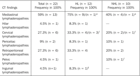
pres-Table 1 Computed tomographyfindings in the total of cases of lymphoma categorized by histological subtype in lymphadenopathies.
CT findings
Mediastinal lymphadenopathies Hilar
lymphadenopathies Cervical
lymphadenopathies Periceliac lymphadenopathies Retroperitoneal lymphadenopathies Pelvic
lymphadenopathies Inguinal
lymphadenopathies
Total (n = 22) Frequency in 100%
59% (n = 13)
4.5% (n = 1)
27.3% (n = 6)
9% (n = 2)
27.3% (n = 6)
4.5% (n = 1)
4.5% (n=1)
HL (n = 12) Frequency in 100%
75% (n = 9)/(n = 1)*
8.3% (n = 1)
33.3% (n = 4)/(n = 3)†
8.3% (n = 1)
33.3% (n = 4)
—
8.3% (n = 1)‡
NHL (n = 10) Frequency in 100%
40% (n = 4)/(n = 1)*
—
20% (n = 2)/(n = 1)†
10% (n = 1)
20% (n = 2)
10% (n = 1)†
—
HL, Hodgkin lymphoma; NHL, non-Hodgkin lymphoma; *Heterogeneous; †Bilateral; ‡Unilateral.
Hepatomegaly as an isolate finding was observed only in the HL group (n = 2).
Small bowel parietal thickening (n = 4) was found in four patients, all of them in the NHL group, 50% of Burkitt subtype. This alteration was associated exclusively with hepatosplenomegaly in three of the four cases (75%), and was found as a single manifestation in only one case (Figure 4). Pleural effusion (n = 3), pericardial ef-fusion (n = 1), and ascites (n = 1) were found only in the NHL group.
Pulmonary involvement represented by nodule(s) (n = 2) was found only in the NHL group. Ground-glass opacities (n = 2) were equivalent in both groups (Figure 5). Associated infection was not found in any of the cases.
Only one unilateral mass was found in the tonsil, associated with cervical lym-phadenopathy, in the HL group, nodular sclerosis subtype (Figure 6).
Bone lesions, in the most severe case of NHL group (large B cell subtype) were
Table 2 Extranodal findings on computed tomography in the total of cases of lymphoma categorized by histological subtype.
CT findings
Hepatosplenomegaly Hepatomegaly Thickening of intestinal loop
Ascites Pleural effusion Pericardial effusion Pulmonary nodule(s) Pulmonary ground-glass opacity
Tonsil mass Mixed bone lesions Renal nodules
Total (n = 22) Frequency in 100%
50% (n = 11) 9% (n = 2)
18.2% (n = 4) 4.5% (n = 1) 13.6% (n = 3) 4.5% (n = 1) 9% (n = 2) 9% (n = 2)
4.5% (n = 1) 4.5% (n = 1) 4.5% (n = 1)
HL (n = 12) Frequency in 100%
50% (n = 6) 16.7% (n = 2)
— — — — —
8.3% (n = 1)
8.3% (n = 1)† —
8.3% (n = 1)‡
NHL (n=10) Frequency in 100%
50% (n = 5 )/(n = 1)* —
40% (n = 4) Burkitt (n = 2)
10% (n = 1) 30% (n = 3)/(n = 2)† 10% (n = 1) 20% (n = 2) 10% (n = 1
—
10% (n = 1) —
HL, Hodgkin lymphoma; NHL, non-Hodgkin lymphoma; * Heterogeneous splenomegaly; †Unilateral; ‡Bilateral.
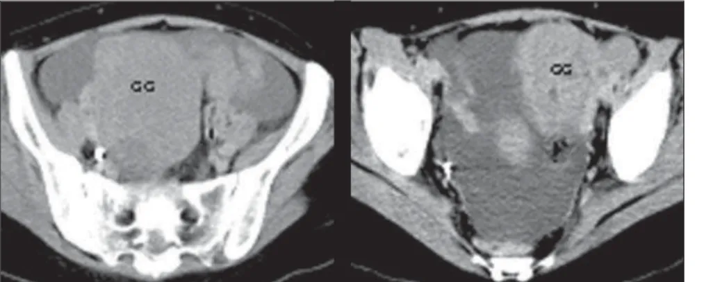
Figure 2. Heterogeneous, bilateral pelvic lymphadenopathy (GG) at left (NHL, large B-cell subtype). Figure 3. Hepatosplenomegaly with spleen pre-senting hypodense nodules (NHL, large B-cell sub-type).
Figure 4. Parietal thickening of small intestinal loop (arrow); note lymphadenopathy (GG) at right (NHL, Burkitt subtype).
Figure 5. Ground-glass opacity (double arrow) and nodule (arrow) in the lower lobe of the left lung (NHL, large B-cell subtype).
ence of necrosis and/or calcification. The other lymph node chains affected showed variable presentations, highlighting espe-cially the pelvic ones in the most severe case (Figure 2).
management of this disease. According to the Ann Arbor System, the staging itself justifies the use of imaging methods, since it is based on a systematized analysis of the disease extent(4,6).
Computed tomography, due the fast images acquisition in comparison with magnetic resonance imaging, allows a rapid and comprehensive staging, which is essential for definition of the clinical on-cological treatment. Reports in the litera-ture describe, for example, changes in the planning of up to 10% of cases, based on data from computed tomography(6), as well
as the identification of non-suspect sites of intrathoracic diseases or the elucidation of questionable radiological abnormalities which eventually might change therapeu-tic decisions(5).
As regards the frequency of general imaging findings, our data are consistent with the literature reviewed(3,7).
The pattern of thoracic involvement in patients with lymphoma is variable, and, at the moment of the diagnosis this involve-ment is more frequent in HL than in NHL, with presence in up to 11.6% of cases of HL. In these patients, the pulmonary in-volvement is associated with the presence of the disease in hilar or mediastinal lymph node chains. In cases with unilateral hilar adenopathy, parenchymal involvement, when present, occurs in the ipsilateral lung8,9). The pulmonary disease, without
lymph node involvement is more frequent in recurrent lymphomas. Pulmonary in-volvement is frequently asymptomatic and symptoms, when present, are usually non-specific(8,10). One characteristic of HL in the
lung is the dissemination along the lym-phatic route(10) and, in the mediastinum, the
dissemination contiguously from a lymph node group to the next, with initial presen-tation in about 65%–80% of patients with abnormal chest x-ray(5,10). In other studies(5)
reporting a 98% frequency in patients with intrathoracic disease, we have observed a prevalence of mediastinal lymphadenopa-thy in relation to other sites, in the HL group, inclusive.
We have not identified calcifications which rarely are seen before the therapy, even in cases of follow-up; but areas with necrosis were detected in both subtypes(7).
Differently from carcinomas, lymphoma
Figure 6. Mass in the tonsil at right (M) associated with unilateral lymphadenopathy (G) (HL, nodular sclerosis subtype).
Figure 7. Blastic lesion in the sacrum at right (arrow), and lytic lesion in the ischiopubic branch at left (NHL, large B-cell subtype).
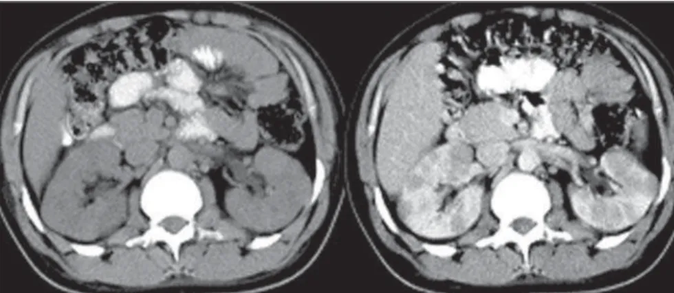
Figure 8. Bilateral, solid renal nodules - pre-contrast at left, and post-contrast at right. Note hypodense nodules. Hepatomegaly (HL, lymphoblastic).
multiple and mixed (lytic and blastic as-pect) (Figure 7).
There was one case of renal involve-ment characterized by multiple and bilat-eral solid nodules (Figure 8) in the HL sub-type.
DISCUSSION
masses are more likely to displace adjacent structures than invade them(5), just as the
cases of vascular compression observed in the present study. Approximately one third of patients with thoracic disease present unilateral or bilateral hilar lymphadenopa-thy(5), a finding observed in only one case
in our study.
Contrarily to HL, NHL tends to mani-fest as a single, bulky lymph node mass, and not as individually increased lymph nodes. According to our results, in the chest, other sites are usually involved, such as pulmonary parenchyma, pleura and peri-cardium(2,10).
In patients with mediastinal HL and NHL mediastinal, a secondary pulmonary involvement usually is seen later with the progress of the disease. Only 12% of pa-tients with HL and 4% of those with NHL present pulmonary involvement as a pri-mary manifestation of the disease. The most frequent patterns observed on com-puted tomography studies are parenchymal consolidation, nodules or masses, and lym-phatic dissemination. Among these pat-terns, nodules are the most frequent ones (50% to 90% of cases), and can be solitary or, most frequently, multiple.
Cavitation is rare (< 10%), but may oc-cur, especially in the HL subtype. Another aspect described is the presence of ground-glass halo surrounding a nodule (halo sign) or isolatedly, due to alveolar septa infiltra-tion by neoplastic cells, with preserved al-veolar spaces(10). In the present study, only
one case of nodule presented this pattern, and, when in association with ground-glass opacity, there was a random distribution.
The protocol for chest examination must include the base of the neck as a rou-tine, to discard head & neck involve-ment(5,7,11). Cervical lymphadenopathies
were the second main finding in general frequency order, predominating in the HL group. Lymphoma is the second most fre-quent malignant neoplasm in the neck, besides being the most common extrala-ryngeal malignant neoplasm. NHL is one of the most frequent malignant neoplasms in children. Neck lymphoma usually in-volves the adenoids as well as the lingual and pharyngeal tonsils (Waldeyer’s ring); the unilateral tonsillar increase in children is highly suspect for malignancy. Cervical
lymph nodes, especially internal jugular and spinal accessory lymph nodes which generally also are involved. In imaging studies groups of non-necrotic lymph nodes are observed(5,11). In the case of
tonsilar mass, we observed association with cervical lymphadenopathies, some of them with a hypodense nucleus.
The greatest part of NHLs in the child-hood is extranodal, in contrast to both the NHL in adults and Hodgkin´s disease at any age. The most frequent primary site of NHL in children is the abdomen; particu-larly the ileocecal region(3), literally in
agreement, and according to our results, predominantly associated with hepatosple-nomegaly.
Common findings of computed tomog-raphy in all the types of small bowel lym-phoma are: intestinal wall thickening, sometimes nodular in a diffuse or focal distribution, and also distinct lymph node masses in the mesenteric folds of the in-volved segment; in some cases, the CT shows a single focal, frequently cecal, co-lonic mass.
The evaluation of the liver and spleen is an essential part of the computed tomog-raphy study. Although an organ enlarge-ment is an indicator of its involveenlarge-ment, this is not a reliable diagnostic criterion, and the sensitivity for detection of focal lesion in dubious cases of lymphomas is signifi-cantly increased with sequential CT slices obtained after contrast bolus injection(12).
Secondary splenic involvement is frequent both in HL and NHL, and malignant splenic neoplasm is the most common. It is estimated that at the moment of the di-agnosis there is splenic involvement in one-fourth to one-third of patients with HL or NHL. In patients with NHL, the splenic involvement is associated with infiltration of paraaortic lymph nodes in approximately 70% of them. Homogeneous organ en-largement, miliary nodules, multifocal le-sions between 1 cm and 10 cm, and soli-tary mass are some forms of presentation. In cases of necrosis in large lesions, an ir-regular, cystic pattern is observed, and a case of a patient with fever associated with lymphoma mimicking splenic abscess is reported in the literature(5). In the present
study, multiple lesions did not present pe-ripheral contrast enhancement, which in
association with clinical data, characterized the disease manifestation.
Most frequently, in NHL, the small bowel mesentery and omentum are in-volved by a tumor disseminated via the intraperitoneal pathway. The four general patterns of computed tomography imaging are: rounded masses, cake-like masses, ill-defined masses, and stellate mesentery. Rounded masses are more frequent in NHL, basically due to the lymphadenopa-thy, and not to the intraperitoneal dissemi-nation(5,13).The presence of ascites suggests
this forma of dissemination.
Bone lymphoma causes diffuse or focal osseous pain and usually corresponds to dissemination from any other primary site. Manifestations of systemic dissemination, such as anaplastic lymphoma of the skin, may occur in association with alterations related to the local tumor(2,14). The case of
bone involvement identified in the present study was exactly the one with more exten-sive systemic manifestation (large B-cell NHL), where chest, abdomen and pelvis presented with extensive lesions.
CONCLUSION
Computed tomography is an invaluable tool for the diagnosis, staging and follow-up of lymphomas, with alert findings such as lymph node masses (especially the me-diastinal ones), hepatosplenomegaly, uni-lateral tonsilar mass, and parietal thicken-ing of intestinal loop.
REFERENCES
1. Johnston JM, Sakamoto K, Windle ML, Cripe TP, Gross S, Coppes MJ. Non-Hodgkin lymphoma. Acesso em: 10/6/2005. Disponível em: http:// www.emedicine.com/ped/topic1343.htm 2. Juhl JH. Linfoma. In: Juhl JH, editor.
Interpreta-ção radiológica. 7ª ed. Rio de Janeiro: Guanabara Koogan, 2000;200–215.
3. Kirks DR, Griscon NT. Diagnóstico por imagem em pediatria e neonatologia. 3ª ed. São Paulo: Revinter, 2003.
4. Associação Brasileira de Linfoma e Leucemia – Abrale. Linfoma de Hodgkin. Acesso em: 15/6/ 2005. Disponível em: http://www.abrale.com.br 5. Glazer HS, Semenkovich JW, Gutierrez FR. Mediastino. In: Lee JKT, Sagel SS, Stanley RJ, Heiken JP, editores. Tomografia computadorizada do corpo em correlação com ressonância magné-tica. 3ª ed. Rio de Janeiro: Guanabara Koogan, 2001;263–268.
6. Bernard A, Murphy SB, Melvin S. Neoplasias e estruturas semelhantes a neoplasias. In: Behrman RE, Kliegman RM, Nelson WE, editores. Tratado de pediatria. 15ª ed. Rio de Janeiro: Guanabara Koogan, 2000;1977–1822.
7. Effmann EL. The mediastinum. In: Kuhn JP, Slovis TL, Haller JO, editors. Caffey’s pediatric diagnostic imaging. 10th ed. Philadelphia: Mosby, 2004;1649–1690.
8. Guermazi A, Brice P, de Kerviler EE, et al. Extra-nodal Hodgkin disease: spectrum of disease. RadioGraphics 2001;21:161–179.
9. Fishman EK, Kuhlman JE, Jones RJ. CT of lym-phoma: spectrum of disease. RadioGraphics 1991;11:647–669.
10. Marchiori E, Valiante PM, Gutierrez ALTM, Bo-danese L, Souza Jr AS. Linfomas pulmonares: correlação da tomografia computadorizada de alta resolução com a anatomopatologia. Radiol Bras 2002;35:1–6.
11. Dunleavy KM, Waes CV, Kass E, Grill R. Lym-phomas of the head and neck. [update 2004 Aug 19]. Acesso em: 10/6/2005. Disponível em: http:/ /www.emedicine.com/ent/topic742.htm 12. Cohan RH, Baber ME, Cooper C, Moore JO,
Dunnick NR. Retroperitoneal cavity. In: Wegener OH, editor. Whole body computed tomography. 2nd ed. Oxford: Blackwell Sci Publ, 1993;471– 477.
13. Byun JH, Ha HK, Kim AY, et al. CT findings in peripheral T-cell lymphoma involving the gas-trointestinal tract. Radiology 2003;227:59–67. 14. Mulligan ME. Lymphoma, bone. [update 2005
Jan 5]. Acesso em: 10/6/2005. Disponível em: http://www.emedicine.com/radio/topic819.htm 15. Shirkhoda A. Kidney, lymphoma. [update 2005