A PTK7/Ror2 Co-Receptor Complex Affects
Xenopus
Neural Crest Migration
Martina Podleschny☯, Anita Grund☯, Hanna Berger, Erik Rollwitz, Annette Borchers*
Faculty of Biology, Molecular Embryology, Philipps-Universität Marburg, 35043 Marburg, Germany
☯These authors contributed equally to this work. *[email protected]
Abstract
Neural crest cells are a highly migratory pluripotent cell population that generates a wide array of different cell types and failure in their migration can result in severe birth defects and malformation syndromes. Neural crest migration is controlled by various means includ-ing chemotaxis, repellent guidance cues and cell-cell interaction. Non-canonical Wnt PCP (planar cell polarity) signaling has previously been shown to control cell-contact mediated neural crest cell guidance. PTK7 (protein tyrosine kinase 7) is a transmembrane pseudoki-nase and a known regulator of Wnt/PCP signaling, which is expressed inXenopusneural crest cells and required for their migration. PTK7 functions as a Wnt co-receptor; however, it remains unclear by which means PTK7 affects neural crest migration. Expressing fluores-cently labeled proteins inXenopusneural crest cells we find that PTK7 co-localizes with the Ror2 Wnt-receptor. Further, co-immunoprecipitation experiments demonstrate that PTK7 interacts with Ror2. The PTK7/Ror2 interaction is likely relevant for neural crest migration, because Ror2 expression can rescue the PTK7 loss of function migration defect. Live cell imaging of explanted neural crest cells shows that PTK7 loss of function affects the forma-tion of cell protrusions as well as cell motility. Co-expression of Ror2 can rescue these defects.In vivoanalysis demonstrates that a kinase dead Ror2 mutant cannot rescue PTK7 loss of function. Thus, our data suggest that Ror2 can substitute for PTK7 and that the sig-naling function of its kinase domain is required for this effect.
Introduction
Neural crest (NC) cells are a highly migratory pluripotent cell population that gives rise to a wide range of derivatives contributing to many tissues and organs. NC cells develop along the ante-rior-posterior axis of the vertebrate embryo at the border region between the epidermis and the neural plate. After undergoing an epithelial to mesenchymal transition NC cells migrate and col-onize almost all tissues of the embryo. In this respect NC migration is very similar to cancer cell invasion and metastasis dissemination, which is also mirrored by the conservation of molecules and signaling pathways involved in both processes [1,2,3]. During migration NC cells follow pre-cise pathways and encounter various molecular microenvironments, which guide them to their
OPEN ACCESS
Citation:Podleschny M, Grund A, Berger H, Rollwitz E, Borchers A (2015) A PTK7/Ror2 Co-Receptor Complex AffectsXenopusNeural Crest Migration. PLoS ONE 10(12): e0145169. doi:10.1371/journal. pone.0145169
Editor:Zoha Kibar, CHU Sainte Justine and University of Montreal, CANADA
Received:January 8, 2015
Accepted:November 30, 2015
Published:December 17, 2015
Copyright:© 2015 Podleschny et al. This is an open access article distributed under the terms of the
Creative Commons Attribution License, which permits unrestricted use, distribution, and reproduction in any medium, provided the original author and source are credited.
Data Availability Statement:All relevant data are within the paper and its Supporting Information files.
Funding:This work was funded by Deutsche Forschungsgemeinschaft, grant number BO 1978/ 4-2. The funders had no role in study design, data collection and analysis, decision to publish, or preparation of the manuscript.
final destinations and determine their terminal differentiation [4]. Therefore, NC migration needs to be tightly controlled to ensure the development of multiple organs and tissues. Indeed, failure results in severe birth defects and malformation syndromes–so called neurocristopathies [5,6,7]. Thus, understanding the molecular mechanism that control NC migration will also pro-vide insight into pathological conditions like neurocristopathies or the development of cancer.
Non-canonical–ß-catenin-independent–Wnt signaling contributes significantly to the reg-ulation of NC migration [8,9,10,11,12,13,14]. NC cells show collective cell migration and form streams of migrating cells directed for example by repellent guidance cues and chemo-attrac-tants [2,4]. In addition, NC cell directionality is achieved by communication of NC cells with each other. One mechanism, which was proposed for the directional migration of cranial NC cells is contact inhibition of locomotion. This phenomenon was discovered by Abercrombie [15,16] and describes how two colliding cells change their cell polarization upon cell-cell-con-tact and migrate in opposite directions. Recent work showed that non-canonical planar cell polarity (PCP) Wnt signaling is required for contact inhibition of locomotion and directional collective NC migration [8,9,10,17,18]. PCP signaling was first discovered in the fly where it regulates the orientation of hairs in the wing or ommatidia organization in the eye [19,20]. In vertebrates PCP signaling is necessary for inner ear patterning, ciliary beating and tissue move-ments contributing to axis elongation and neural tube closure [21,22]. In all these biological systems PCP proteins become asymmetrically localized to the plasma membrane thereby estab-lishing a polarity in the plane of an epithelium. In moving tissues the situation is more compli-cated. In migrating NC cells PCP signaling seems to determine the formation of cellular protrusions by asymmetrically regulating the activity of small GTPases of the Rho family [14,18,23]. PCP proteins including Frizzled and Dishevelled are localized to the site of cell con-tact thereby leading to a local activation of Rho and inhibition of Rac [8,9,17,24]. Thus, cell protrusions collapse and cells migrate in opposite directions. Thereby, PCP signaling provides a means of NC cell dispersion, which in combination with other repellent, attractant and adhe-sive cues contributes to controlled migration of NC cells [23,25,26,27]. Although, the contribu-tion of PCP signaling to NC migracontribu-tion has been acknowledged in different vertebrate systems the molecular components localizing to cell-contact sites and the signaling pathways leading to cytoskeletal remodeling remain to be characterized.
Protein tyrosine kinase 7 (PTK7, also known as Colon Carcinoma Kinase-4, CCK-4) a known regulator of planar cell polarity may be one of the players involved in cell-contact-medi-ated NC cell guidance. PTK7 is an evolutionary conserved transmembrane protein with extra-cellular immunoglobulin domains and an intraextra-cellular kinase homology domain, which lacks catalytic activity [28,29]. InXenopusPTK7 is required for NC migration [13]. As PTK7 inter-acts with Frizzled7 and recruits Dishevelled to the plasma membrane [13,30], it could play a role in cell-contact-mediated NC cell guidance. Recent publications suggest that PTK7 affects multiple Wnt signaling pathways. Loss of function analysis in mice, zebrafish andXenopus
hypothesis and suggests that PTK7 is a versatile co-receptor [43]. Similarly, receptor context may also modulate PTK7 function in Wnt signaling, however, so far interaction with non-canonical Wnt receptors such as Ror2 have not been analyzed.
The receptor-tyrosine kinase Ror2 is an evolutionary conserved Wnt receptor that has been shown to activate ß-catenin-independent Wnt pathways and to antagonize Wnt/ß-catenin sig-naling [44,45,46,47,48,49,50]. The extracellular part of the Ror2 protein contains one Ig-domain, the Wnt-binding CRD Ig-domain, and one kringle domain; its intracellular part consists of a supposedly active tyrosine-kinase domain and a C-terminal serine/threonine- and proline-rich domain. The latter is the major interface for cytoplasmic interactions e.g. with Dishevelled, Filamin A, CK1 and GSK3ß [51,52,53,54]. In vertebrates, Ror2 was found to form a receptor-complex with Frizzled and is one major co-receptor in Wnt/Frizzled mediated planar cell polarity signaling [55,56] and Wnt-regulated cell movements [52,57,58,59]. In addition, Ror2 can act as a bona fide receptor tyrosine kinase to activate a PI3K-JNK cascade in gastrulating
Xenopusembryos [44]. Ror2 is expressed in the dorsal mesoderm, the neuroectoderm, in pre-migratory and migrating NC cells inXenopusembryos and in the cranial ganglia of tadpoles [57,60]. Thus, Ror2 and PTK7 are co-expressed in migrating NC cells and share a common function in the regulation of canonical and non-canonical Wnt signaling pathways suggesting that they may interact in the regulation of NC migration. Here, we provide evidence supporting this hypothesis. We find that PTK7 and Ror2 co-localize in NC cells and that they interact bio-chemically. Further, Ror2 can rescue the PTK7 loss of function NC migration defect indicating that it can substitute for PTK7.
Materials and Methods
Plasmid cloning
For cloning of the HA-tagged full-length mouse Ror2 (Ror2-HA) the coding sequence of mouseror2was amplified using the following primers: forward5’ACGCTCGAGGTGCATC GGGGCAGGAAAGGGGAC3’and reverse 5’ACGCTCGAGGGCTTCAAGCTGGACATGA GCCG3’. The PCR product was introduced into the XhoI restriction site of the pCS2+/HA vec-tor. For myc-tagged full-length human PTK7 (PTK7-MT), the coding sequence of humanptk7
was amplified using the following primers: forward 5’CACGTGATCGATGCCCTCAGCTC CTTTTCCTGA3’and reverse 5’GACGTGATCGATGCGGCTTGCTGTCCACGGT3’. The PCR product was introduced into the ClaI restriction site of pCS2+/MT. Myc-tagged human PTK7 lacking the intracellular kinase homology domain (hΔkPTK7-MT) was amplified from PTK7-MT using the following primers: forward 5’CACGTGATCGATGCCCTCAGCTC CTTTTCCTGA3’and reverse 5’CCGTATCGATCGGGCTCCTTCTGCAGC3’. The PCR product was introduced into the ClaI restriction site of pCS2+/MT. For the secreted extracellu-lar domain of mouse Ror2 (sRor2-HA) the extra celluextracellu-lar part was amplified using the following primers: forward 5’CAGCTCGAGCCGCAGCATGGCTCGGGGCTGG3’and reverse
5’gctgCTCGAG acacgggggtacgtcacacagttg3’. The PCR product was introduced into the XhoI site of pCS2+/HA. TheXenopusRFP-tagged PTK7 (PTK7-RFP) was cloned using a 3 step PCR with a pair of chimeric primers thereby fusing the RFP-tag in frame to the C-terminus of PTK7.
Xenopus
injection
capped sense mRNA was synthesized using the mMessage Machine Kit (Ambion, Life Tech-nologies). The following published plasmids were used: LacZ [62], mGFP [63], H2B-mcherry [64],XenopusRor2-3I [57], mouse Ror2Δ469-Flag [65] andXenopusRor2-EGFP [60].
For the PTK7 loss of function rescue experiments different combinations of MO and sense mRNA were co-injected. 10 ng of PTK7 MO (a combination of two different MO was used as previously described [30]) or standard control MO (GeneTools) were co-injected with 100 pg
mGFP, mouseRor2, mouseRor2Δ469orXenopus Ror2-3Iin one blastomere of a two-cell stage embryo. In addition 100 pgLacZRNA was injected as a lineage tracer. Cultivation, staging and fixation of embryos were performed as previously described [66]. Embryos were cultured until tadpole stages (stage 26–28) and then further analyzed using ß-galactosidase staining andin situhybridization using the NC markertwist.
Cranial NC explants and time-lapse imaging
For NC cell explantation embryos were microinjected with 50 pgmGFPmRNA and 250 pg
H2B-mcherrymRNA together with 7.5 ng control MO or PTK7 MO in one blastomere at the two-cell stage; for rescue experiments 150 pgRor2RNA were injected. To analyze for co-localization of PTK7 and Ror2 embryos were injected with 400 pg PTK7-RFP and 150 pg Ror2-EGFP. Explantation of NC cells was performed as described [67]. Explanted NC cells were placed in fibronectin coated (1 mg/ml in PBS) chamber slides (Lab-Tek Chambered Cov-erglass (Thermo Scientific)) filled with DFA medium (53 mM NaCl, 5 mM Na2CO3, 4.5 mM
K-Gluconacid, 32 mM Na-Gluconacid, 1 mM MgSO4, 1 mM CaCl2, 0.1% BSA, adjusted with
bicine (1M) to pH 8.3). Explants were dissected into smaller pieces using an eyebrow-knife and incubated for two to four hours at 14°C to ensure adherence of the cells. Subsequently NC migration was observed and analyzed by spinning disk microscopy (AxioObserver Z1, Zeiss, Objective 10x plan apochromat NA 0,45 or 63x plan apochromat NA 1.4 oil). Images were taken every 30 seconds (for imaging at 63x) or every 60 seconds (for imaging at 10x) for a time interval of up to 8 hours.
Image analysis was performed using ImageJ. For cell tracking the MTrackJ plugin [68] was used. Briefly, for each condition 5 cells were tracked using the nuclear H2B fluorescence to fol-low their movement throughout all frames of the movie. Further, to analyze the dispersion of NC cells Delaunay triangulation was used. In short, the nuclei were selected and the Delaunay/ Voronoi diagram plugin was used to draw a Delaunay diagram from the point selections.
Co-immunoprecipitation and Western blot
washed five times with lysis buffer for five minutes at 4°C. The beads were mixed with 6x Laemmli loading buffer (0.35 M Tris (pH 6.8), 30% Glycerin, 10% SDS, 9.3% Dithiothreitol, 0.02% bromphenol blue), denatured at 95°C and loaded on 10% SDS PAGE gels. Detection of proteins by immunoblotting was carried out using different antibodies: goat anti-myc (ab 19234 Abcam, dilution 1:1000), mouse anti-myc (9E10 M5546 Sigma, dilution 1:2000), mouse anti-HA (HA.11 16B12 Covance, dilution 1:1000), goat anti-Ror2 (AF 2064 R&D Systems, dilution 1:1000). As secondary antibodies anti-mouse HRP (sc2005 Santa Cruz, dilution 1:5000) or anti-goat HRP (sc2020 Santa Cruz, dilution 1:10000) were used.
Results
PTK7 and Ror2 localize in NC cells and interact in
co-immunoprecipitation experiments
Published data suggest that PTK7 is a versatile co-receptor, however, its interaction partners in migrating NC cells have not been determined yet. To analyze if PTK7 may interact with the Ror2 receptor, we first checked for co-localization in migrating NC cells. RFP-labeled PTK7 and GFP-labeled Ror2 were expressed inXenopusNC cells, which were explanted at premigra-tory stage 17 and monitored using live-cell imaging. Interestingly, distinct areas of the cell membrane could be distinguished where either PTK7, Ror2 or a combination of both proteins were localized (Fig 1A). The fact that PTK7 and Ror2 co-localize at the cell membrane suggests that these proteins may form a co-receptor complex.
In order to determine a potential biochemical interaction between PTK7 and Ror2 we tested for co-immunoprecipitation. Full-length myc-tagged PTK7 (PTK7-MT) and full-length
HA-tagged Ror2 (Ror2-HA) were overexpressed in MCF7 cells. Precipitation of PTK7 using myc-antibodies resulted in co-precipitation of Ror2, while EGFR and TGFßR1 were not co-precipitated (Fig 1B and 1C;S1 Fig). Conversely, precipitation of Ror2 using HA-antibodies co-precipitated PTK7 (Fig 1C), but not EGFR or TGFßR1 (S1 Fig). As the intracellular kinase-homology domain of PTK7 is also required for its function [13,30], we further tested if full-length Ror2 also interacts with a PTK7 construct lacking this domain (ΔkPTK7-HA). Indeed, we found that Ror2 co-precip-itatesΔkPTK7 and vice versa (Fig 1C). Thus, these data suggest that PTK7 and Ror2 interact and that this interaction does not require the kinase homology domain of PTK7.
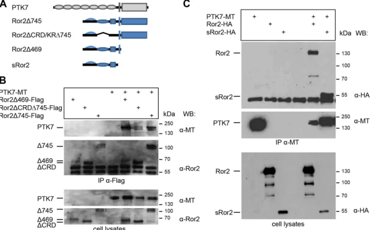
To determine which Ror2 domains are required for PTK7/Ror2 interaction different Ror2 deletion mutants were used (Fig 2A). Ror2Δ745 has a deletion of the intracellular serine/threo-nine-rich domains. The Ror2ΔCRD/KRΔ745 mutant lacks additionally the extracellular friz-zled-like cysteine-rich domain (CRD) required for Wnt binding and the kringle domain (KR). In the Ror2Δ469 construct major parts of the intracellular domain including the tyrosine-kinase domain, the serine/threonine-rich and the proline-rich domains have been deleted. Finally, the sRor2 lacks the entire intracellular and transmembrane domains. The FLAG-tagged Ror2 deletion constructs were expressed with full-length myc-FLAG-tagged PTK7 and co-precipitations were performed using anti-FLAG antibodies. All Ror2 deletion mutants were able to co-precipitate full-length PTK7 (Fig 2B and 2Cand data not shown). Conversely, if PTK7 was precipitated using anti-myc antibodies sRor2 was co-precipitated (Fig 2C). Thus, the PTK7/Ror2 interaction is likely mediated by the extracellular domains of these proteins, but does not require the CRD and kringle domains of Ror2.
PTK7 loss of function inhibits cell motility of explanted NC cells
Fig 1. PTK7 and Ror2 co-localize in NC cells and co-precipitate independent of the kinase-homology domain of PTK7. ACo-localization of PTK7 and Ror2. NC cells co-expressing Ror2-EGFP and PTK7-RFP show distinct areas of co-localization (yellow) as well as membrane areas where only PTK7-RFP (red) or Ror2-EGFP (green) is localized. The right panel shows a higher magnification of the single cell in the left panel (indicated by a dashed square), scale bar = 10μm.B,CCo-immunoprecipitation of PTK7 and Ror2. Full-length myc-tagged PTK7 (PTK7-MT) and a myc-tagged PTK7 deletion construct lacking the kinase homology domain (ΔkPTK7-MT) as well as a full-length HA-tagged Ror2 construct were expressed in MCF7 cells.BConstructs and their protein domains are depicted in the top panel. Abbreviations are as follows: IG (immunoglobulin domain), CRD (cysteine-rich domain), K (kringle domain), TM (transmembrane domain), KH (kinase homology domain), TK (tyrosine kinase), S/T (serine/threonine-rich domain), P (proline-rich domain).CImmunoprecipitation experiment; the cell transfection scheme is indicated at the top. Co-immunoprecipitation was carried out using either anti-myc (IPα-MT, upper panel) or anti-HA antibodies (IPα-HA, middle panel). The respective cell lysates
are shown in the bottom panel. Antibodies used for Western blotting and molecular weights are indicated at the right.
been shown to be required for NC migration [13], but its function on a cellular level remains elusive. Therefore, we used live-cell imaging to further analyze the migration behavior of explanted control and PTK7 morphant NC cells. Membrane-targeted GFP (mGFP) and cherry-labeled Histone2B (H2B-mcherry) were co-expressed to visualize cell shape and polar-ity. At the start of the experiment (0 hours), control NC cells formed a cell cluster where the leading-edge cells started to get polarized (Fig 3A, upper panel,S1 Movie). After one hour the leading cells left the main cell cluster and then dispersed rapidly during the 5 hour time-span of the experiment. In contrast, PTK7 MO injected NC cells adopted a more roundish shape and did not disperse as well as control cells (Fig 3 A, lower panel,S1 Movie). The dispersion of NC cells can be seen in the time series (Fig 3A), where–although explant size in the different experimental conditions is comparable at the beginning of the time-series–the area of cell spreading is significantly larger for the controls than the MO-injected explants after 5 hours of imaging. As it is difficult to determine the exact initial cell number and as the cell dissociation may not occur homogenously, we used cell tracking and Delaunay triangulation as more sophisticated methods to measure cell dispersion (Fig 3B and 3C). Delaunay triangulation determines the two closest cell neighbors and the area of the formed triangles is proportional Fig 2. The intracellular domain and the CRD domain of Ror2 are not required for PTK7/Ror2 interaction.Myc-tagged PTK7 (PTK7-MT) together with FLAG- or HA-tagged Ror2 deletions were expressed in MCF7 cells.AThe respective constructs and their protein domains are depicted in the top panel.B Immunoprecipitation using anti-FLAG antibodies; the cell transfection scheme is indicated at the top. Co-precipitated PTK7 was detected using anti-myc antibodies. Immunoprecipitated PTK7 is shown in the top panel, immunoprecipitated Ror2 constructs in the middle panel and cell lysates in the bottom panel. Antibodies used for Western blotting and molecular weights are indicated at the right.CImmunoprecipitation using anti-myc antibodies; the cell transfection scheme is indicated at the top. Co-precipitated full-length Ror2 and sRor2 were detected using anti-HA antibodies (top panel). Immunoprecipitated PTK7 is shown in the middle panel and cell lysates in the bottom panel. Antibodies used for Western blotting and molecular weights are indicated at the right.
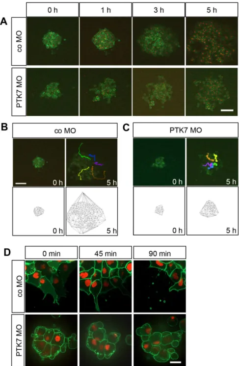
Fig 3. Loss of function of PTK7 affects NC cell shape and inhibits migration of explanted NC. ATime series showing explanted NC cells injected with 7.5 ng control or PTK7 MO in combination with 50 pgmGFP RNA and 250 pgH2B-mcherry. Cranial NC explants were excised at stage 16–17 and explanted on a fibronectin matrix and incubated until they had stably adhered to the matrix. NC migration was monitored for 5 hours using spinning disk microscopy (10x objective NA 0.45). Images for representative explants injected either with control or PTK7 MO are shown at the start of the experiment (0 h) or after 1, 3 or 5 hours; scale bar = 50μm.BCell tracking and Delaunay triangulation for explants injected with 7.5 ng co MO. The upper panel shows a single frame of the spinning disk movie and the lower panel the Delaunay triangulation at the start of the experiment (0 h) or after 5 hours. Cells were tracked over the whole five-hour time interval using the H2B staining of single nuclei. These tracks are shown for single cells as differently colored lines in the images taken after 5 hours, scale bar = 50μm.CCell tracking and Delaunay triangulation for explants injected with 7.5 ng PTK7 MO.DTime series showing explants injected with 7.5 ng co MO (upper panel) or 7.5 ng PTK7 MO (lower panel) at a higher magnification. Injected NC cells were explanted at stage 17, cultured for 1.5 hours and imaged with a 63x objective (NA 1.4); scale bar = 20μm. Images are shown at the start of the experiment (0 min) and after 45 and 90 minutes.
to the cell dispersion. The time series as well as cell tracking and Delaunay triangulation all show that control cells dispersed efficiently during the 5 hours of imaging (Fig 3B). In contrast, in explants where PTK7 protein expression was knocked down, the NC cells had difficulties to detach from the main cluster and did not disperse as well as the control NC cells (Fig 3C). Higher magnification shows that PTK7 morphant cells do not form extensive protrusions like control cells, but adopt a more roundish shape. This shape change does not correlate with cell death as determined by Propidium iodine staining (S2 Fig). Some of these explants are still able to form protrusion, but others adopt a cell movement reminiscent of blebbing (Fig 3D,S2 Movie). Thus, PTK7 loss of function has severe effects on NC cell shape and motility.
Ror2 rescues the PTK7 loss of function NC migration defect and this
requires the kinase domain of Ror2
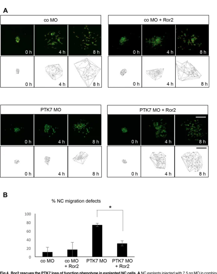
As PTK7 and Ror2 are both non-canonical Wnt receptors, which share functions in Wnt sig-naling, we analyzed if Ror2 can rescue the PTK7 loss of function NC migration defect. To this end NC cells injected either with co MO or PTK7 MO alone or in combination with Ror2 were explanted and their migration was analyzed by time-lapse imaging. While the majority of PTK7 MO injected NC cells showed migration defects, NC cells, which were co-injected with the PTK7 MO and Ror2 showed normal NC migration behavior (Fig 4,S3 Movie). Defects in protrusion formation as well as cell motility were rescued by co-expression of Ror2 (Fig 4A and 4B,S3 Movie) indicating that Ror2 expression can substitute for PTK7 in NC cells.
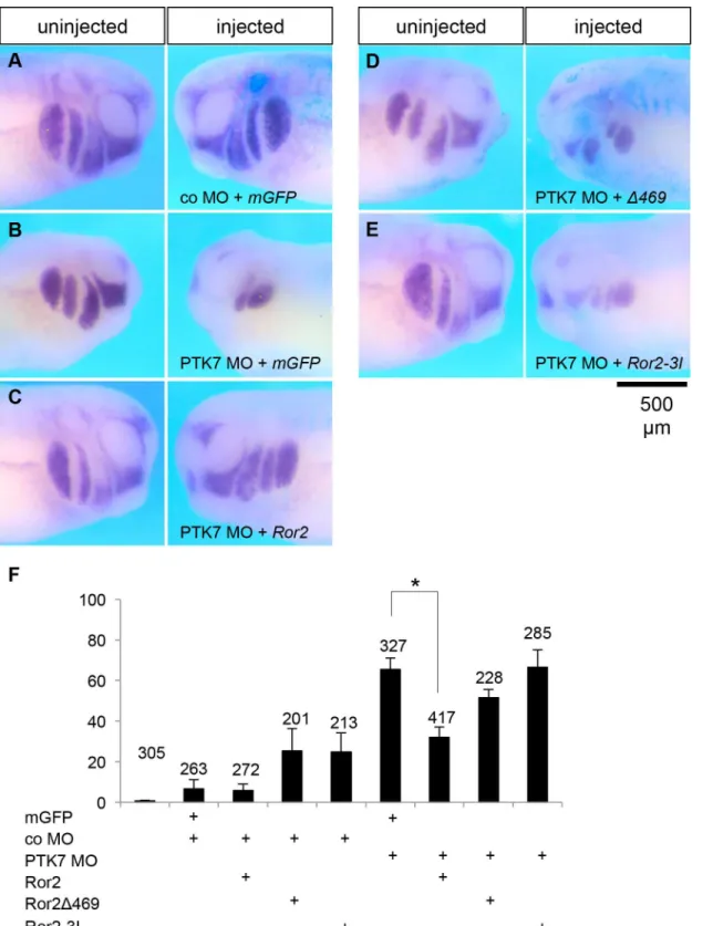
Similar results were obtained inin vivoexperiments where NC migration was analyzed at tadpole stages using twistin situhybridization. Embryos injected with co MO were unaffected, while the majority of embryos injected with the PTK7 MO showed severe NC migration defects (Fig 5A, 5B and 5F). Co-injection ofRor2RNA with the PTK7 MO significantly decreased the percentage of embryos showing NC migration defects (Fig 3C and 3F). In contrast co-injection of a deletion mutant ofRor2lacking major parts of the intracellular domain,Ror2Δ469(Fig 2A and 2F) did not show a significant rescue defect (Fig 3D and 3F). Interestingly, a kinase dead mutant of Ror2 (Ror2-3I), where three lysines at position 504 (in the putative ATP-binding motif), 507 and 509 were all replaced with isoleucine [57] to abolish the catalytic activity, was not able to rescue the PTK7 morphant NC migration defect. Thus, these results suggest that Ror2 can functionally replace PTK7 and that the kinase function of Ror2 is required for this activity.
Discussion
Fig 4. Ror2 rescues the PTK7 loss of function phenotype in explanted NC cells. ANC explants injected with 7.5 ng MO in combination with 50 pgmGFP RNA, 250 pgH2B-mcherryand 150 pgRor2RNA. Time-lapse images (upper panel) and Delaunay triangulations (lower panel) at the start of the experiment (0 h) and after 4 or 8 hours are shown for the different conditions.BGraph summarizing percentage of migration defects of 3 independent experiments (total of 39 explants). Standard error of the means are shown. Asterisks indicates a p-value in a Student’s t-test<0.05. Scale bars = 200μm.
interaction is direct or for example mediated by Wnt ligands or Frizzled co-receptors. Since interaction of both PTK7 and Ror2 with Frizzled receptors has been shown [13,36,50,55], members of the Frizzled receptor family may also contribute to the PTK7/Ror2 interaction. In addition, PTK7 [35,36] and Ror2 interact with different members of the Wnt protein family, although in the case of Ror2 only non-canonical Wnts such as Wnt-5a were found to activate downstream signaling [49,50,57,71]. Thus, although the exact molecular composition of a PTK7/Ror2 receptor complex remains yet to be analyzed, the formation and composition may also affect the selective binding to members of the Wnt family and thereby modulate intracellu-lar signaling events.
The downstream signaling events of the PTK7/Ror2 complex are currently unclear, but may be mediated by an activation of c-Jun N-terminal kinase (JNK). Previously, we have shown that PTK7 overexpression inXenopusectodermal explants leads to a nuclear localization of phosphorylated JNK [13] and this is not observed if a kinase deletion mutant of PTK7 (ΔkPTK7) is overexpressed (data not shown). Ror2 has been shown to have a similar function [44,46,53]. Therefore, possibly these molecules collaborate to recruit Dishevelled and to activate JNK via the PCP pathway [72] and thereby enable the migration of NC cells [8,9,17,18,25]. Supporting this hypothesis, Ror2 overexpression can rescue the PTK7 loss of function NC migration defect possibly by compensating for PTK7. Consistently, this rescue effect requires the kinase domain of Ror2, which is necessary for JNK activation [44]. In the context of mesodermal convergent extension it was previously shown that Ror2 activates a sig-naling cascade including PI3K, the small GTPase cdc42, MKK7, JNK and c-jun and ATF2 tran-scription factors [44]. For PTK7 the mechanism of JNK activation remains unclear, but may involve the adaptor protein RACK1, which is required for PTK7-mediated Dishevelled recruit-ment [30]. As RACK1 plays a role in PKC-mediated JNK phosphorylation [73], PTK7 may activate JNK via RACK1 or Dishevelled itself [72]. JNK is involved in a multitude of processes and can activate the transcription factors jun, fos and ATF2 [74]. Like Ror2, PTK7 is also able to activate ATF2-mediated transcription as shown by activation of an ATF2 luciferase reporter inXenopuslysates [35,75]. Thus, one of the common signaling outcomes of PTK7 and Ror2 may be the transcription of ATF2-dependent target genes, which has previously also been acknowledged as a readout for a bona fide activator of non-canonical Wnt signaling [75].
migration. We have shown that the tyrosine kinase domain of Ror2 is required to rescue PTK7 loss of function. However, as the tyrosine kinase activity of Ror2 is required for both activation of JNK and inhibition of canonical Wnt signaling [44,78], Ror2 is capable to compensate for both putative functions of PTK7. Therefore, it remains to be seen which one of these or if possi-bly even a combination of both is required. In summary, PTK7 and Ror2 share signaling func-tions that may be enhanced by the combination of both molecules and that may allow for example Ror2 to compensate for PTK7.
A possible interaction of PTK7 with Ror2 is likely not limited to NC cells, as PTK7 and Ror2 have also been implicated in tumor development and progression. PTK7 and Ror2 expression is frequently deregulated in a variety of cancers [79,80,81,82,83,84,85]. Several stud-ies indicate an association of Ror2, in some cases downstream of Wnt5a, with tumor invasive-ness and metastasis [82,84,86,87,88]. However, it has been shown that epigenetic silencing of Ror2 in colon cancer promotes cell proliferation and tumor growth [89], suggesting that depending on the cellular context Ror2 could also act as a tumor-suppressor. Similarly, contra-dictory results are also seen for the function of PTK7. PTK7 is expressed in acute myeloid leu-kemia, where it promotes cell migration and leads to a poor clinical outcome [90]. Additional studies found migration and invasion promoting functions of PTK7 in lung cancer, intrahepa-tic cholangiocarcinoma, glioma and prostate cancer [42,91,92,93]. In contrast, another publica-tion shows that PTK7 is a target of membrane type-1 matrix metalloproteinase and that PTK7 expression inhibits cell invasion [94]. Thus, the function of PTK7 and Ror2 may depend on tumor context and ultimately on receptor context.
Supporting Information
S1 Fig. PTK7 and Ror2 co-precipitate each other but not EGFR or TGFß1R. AFull-length
myc-tagged PTK7 (PTK7-MT) was co-expressed with Ror2-EGFP, EGFR-EGFP [95] or TGFß1R-EGFP (kind gift of A. Menke, Molecular Oncology of Solid Tumors, Giessen, Ger-many) as indicated in MCF7 cells. Cell lysates were precipitated using anti-myc antibodies (IP α-MT, upper panel). Precipitates are shown in the upper panels, cell lysates in the lower panel. Antibodies used for Western blotting and molecular weights are indicated at the right.B Full-length HA-tagged Ror2 was co-expressed with PTK7-MT, EGFR-EGFP or TGFß1R-GFP as indicated in MCF7 cells and cell lysates were precipitated using anti-HA antibodies (IPα-HA, upper panel). Precipitates are shown in the three upper panels and lysates in the two lower pan-els. Antibodies used for Western blotting and molecular weights are indicated at the right;
marks unspecific bands,Ror2 signal remaining from previous anti-HA staining, which was
only partially removed by blot stripping. (TIF)
S2 Fig. Propidium Iodine (PI) staining to determine the viability of PTK7 loss of function
NC cells.NC explants injected with 7.5 ng control MO or PTK7 MO in combination with
50 pg mGFP were treated with PI (10μg/ml) to test for the viability of the explanted NC cells. Cell membrane integrity prevents PI staining of viable cells, while it can stain nucleic acids of apoptotic cells (red). In PTK7 morphant cells few PI-positive cells appear after 3 hours com-pared to 6 hours in controls (one example each is marked by a red arrow). Round-shaped PTK7 morphant NC cells appear early in the experiment and keep moving/blebbing for up to a couple of hours (white arrows, numbers indicate specific cells during the course of the experi-ment). Thus, the roundish cell shape is not necessarily an indication of cell death. Dashed squares show higher magnifications of specific cells. Scale bar = 50μm.
S1 Movie. Time-lapse movie showing the migration of NC cells injected with control MO
(left) in comparison to an explant injected with PTK7 MO.Images were taken using a
Spin-ning Disk microscope with a 10x plan apochromat objective NA (0.45). (AVI)
S2 Movie. Higher magnification using a 63x plan apochromat objective NA 1.4) of control MO (left) and PTK7 MO injected (right) NC explants.
(AVI)
S3 Movie. Time-lapse movie showing that Ror2 expression rescues the PTK7 morphant
phenotype.Explants injected with control MO (upper panel, left), control MO and Ror2 RNA
(upper panel, right), PTK7 MO (lower panel, left) or PTK7 MO and Ror2 RNA (loer panel, right) are shown over a time interval of 8 hours and 20 minutes.
(AVI)
Acknowledgments
We would like to especially thank Alexandra Schambony for constructs and helpful discussions and comments on the manuscript. We thank Iryna Shnitsar for cloning of the PTK7-RFP and Jubin Kashef and Andre Mencke for supplying plasmids. Further, we thank Ingrid Bohl-Maser and Christiane Rohrbach for technical assistance and Melanie Bernhardt for taking excellent care of ourXenopuscolony.
Author Contributions
Conceived and designed the experiments: AB MP AG HB. Performed the experiments: MP AG HB ER. Analyzed the data: AB MP AG HB ER. Contributed reagents/materials/analysis tools: AB. Wrote the paper: AB MP AG.
References
1. Powell DR, Blasky AJ, Britt SG, Artinger KB (2013) Riding the crest of the wave: parallels between the neural crest and cancer in epithelial-to-mesenchymal transition and migration. Wiley interdisciplinary reviews Systems biology and medicine 5: 511–522. doi:10.1002/wsbm.1224PMID:23576382 2. Kuriyama S, Mayor R (2008) Molecular analysis of neural crest migration. Philos Trans R Soc Lond B
Biol Sci.
3. Theveneau E, Mayor R (2012) Neural crest delamination and migration: from epithelium-to-mesen-chyme transition to collective cell migration. Developmental biology 366: 34–54. doi:10.1016/j.ydbio. 2011.12.041PMID:22261150
4. Theveneau E, Mayor R (2011) Collective cell migration of the cephalic neural crest: the art of integrating information. Genesis 49: 164–176. doi:10.1002/dvg.20700PMID:21157935
5. Snider TN, Mishina Y (2014) Cranial neural crest cell contribution to craniofacial formation, pathology, and future directions in tissue engineering. Birth defects research Part C, Embryo today: reviews 102: 324–332.
6. Zhang D, Ighaniyan S, Stathopoulos L, Rollo B, Landman K, Hutson J, et al. (2014) The neural crest: a versatile organ system. Birth defects research Part C, Embryo today: reviews 102: 275–298. 7. Bolande RP (1997) Neurocristopathy: its growth and development in 20 years. Pediatric pathology &
laboratory medicine: journal of the Society for Pediatric Pathology, affiliated with the International Pae-diatric Pathology Association 17: 1–25.
8. Carmona-Fontaine C, Matthews HK, Kuriyama S, Moreno M, Dunn GA, Parsons M, et al. (2008) Con-tact inhibition of locomotion in vivo controls neural crest directional migration. Nature 456: 957–961. doi:10.1038/nature07441PMID:19078960
10. Theveneau E, Steventon B, Scarpa E, Garcia S, Trepat X, Streit A, et al. (2013) Chase-and-run between adjacent cell populations promotes directional collective migration. Nature cell biology 15: 763–772. doi:10.1038/ncb2772PMID:23770678
11. Banerjee S, Gordon L, Donn TM, Berti C, Moens CB, Burden SJ, et al. (2011) A novel role for MuSK and non-canonical Wnt signaling during segmental neural crest cell migration. Development 138: 3287–3296. doi:10.1242/dev.067306PMID:21750038
12. Ulmer B, Hagenlocher C, Schmalholz S, Kurz S, Schweickert A, Kohl A, et al. (2013) Calponin 2 acts as an effector of noncanonical Wnt-mediated cell polarization during neural crest cell migration. Cell reports 3: 615–621. doi:10.1016/j.celrep.2013.02.015PMID:23499442
13. Shnitsar I, Borchers A (2008) PTK7 recruits dsh to regulate neural crest migration. Development 135: 4015–4024. doi:10.1242/dev.023556PMID:19004858
14. Mayor R, Theveneau E (2014) The role of the non-canonical Wnt-planar cell polarity pathway in neural crest migration. The Biochemical journal 457: 19–26. doi:10.1042/BJ20131182PMID:24325550 15. Abercrombie M, Heaysman JE (1953) Observations on the social behaviour of cells in tissue culture. I.
Speed of movement of chick heart fibroblasts in relation to their mutual contacts. Experimental cell research 5: 111–131. PMID:13083622
16. Abercrombie M, Heaysman JE (1954) Observations on the social behaviour of cells in tissue culture. II. Monolayering of fibroblasts. Exp Cell Res 6: 293–306. PMID:13173482
17. Theveneau E, Marchant L, Kuriyama S, Gull M, Moepps B, Parsons M, et al. (2010) Collective chemo-taxis requires contact-dependent cell polarity. Developmental cell 19: 39–53. doi:10.1016/j.devcel. 2010.06.012PMID:20643349
18. Mayor R, Carmona-Fontaine C (2010) Keeping in touch with contact inhibition of locomotion. Trends in cell biology 20: 319–328. doi:10.1016/j.tcb.2010.03.005PMID:20399659
19. Axelrod JD, McNeill H (2002) Coupling planar cell polarity signaling to morphogenesis. ScientificWorld-Journal 2: 434–454. PMID:12806028
20. Klein TJ, Mlodzik M (2005) Planar cell polarization: an emerging model points in the right direction. Annu Rev Cell Dev Biol 21: 155–176. PMID:16212491
21. Vladar EK, Antic D, Axelrod JD (2009) Planar cell polarity signaling: the developing cell's compass. Cold Spring Harbor perspectives in biology 1: a002964. doi:10.1101/cshperspect.a002964PMID: 20066108
22. Wallingford JB (2012) Planar cell polarity and the developmental control of cell behavior in vertebrate embryos. Annual review of cell and developmental biology 28: 627–653. doi: 10.1146/annurev-cellbio-092910-154208PMID:22905955
23. Theveneau E, Mayor R (2010) Integrating chemotaxis and contact-inhibition during collective cell migration: Small GTPases at work. Small GTPases 1: 113–117. PMID:21686264
24. Clay MR, Halloran MC (2013) Rho activation is apically restricted by Arhgap1 in neural crest cells and drives epithelial-to-mesenchymal transition. Development 140: 3198–3209. doi:10.1242/dev.095448 PMID:23804498
25. Theveneau E, Mayor R (2013) Collective cell migration of epithelial and mesenchymal cells. Cellular and molecular life sciences: CMLS 70: 3481–3492. doi:10.1007/s00018-012-1251-7PMID:23314710 26. Becker SF, Mayor R, Kashef J (2013) Cadherin-11 mediates contact inhibition of locomotion during
Xenopus neural crest cell migration. PloS one 8: e85717. doi:10.1371/journal.pone.0085717PMID: 24392028
27. Carmona-Fontaine C, Theveneau E, Tzekou A, Tada M, Woods M, Page KM, et al. (2011) Complement fragment C3a controls mutual cell attraction during collective cell migration. Developmental cell 21: 1026–1037. doi:10.1016/j.devcel.2011.10.012PMID:22118769
28. Miller MA, Steele RE (2000) Lemon encodes an unusual receptor protein-tyrosine kinase expressed during gametogenesis in Hydra. Dev Biol 224: 286–298. PMID:10926767
29. Kroiher M, Miller MA, Steele RE (2001) Deceiving appearances: signaling by "dead" and "fractured" receptor protein-tyrosine kinases. Bioessays 23: 69–76. PMID:11135311
30. Wehner P, Shnitsar I, Urlaub H, Borchers A (2011) RACK1 is a novel interaction partner of PTK7 that is required for neural tube closure. Development 138: 1321–1327. doi:10.1242/dev.056291PMID: 21350015
31. Lu X, Borchers AG, Jolicoeur C, Rayburn H, Baker JC, Tessier-Lavigne M (2004) PTK7/CCK-4 is a novel regulator of planar cell polarity in vertebrates. Nature 430: 93–98. PMID:15229603
33. Paudyal A, Damrau C, Patterson VL, Ermakov A, Formstone C, Lalanne Z, et al. (2010) The novel mouse mutant, chuzhoi, has disruption of Ptk7 protein and exhibits defects in neural tube, heart and lung development and abnormal planar cell polarity in the ear. Bmc Developmental Biology 286: 20970–20976.
34. Hayes M, Naito M, Daulat A, Angers S, Ciruna B (2013) Ptk7 promotes non-canonical Wnt/PCP-medi-ated morphogenesis and inhibits Wnt/beta-catenin-dependent cell fate decisions during vertebrate development. Development 140: 1807–1818. doi:10.1242/dev.090183PMID:23533179
35. Peradziryi H, Kaplan NA, Podleschny M, Liu X, Wehner P, Borchers A, et al. (2011) PTK7/Otk interacts with Wnts and inhibits canonical Wnt signalling. The EMBO journal 30: 3729–3740. doi:10.1038/ emboj.2011.236PMID:21772251
36. Linnemannstons K, Ripp C, Honemann-Capito M, Brechtel-Curth K, Hedderich M, Wodarz A (2014) The PTK7-related transmembrane proteins off-track and off-track 2 are co-receptors for Drosophila Wnt2 required for male fertility. PLoS genetics 10: e1004443. doi:10.1371/journal.pgen.1004443 PMID:25010066
37. Puppo F, Thome V, Lhoumeau AC, Cibois M, Gangar A, Lembo F, et al. (2011) Protein tyrosine kinase 7 has a conserved role in Wnt/beta-catenin canonical signalling. EMBO reports 12: 43–49. doi:10. 1038/embor.2010.185PMID:21132015
38. Bin-Nun N, Lichtig H, Malyarova A, Levy M, Elias S, Frank D (2014) PTK7 modulates Wnt signaling activity via LRP6. Development 141: 410–421. doi:10.1242/dev.095984PMID:24353057 39. Andreeva A, Lee J, Lohia M, Wu X, Macara IG, Lu X (2014) PTK7-Src signaling at epithelial cell
con-tacts mediates spatial organization of actomyosin and planar cell polarity. Developmental cell 29: 20– 33. doi:10.1016/j.devcel.2014.02.008PMID:24703874
40. Golubkov VS, Strongin AY (2014) Downstream signaling and genome-wide regulatory effects of PTK7 pseudokinase and its proteolytic fragments in cancer cells. Cell communication and signaling: CCS 12: 15. doi:10.1186/1478-811X-12-15PMID:24618420
41. Chen R, Khatri P, Mazur PK, Polin M, Zheng Y, Vaka D, et al. (2014) A meta-analysis of lung cancer gene expression identifies PTK7 as a survival gene in lung adenocarcinoma. Cancer research 74: 2892–2902. doi:10.1158/0008-5472.CAN-13-2775PMID:24654231
42. Jin J, Ryu HS, Lee KB, Jang JJ (2014) High expression of protein tyrosine kinase 7 significantly associ-ates with invasiveness and poor prognosis in intrahepatic cholangiocarcinoma. PloS one 9: e90247. doi:10.1371/journal.pone.0090247PMID:24587299
43. Peradziryi H, Tolwinski NS, Borchers A (2012) The many roles of PTK7: a versatile regulator of cell-cell communication. Archives of biochemistry and biophysics 524: 71–76. doi:10.1016/j.abb.2011.12.019 PMID:22230326
44. Schambony A, Wedlich D (2007) Wnt-5A/Ror2 regulate expression of XPAPC through an alternative noncanonical signaling pathway. Dev Cell 12: 779–792. PMID:17488628
45. Mikels AJ, Nusse R (2006) Purified Wnt5a protein activates or inhibits beta-catenin-TCF signaling depending on receptor context. Plos Biology 4: 570–582.
46. Nomachi A, Nishita M, Inaba D, Enomoto M, Hamasaki M, Minami Y (2008) Receptor tyrosine kinase Ror2 mediates Wnt5a-induced polarized cell migration by activating c-Jun N-terminal kinase via actin-binding protein filamin A. J Biol Chem 283: 27973–27981. doi:10.1074/jbc.M802325200PMID: 18667433
47. Grumolato L, Liu GZ, Mong P, Mudbhary R, Biswas R, Arroyave R, et al. (2010) Canonical and nonca-nonical Wnts use a common mechanism to activate completely unrelated coreceptors. Genes & Devel-opment 24: 2517–2530.
48. Winkel A, Stricker S, Tylzanowski P, Seiffart V, Mundlos S, Gross G, et al. (2008) Wnt-ligand-depen-dent interaction of TAK1 (TGF-beta-activated kinase-1) with the receptor tyrosine kinase Ror2 modu-lates canonical Wnt-signalling. Cell Signal 20: 2134–2144. doi:10.1016/j.cellsig.2008.08.009PMID: 18762249
49. Billiard J, Way DS, Seestaller-Wehr LM, Moran RA, Mangine A, Bodine PV (2005) The orphan receptor tyrosine kinase Ror2 modulates canonical Wnt signaling in osteoblastic cells. Molecular endocrinology 19: 90–101. PMID:15388793
50. Oishi I, Suzuki H, Onishi N, Takada R, Kani S, Ohkawara B, et al. (2003) The receptor tyrosine kinase Ror2 is involved in non-canonical Wnt5a/JNK signalling pathway. Genes to cells: devoted to molecular & cellular mechanisms 8: 645–654.
52. Nishita M, Yoo SK, Nomachi A, Kani S, Sougawa N, Ohta Y, et al. (2006) Filopodia formation mediated by receptor tyrosine kinase Ror2 is required for Wnt5a-induced cell migration. J Cell Biol 175: 555– 562. PMID:17101698
53. Witte F, Bernatik O, Kirchner K, Masek J, Mahl A, Krejci P, et al. (2010) Negative regulation of Wnt sig-naling mediated by CK1-phosphorylated Dishevelled via Ror2. Faseb J 24: 2417–2426. doi:10.1096/ fj.09-150615PMID:20215527
54. Yamamoto H, Yoo SK, Nishita M, Kikuchi A, Minami Y (2007) Wnt5a modulates glycogen synthase kinase 3 to induce phosphorylation of receptor tyrosine kinase Ror2. Genes to cells: devoted to molecu-lar & cellumolecu-lar mechanisms 12: 1215–1223.
55. Yamamoto S, Nishimura O, Misaki K, Nishita M, Minami Y, Yonemura S, et al. (2008) Cthrc1 selectively activates the planar cell polarity pathway of Wnt signaling by stabilizing the Wnt-receptor complex. Dev Cell 15: 23–36. doi:10.1016/j.devcel.2008.05.007PMID:18606138
56. Gao B, Song H, Bishop K, Elliot G, Garrett L, English MA, et al. (2011) Wnt signaling gradients establish planar cell polarity by inducing Vangl2 phosphorylation through Ror2. Developmental cell 20: 163– 176. doi:10.1016/j.devcel.2011.01.001PMID:21316585
57. Hikasa H, Shibata M, Hiratani I, Taira M (2002) The Xenopus receptor tyrosine kinase Xror2 modulates morphogenetic movements of the axial mesoderm and neuroectoderm via Wnt signaling. Development 129: 5227–5239. PMID:12399314
58. He F, Xiong W, Yu X, Espinoza-Lewis R, Liu C, Gu S, et al. (2008) Wnt5a regulates directional cell migration and cell proliferation via Ror2-mediated noncanonical pathway in mammalian palate develop-ment. Development 135: 3871–3879. doi:10.1242/dev.025767PMID:18948417
59. Yamada M, Udagawa J, Matsumoto A, Hashimoto R, Hatta T, Nishita M, et al. (2010) Ror2 is required for midgut elongation during mouse development. Developmental dynamics: an official publication of the American Association of Anatomists 239: 941–953.
60. Feike AC, Rachor K, Gentzel M, Schambony A (2010) Wnt5a/Ror2-induced upregulation of xPAPC requires xShcA. Biochem Biophys Res Commun.
61. Nieuwkoop PD, Faber J, Hubrecht Laboratory Utrecht. (1956) Normal table of Xenopus laevis (Daudin): a systematical and chronological survey of the development from the fertilized egg till the end of meta-morphosis. Amsterdam: North-Holland Pub. Co. 243 p. p.
62. Smith WC, Harland RM (1991) Injected Xwnt-8 RNA acts early in Xenopus embryos to promote forma-tion of a vegetal dorsalizing center. Cell 67: 753–765. PMID:1657405
63. Kim SH, Yamamoto A, Bouwmeester T, Agius E, Robertis EM (1998) The role of paraxial protocadherin in selective adhesion and cell movements of the mesoderm during Xenopus gastrulation. Development 125: 4681–4690. PMID:9806917
64. Kashef J, Kohler A, Kuriyama S, Alfandari D, Mayor R, Wedlich D (2009) Cadherin-11 regulates protru-sive activity in Xenopus cranial neural crest cells upstream of Trio and the small GTPases. Genes Dev 23: 1393–1398. doi:10.1101/gad.519409PMID:19528317
65. Sammar M, Stricker S, Schwabe GC, Sieber C, Hartung A, Hanke M, et al. (2004) Modulation of GDF5/ BRI-b signalling through interaction with the tyrosine kinase receptor Ror2. Genes to cells: devoted to molecular & cellular mechanisms 9: 1227–1238.
66. Borchers A, David R, Wedlich D (2001) Xenopus cadherin-11 restrains cranial neural crest migration and influences neural crest specification. Development 128: 3049–3060. PMID:11688555 67. Borchers A, Epperlein HH, Wedlich D (2000) An assay system to study migratory behavior of cranial
neural crest cells in Xenopus. Dev Genes Evol 210: 217–222. PMID:11180825
68. Meijering E, Dzyubachyk O, Smal I (2012) Methods for cell and particle tracking. Methods in enzymol-ogy 504: 183–200. doi:10.1016/B978-0-12-391857-4.00009-4PMID:22264535
69. Ossipova O, Sokol SY (2011) Neural crest specification by noncanonical Wnt signaling and PAR-1. Development 138: 5441–5450. doi:10.1242/dev.067280PMID:22110058
70. Barclay AN (2003) Membrane proteins with immunoglobulin-like domains—a master superfamily of interaction molecules. Seminars in immunology 15: 215–223. PMID:14690046
71. Liu Y, Rubin B, Bodine PV, Billiard J (2008) Wnt5a induces homodimerization and activation of Ror2 receptor tyrosine kinase. Journal of cellular biochemistry 105: 497–502. doi:10.1002/jcb.21848PMID: 18615587
72. Boutros M, Paricio N, Strutt DI, Mlodzik M (1998) Dishevelled activates JNK and discriminates between JNK pathways in planar polarity and wingless signaling. Cell 94: 109–118. PMID:9674432
74. Weston CR, Davis RJ (2002) The JNK signal transduction pathway. Current opinion in genetics & development 12: 14–21.
75. Ohkawara B, Niehrs C (2011) An ATF2-based luciferase reporter to monitor non-canonical Wnt signal-ing in Xenopus embryos. Dev Dyn 240: 188–194. doi:10.1002/dvdy.22500PMID:21128306 76. Medina A, Reintsch W, Steinbeisser H (2000) Xenopus frizzled 7 can act in canonical and
non-canoni-cal Wnt signaling pathways: implications on early patterning and morphogenesis. Mech Dev 92: 227– 237. PMID:10727861
77. de Melker AA, Desban N, Duband JL (2004) Cellular localization and signaling activity of beta-catenin in migrating neural crest cells. Developmental dynamics: an official publication of the American Associ-ation of Anatomists 230: 708–726.
78. Mikels A, Minami Y, Nusse R (2009) Ror2 receptor requires tyrosine kinase activity to mediate Wnt5A signaling. The Journal of biological chemistry 284: 30167–30176. doi:10.1074/jbc.M109.041715 PMID:19720827
79. Easty DJ, Mitchell PJ, Patel K, Florenes VA, Spritz RA, Bennett DC (1997) Loss of expression of recep-tor tyrosine kinase family genes PTK7 and SEK in metastatic melanoma. Int J Cancer 71: 1061–1065. PMID:9185712
80. Endoh H, Tomida S, Yatabe Y, Konishi H, Osada H, Tajima K, et al. (2004) Prognostic model of pulmo-nary adenocarcinoma by expression profiling of eight genes as determined by quantitative real-time reverse transcriptase polymerase chain reaction. J Clin Oncol 22: 811–819. PMID:14990636 81. Muller-Tidow C, Schwable J, Steffen B, Tidow N, Brandt B, Becker K, et al. (2004) High-throughput
analysis of genome-wide receptor tyrosine kinase expression in human cancers identifies potential novel drug targets. Clin Cancer Res 10: 1241–1249. PMID:14977821
82. O'Connell MP, Fiori JL, Xu M, Carter AD, Frank BP, Camilli TC, et al. (2010) The orphan tyrosine kinase receptor, ROR2, mediates Wnt5A signaling in metastatic melanoma. Oncogene 29: 34–44. doi:10. 1038/onc.2009.305PMID:19802008
83. Saha S, Bardelli A, Buckhaults P, Velculescu VE, Rago C, St Croix B, et al. (2001) A phosphatase associated with metastasis of colorectal cancer. Science 294: 1343–1346. PMID:11598267 84. Wright TM, Brannon AR, Gordan JD, Mikels AJ, Mitchell C, Chen S, et al. (2009) Ror2, a
developmen-tally regulated kinase, promotes tumor growth potential in renal cell carcinoma. Oncogene 28: 2513– 2523. doi:10.1038/onc.2009.116PMID:19448672
85. Ataseven B, Gunesch A, Eiermann W, Kates RE, Hogel B, Knyazev P, et al. (2014) PTK7 as a potential prognostic and predictive marker of response to adjuvant chemotherapy in breast cancer patients, and resistance to anthracycline drugs. OncoTargets and therapy 7: 1723–1731. doi:10.2147/OTT.S62676 PMID:25336969
86. Enomoto M, Hayakawa S, Itsukushima S, Ren DY, Matsuo M, Tamada K, et al. (2009) Autonomous regulation of osteosarcoma cell invasiveness by Wnt5a/Ror2 signaling. Oncogene 28: 3197–3208. doi:10.1038/onc.2009.175PMID:19561643
87. Kobayashi M, Shibuya Y, Takeuchi J, Murata M, Suzuki H, Yokoo S, et al. (2009) Ror2 expression in squamous cell carcinoma and epithelial dysplasia of the oral cavity. Oral Surg Oral Med Oral Pathol Oral Radiol Endod 107: 398–406. doi:10.1016/j.tripleo.2008.08.018PMID:19217015
88. Morioka K, Tanikawa C, Ochi K, Daigo Y, Katagiri T, Kawano H, et al. (2009) Orphan receptor tyrosine kinase ROR2 as a potential therapeutic target for osteosarcoma. Cancer Sci 100: 1227–1233. doi:10. 1111/j.1349-7006.2009.01165.xPMID:19486338
89. Lara E, Calvanese V, Huidobro C, Fernandez AF, Moncada-Pazos A, Obaya AJ, et al. (2010) Epige-netic repression of ROR2 has a Wnt-mediated, pro-tumourigenic role in colon cancer. Mol Cancer 9: 170. doi:10.1186/1476-4598-9-170PMID:20591152
90. Prebet T, Lhoumeau AC, Arnoulet C, Aulas A, Marchetto S, Audebert S, et al. (2010) The cell polarity PTK7 receptor acts as a modulator of the chemotherapeutic response in acute myeloid leukaemia and impairs clinical outcome. Blood.
91. Gartner S, Gunesch A, Knyazeva T, Wolf P, Hogel B, Eiermann W, et al. (2014) PTK 7 is a transforming gene and prognostic marker for breast cancer and nodal metastasis involvement. PloS one 9: e84472. doi:10.1371/journal.pone.0084472PMID:24409301
92. Liu Q, Zhang C, Yuan J, Fu J, Wu M, Su J, et al. (2014) PTK7 regulates Id1 expression in CD44-high gli-oma cells. Neuro-oncology.
94. Golubkov VS, Chekanov AV, Cieplak P, Aleshin AE, Chernov AV, Zhu W, et al. (2010) The Wnt/planar cell polarity (PCP) protein tyrosine kinase-7 (PTK7) is a highly efficient proteolytic target of membrane type-1 matrix metalloproteinase (MT1-MMP): implications in cancer and embryogenesis. J Biol Chem. 95. Carter RE, Sorkin A (1998) Endocytosis of functional epidermal growth factor receptor-green