RESEARCH ARTICLE
Entamoeba histolytica
Trophozoites and
Lipopeptidophosphoglycan Trigger Human
Neutrophil Extracellular Traps
Eva E. Ávila1, Norma Salaiza2☯, Julieta Pulido1☯, Mayra C. Rodríguez1, César
Díaz-Godínez3, Juan P. Laclette3, Ingeborg Becker2*, Julio C. Carrero3
*
1Department of Biology, Division of Exact and Natural Sciences, Universidad de Guanajuato, 36050, Guanajuato, México,2Department of Experimental Medicine, Medical Faculty, Universidad Nacional Autónoma de México, 04510, México D.F., México,3Department of Immunology, Instituto de Investigaciones Biomédicas, Universidad Nacional Autónoma de México, 04510, México D.F., México
☯These authors contributed equally to this work.
*carrero@unam.mx(JCC);ingeborg_becker@hotmail.com(IB)
Abstract
Neutrophil defense mechanisms include phagocytosis, degranulation and the formation of extracellular traps (NET). These networks of DNA are triggered by several immune and microbial factors, representing a defense strategy to prevent microbial spread by trapping/ killing pathogens. This may be important againstEntamoeba histolytica, since its large size hinders its phagocytosis. The aim of this study was to determine whetherE.histolyticaand their lipopeptidophosphoglycan (EhLPPG) induce the formation of NETs and the outcome of their interaction with the parasite. Our data show that live amoebae and EhLPPG, but not fixed trophozoites, induced NET formation in a time and dose dependent manner, starting at 5 min of co-incubation. Although immunofluorescence studies showed that the NETs contain cathelicidin LL-37 in close proximity to amoebae, the trophozoite growth was only affected when ethylene glycol tetra-acetic acid (EGTA) was present during contact with NETs, suggesting that the activity of enzymes requiring calcium, such as DNases, may be important for amoeba survival. In conclusion,E.histolyticatrophozoites and EhLPPG inducein vitroformation of human NETs, which did not affect the parasite growth unless a chelating agent was present. These results suggest that NETs may be an important factor of the innate immune response during infection withE.histolytica.
Introduction
Maternal and child undernutrition, highly prevalent in low- and middle-income countries, account for about 35% of deaths for children younger than 5 years[1]. The limitation of nutri-ents negatively impacts the immune response, predisposing to infectious diseases, among them amoebiasis and other diarrheal infections[2]. Amoebiasis caused by the protozoan parasite Ent-amoeba histolyticais ranked as the third leading parasite-associated cause of human mortality worldwide, behind malaria and schistosomiasis[3], and the second leading cause of intestinal
a11111
OPEN ACCESS
Citation:Ávila EE, Salaiza N, Pulido J, Rodríguez MC, Díaz-Godínez C, Laclette JP, et al. (2016)
Entamoeba histolyticaTrophozoites and
Lipopeptidophosphoglycan Trigger Human Neutrophil Extracellular Traps. PLoS ONE 11(7): e0158979. doi:10.1371/journal.pone.0158979
Editor:Nades Palaniyar, The Hospital for Sick Children and The University of Toronto, CANADA
Received:March 19, 2016
Accepted:June 24, 2016
Published:July 14, 2016
Copyright:© 2016 Ávila et al. This is an open access article distributed under the terms of the
Creative Commons Attribution License, which permits unrestricted use, distribution, and reproduction in any medium, provided the original author and source are credited.
Data Availability Statement:All relevant data are within the paper and its Supporting Information files.
parasitosis behind cryptosporidiosis[4]. Thus,E.histolyticawas found responsible for 55,500 deaths worldwide in 2010 and it is estimated to account for 10 million cases of dysentery and liver abscesses every year. Tissue invasion of the intestine or liver byE.histolyticais associated with the induction of a strong inflammatory response characterized by the recruitment of a large number of neutrophils in the early stages[5–8]. Usually, large neutrophil infiltrates can be
found surrounding trophozoites, which show no evidence of apparent damage. Therefore, the role of neutrophils in amoebiasis has always been controversial, since some groups claim that these granulocytes participate in the resolution of infection[9–14], whereas other groups
sug-gest that they are involved in tissue damage[15–20]. However, humans with a mutation in the
leptin receptor (Q223R) have increased susceptibility to amoebiasis, likely due to impaired che-motaxis and reduced gut infiltration of neutrophils, suggesting a contribution of these cells in eliminatingE.histolyticain natural infection[21].
One of the innate immune mechanisms exerted by neutrophils is the formation of extracel-lular traps of DNA, known as NETs. NETs are complex weblike structures of decondensed chromatin decorated with granular and cytoplasmic proteins that arise from the release of the neutrophil nuclear contents under several stimulating conditions[22–25]. Among other
pro-teins, human NETs contain cathelicidin (LL-37), a cationic antimicrobial peptide present in the specific granules and produced after the C terminal cleavage of the human cationic antimi-crobial Protein 18 (hCAP-18) by serine proteases[26]. It has been described that NETs are able to trap both gram positive and negative bacteria, as well as fungi, viruses and parasites, killing or inhibiting their growth, preventing the spread of infections and thus contributing to the establishment of a protective immune response against pathogens[27]. However, conflicting reports have arisen as consequence of using different techniques to assess microbial killing, such as counting of plated colonies, where NETs are able to clump the microbes without killing them. Furthermore, the excessive development of NETs has recently begun to be associated with autoimmune and vasculitic diseases, contributing in general to the pathology of some dis-eases associated with microbial infections[25].
NET formation has been described to occur in response to several human protozoan parasites. Thus, these structures were identified in blood smears of children with uncomplicated malaria infected withPlasmodium falciparumand appeared to correlate with the presence of antinuclear antibodies, predictive of autoimmunity[28]. On the other hand, NET formation has been reported in response toex vivostimulation withLeishmania amazonensisamastigotes and pro-mastigotes ofL.amazonensis,L.major,L.chagasiandL.donovanipromastigotes. As a result of NET-parasite interaction,L.amazonensispromastigotes were killed, whereasL.donovani sur-vived, andL.mexicanasequestered by NETs delayed the recruitment other immune cells contrib-uting to the persistence of skin lesions in mice[29–31]. NETs are also triggered withToxoplasma gondiiinfections, killing approximately 25% of the entangled parasites, which suggests a protec-tive role to contain the infection[32]. Recently, anin vitroinduction of NETs, dependent on the signaling through toll-like receptors (TLRs), was reported forTrypanosoma cruzi[33]. Although the NETs were unable to kill the parasite, they did decrease the number of infected cells and the number of released trypomastigote forms. Taken together, the role of NETs in parasitic infections remains unclear and further studies are warranted. In this context, the role of NETs in the viabil-ity ofE.histolyticaand the pathogenesis of amoebiasis has not been characterized.
Results
E
.
histolytica
trophozoites trigger NET formation in human neutrophils
Incubation ofEntamoeba histolyticawith human neutrophils (ratio 1:20) trigger their rapid ejection of 7-aminoactinomycin D (7AAD)-stained thin filaments being ejected from
neutrophils. These fibers were clearly observed beginning after 5 min of incubation and increasing in number and density over time, showing a tangle of chunky meshworks that cover almost the entire field of vision at 60 min post-incubation (Fig 1A). Release of DNA was not observed in unstimulated neutrophils within the hour of incubation (Fig 1A). Furthermore, amoeba-induced NET formation was also dose-dependent, since the increase of the trophozo-ite:neutrophil ratio (1:10) resulted in higher number of NET formations (data not shown).
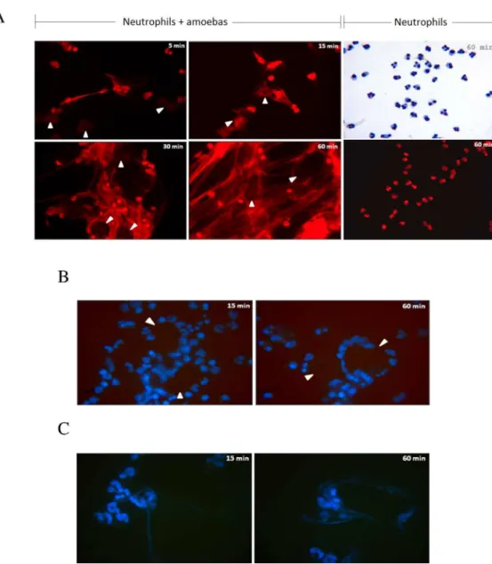
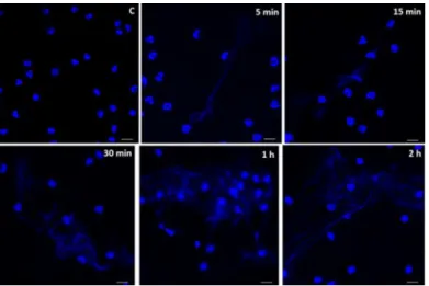
Fig 1.E. histolyticatrophozoites induce the formation of human neutrophil extracellular traps.A) Human neutrophils isolated by positive selection from peripheral heparinized blood were incubated withE.
histolyticaHM1:IMSS trophozoites (ratio 1 amoeba to 20 neutrophils) and the release of NETs monitored with 7AAD stain at 5, 15, 30 and 60 mins. Spider web-like fibers are observed as rapid as 5 min of incubation and increase in number and density over time. The networks were initially seen projected out of neutrophils toward amoebas (5 and 15 mins), and later gradually increase in number until completely cover the trophozoites that seem to be snared in the mesh (30 and 60 mins). Neutrophils in the absence of amoebas and incubated for 60 min are shown stained with Giemsa or 7ADD.B) Incubation of isolated human neutrophils with formaldehyde-fixed trophozoites did not induce NETs at 15 and 60 min. In A and B, trophozoites location is indicated by white head-arrows; magnification 40X. C) Incubation of isolated human neutrophils with fresh trophozoites whole extract induces scarce NETs at 15 and 60 min. B and C were stained with Hoechst.
doi:10.1371/journal.pone.0158979.g001
NET networks were seen in close contact with trophozoites, surrounding them after 30 min, suggesting that trophozoites were trapped in these structures (Fig 1A, see arrows at 30 and 60 min). However, amoebic trophozoites showed no apparent morphological or size changes within the 60 min of their interaction, and additionally retained their ability to phagocytose PMNs (S1 Fig).
To analyze the role of NETs on trophozoite viability and integrity in NET formation, human neutrophils were incubated at the same ratio with previously paraformaldehyde-fixed trophozoites or with fresh whole extracts. It is noteworthy that fixed trophozoites did not trig-ger NET formation during the hour of their co-incubation, despite that most of the neutrophils interacted with the surface of the fixed trophozoites (Fig 1B). Whole extracts from trophozoites at 1 mg/mL triggered scarce NET formation that increased slightly over time (Fig 1C). These results indicate thatE.histolyticainduced NET formation seems dependent on the trophozoite integrity.
Human NETs are unable to kill
E
.
histolytica
trophozoites
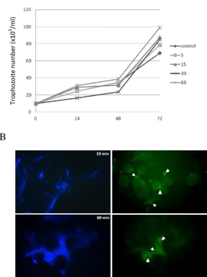
In order to analyze the role of human NETs on the viability ofE.histolytica, trophozoites were co-incubated with human neutrophils (ratio 1:20) for 5, 15, 30 and 60 min, washed and cul-tured for additional 72 h in fresh TYI-S-33 medium. Counting of viable trophozoites every 24 h showed that the NET formation from human neutrophils did not affect the viability and growth of amoebas at any of the co-incubation times tested (Fig 2A). A slight decrease was observed in the growth curves of trophozoites after 24 and 48 h of their exposure to NETs for 30 min, but the differences were not statistically significant. In fact, trophozoites exposed to NETs tended to grow better than control trophozoites at 72 h, albeit the differences were not statistically significant (Fig 2A).
Since cathelicidin LL-37 has previously been shown to affect the integrity ofE.histolytica
trophozoites[34], we analyzed whether cathelicidin LL-37 formed part of the antimicrobial peptide (AMP) in the NETs induced byE.histolyticatrophozoites. The immunofluorescence results show that the NETs induced byE.histolyticacontain cathelicidin LL-37, clearly visible at sites of interaction between neutrophils and amoebas as early as 15 min after the co-incuba-tion, which increases over time. After 60 min, a network tangle containing cathelicidin LL-37 was found surrounding the trophozoites (Fig 2B, white arrows; Upper and lower panels). These results suggest thatE.histolyticatrophozoites could be resistant to the cathelicidin LL-37 and other antimicrobial peptides found within human NETs inducedin vitroby the parasite.
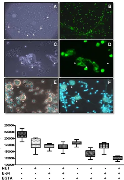
(EGTA) was analyzed on the viability of amoeba exposed to PMA-induced NETs. Both E64 and EGTA were used in order to inhibit possibleE.histolyticasecreted/excreted cysteine prote-ases and DNprote-ases, respectively, which could degrade AMPs or DNA that constitute NET. The results show that E-64 did not affect the growth of trophozoites within NETs, which was simi-lar to controls. In contrast, the addition of EGTA, a chelator of divalent ions, had a deleterious effect on the growth of amoeba within the NET, alone or combined with E-64 (p<0.001;Fig 3,
bottom graphic). This result suggests that a trophozoite-associated DNase activity may be responsible for the resistance ofE.histolyticato the deleterious effect of human NETs.
Fig 2. Trophozoites induced NETs are unable to inhibit parasite growth despite containing anti-microbial cathelicidin LL-37.A) Growth kinetic of trophozoites co-incubated with NETs for the indicated times (5 to 60 min) and thereafter cultured in fresh TYI-S-33 medium. Live trophozoites were counted every 24 h under light microscope using Trypan blue. Data shown at each time is the mean±SD of three independent assays. B) Immunofluorescence assay upon NETs induced by amoebas at 15 and 60 min (left panels) using anti-LL-37 antibody and an anti-rabbit IgG conjugated to FITC (right panels). Magnification 40X.
doi:10.1371/journal.pone.0158979.g002
E
.
histolytica
LPPG can trigger NET formation in human neutrophils
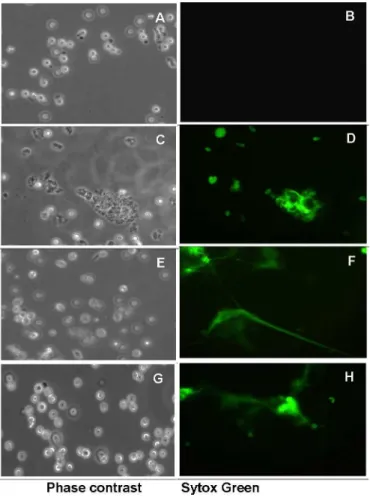
In order to identify some of the molecules of amoeba involved in the formation of NETs, we evaluated whether purifiedE.histolyticalipopeptidophosphoglycan (EhLPPG) triggered NET formation in human neutrophils. Results show that isolated EhLPPG at concentrations ranging from 5 to 15 ng/μL induces the formation of NETs in a dose-dependent manner (Fig 4, panels
D, F, H). Non-treated neutrophils did not change their morphology and did not expulse DNA, remaining non-stained with Sytox Green during the 2 h incubation (Fig 4, panel B), indicating a specific induction of NETs by EhLPPG.
Fig 3. Interaction and growth ofE.histolyticatrophozoites with neutrophil extracellular traps.NETs induced with PMA were incubated 1 h with amoeba trophozoites. Top: A-D, Sytox Green; E and F, Hoechst 33342. A, B at 10X and C-F at 40X objectives at the fluorescence microscope. A and C, phase contrast fields corresponding to B and D, respectively. F, florescence; E, merges phase contrast with fluorescence. Arrow tips,E.histolyticatrophozoites. Bottom: after amoeba-NET interaction, trophozoites were growth in TYI-S-33 culture medium during 72 h and cells harvested were counted under a light microscope. Controls,
trophozoites cultured at the same conditions in the absence of NETs. Amoeba growth after interaction with NETs and EGTA or EGTA + E-64 was significantly different from controls without NETs (p<0.001). No other significant difference (p<0.05) was observed compared to the control without NETs in the absence of other
compounds.
It is noteworthy that EhLPPG-induced NET formation initiates as early as 5 min after expo-sure and increases over time (Fig 5), similarly as observed for trophozoites. At 5 and 15 min of exposure, most NETs consisted of thin DNA fibers, while after 30 min NETs were observed as more complex DNA networks. Untreated neutrophils remained without morphological changes throughout the experiment (panel C).
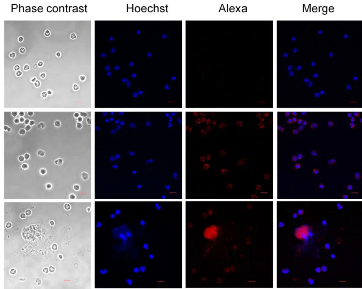
In addition, immunofluorescence analysis show that neutrophil´s cathelicidin LL-37 (Fig 6E and 6F) co-localize with the NETs induced by EhLPPG (Fig 6H and 6I), suggesting that this granule AMP is released bound to the neutrophil DNA ejected upon stimulus with the surface component of amoeba.
PMA-preformed NETs did not affect the infectivity of
E
.
histolytica
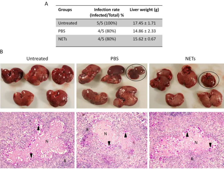
In order to assess whether NETs exert any effect on the infectivity ofE.histolytica, we deter-mined the ability of trophozoites pretreated for 1 h with PMA-induced NETs to develop amoe-bic liver abscesses (ALA) in hamsters, when compared with trophozoites treated with PBS or untreated. Massive development of ALA throughout the liver was observed in all 5 animals of the untreatred group and 4 out 5 animals of the PBS and NETs-treated groups (Fig 7A and 7B). Although one animal did not develop ALA in the group pretreated with NETs, this effect
Fig 4. Induction of neutrophil extracellular traps by different concentrations of
lipopeptidophosphoglycan fromE.histolytica.Human neutrophils were incubated for 2 h at 37°C with 0 to 15 ng/μL of purified EhLPPG. A, C, E and G, phase contrast fields of B, D, F, and H, respectively. Sytox
Green observed at 40X objective in the fluorescence microscope.
doi:10.1371/journal.pone.0158979.g004
appears to be due to the incubation of amoeba with PBS for 1 h, since one animal of that group did not either develop ALA. The hepatomegaly mesured as the average weight of livers, an indi-rect indication of ALA extent, was also similar between groups when compared only animals that developed ALA (Fig 7A). A comparative macroscopic description of infected livers showed similar grades of ALA development in the three groups, with many abscessses of variable size distributed in all lobes (Fig 7B). Similar results were also observed when ALA were analyzed in histological sections. Thus, large abscesses formed by coalescence of many small were observed throughout the liver tissue, containing well-preserved amebas close to the edges of lesions, abundant PMN cell infiltration and necrosis (Fig 7C). No clear differences in the the extent of injuries, population of immune cells recruited to the ALA and number and integrity of amoe-bas were observed between groups. Overall our results suggest thatin vitroobtained human NETs did not affect the pathogenicity ofE.histolyticaevaluated in the model of ALA in hamsters.
Discussion
Neutrophils are the first polymorphonuclear cells recruited to infection sites and are among the first line of defense of the cellular innate immunity[35]. Over a decade ago, networks of extracellular DNA containing granule proteins and histones, known as NETs, were described. NET formation can occur under multiple stimuli including reactive oxygen species[36], anti-bodies and antigen–antibody complexes[37,38], TLR4-activated platelets[39] and microbes
such as many bacteria and some virus, fungi and protozoa[40]. NET formation in response to protozoan parasites has been reported forL.amazonensisamastigotes andL.amazonensis,L.
major,L.chagasi,L.donovaniandL.mexicanapromastigotes[29–31],Toxoplasma gondii
tachyzoites[32],Trypanosoma cruzitrypomastigotes[33] andEimeria arloingisporozoites and oocysts [41]. However, characterization of NETs induced by the protozoan parasiteE. histoly-ticacausing amoebiasis in humans has not been previously described. We here demonstrate that human neutrophils release NETs upon exposure to amoebic trophozoites as well as to its surface molecule EhLPPG. In accordance with other reports on NET formation in the presence of microbes, the trophozoites ofE.histolyticatrigger NET formation rapidly (starting at 5 min)
Fig 5. Kinetics of NET induction by lipopeptidophosphoglycan isolated fromE.histolytica.Induction of neutrophil extracellular traps by 10 ng/μL of EhLPPG during the times indicated. Control, neutrophils
incubated at the same conditions for 2 h in the absence of EhLPPG. Hoechst 33342 stain and confocal microscope observation, bars 10μm.
in a dose and time dependent manner. This effect seemed dependent on amoeba viability, since fixed trophozoites were unable to elicit them. It is noteworthy however that slight and retarded NET formation was induced by totalE.histolyticaextracts as well as by conditioned medium (not shown), suggesting that microbial components present in both preparations can also trig-ger NET formation.
Among the microbial molecular triggers of NETs are the LPS of gram negative bacteria[23] and the LPG ofLeishmania amazonensispromastigotes[29], the most prominent negatively charged surface components involved in the activation of the host innate immune response trough TLR 2 and TLR 4[42,43]. In this study, we showed that purified EhLPPG, a surface mol-ecule structurally similar to Leishmania LPG, was also able to elicit NET formation in a similar manner to viable trophozoites, suggesting that EhLPPG is one of the main parasite molecules triggering traps in human neutrophils. This GPI-anchored proteophosphoglycan[44] is a highly immunogenic molecule directly exposed to the host’s immune system, recognized by
the sera from patients with amoebic liver abscess[45]. The mechanism by which EhLPPG is
Fig 6. Localization of LL-37 in NET induced by LPPG fromE.histolytica.Neutrophils or NET induced by EhLPPG were stained by an anti-LL-37 antibody and an anti-rabbit IgG conjugated to Alexa594 (red). Control PMN, polymorphonuclear leucocytes with only the secondary antibody. PMN, PMN stained with anti-LL-37 and the anti-rabbit IgG conjugated to Alexa594. NET, PMN incubated with LPPG showing the release of NET and colocalization with LL-37. Hoechst 33342 stain and confocal microscope observation, bars 10μm.
doi:10.1371/journal.pone.0158979.g006
able to activate the release of NETs was not addressed in this manuscript and remains
unknown. However, since it was described that EhLPPG is a molecular pattern that triggers the host immune response by signaling through TLR2/TLR4[43], it is conceivable that those pat-tern recognition receptors and the same pathway are responsible for the NET release by EhLPPG. Interestingly, other studies using genetically engineeredL.donovanishowed that NET induction by promastigotes is independent of the parasite surface LPG, suggesting that other parasite molecules are responsible for NET triggering in these species[30]. Studies are underway in our laboratory in order to identify other molecules, in addition to the EhLPPG, that are possibly involved in NET formation.
Amoebic viability assays by growth in fresh medium demonstrated thatE.histolytica tro-phozoites were not significantly affected after co-incubation with the NETs, induced by the amoeba itself or by classical stimuli such as PMA. In some cases, trophozoites grew equally
Fig 7. Development of amoebic liver abscesses (ALA) on hamsters by amoebas pre-treated with NETs.VirulentE.histolyticatrophozoited (1x10⁶) were incubated with 500
μg DNA NET solution (PMA-preformed) or PBS for 1 h at 37°C, and then injected in the portal vein of hamsters.
Untreated trophozoites were used as control of infectivity. Seven days post-challenge, animals were sacrificed, livers escinded and analyzed. A) Table showing the infection rate and weight average of infected livers. B) Upper: Distribution and magnitud of ALA on the liver of the 5 animals of each group. Uninfected livers of groups PBS and NETs are inside ovals. Bottom: Representative histological analysis of ALA from each group stained with PAS. Magnification 20X. Arrows: trophozoites; N: necrosis; Ii: Inflammatory infiltrate, mainly neutrophils.
well or even better than untreated cultures, indicating that at least under the conditions tested
in vitro, amoebas were able to evade the microbicidal effect of human NETs. Similar findings of resistance to NETs were seen in viability or infectivity studies of other parasites such asL.
major[29],L.donovani[30], andL.mexicana[31], as well as in the infectivity onT.cruzi[33]. In the latter case, although the NET did not cause the parasite death, it interfered with the ability ofT.cruzito infect LLC-MK2 cells, suggesting a protective role independent of a direct NET trypanocide effect. A similar effect of human NETs on the infectivity ofE.histolyticacannot be ruled out and is currently being studied by our group. One possible explanation for the inability of amoeba-induced NETs to kill the trophozoitesin vitrowas attributed to the fact that these NETs were atypical and thereafter possibly lacked anti-microbial molecules derived from the granules of neutrophils, including cathelicidin LL-37, an AMP that had previously been shown to decrease the growth ofE.histolyticatrophozoites[34]. Yet this possibility was ruled out since immunofluorescence studies using an antibody against LL-37 showed that this AMP was pres-ent in theE.histolytica- and EhLPPG-induced NETs, and increased with time of exposure. Since treated trophozoites grew well in culture medium we speculate thatE.histolytica pos-sesses evasion mechanisms for NETs, which was also observed when the amoebas were exposed to NETs obtained through other stimuli, such as PMA.
Our study additionally showed that the viability of amoeba showed a slight but significant decrease when treated with EGTA, a chelator used to inhibit some enzymes including DNases. In contrast, the viability was not affected after incubation with E-64, a potent cysteine protease inhibitor, suggestingE.histolyticapossibly secreted/excreted DNases could be involved in the evasion mechanisms of amoeba against human NETsin vitro. The role of DNases in protection against NETs has been reported for many bacteria such as group AStreptococcus[46], Staphylo-coccus aureus[47,48],Streptococcus agalactiae[49],S.pneumoniae[50],Vibrio cholera[51],S.
sanguinis[52],Neisseria gonorrhoeae[53] andS.suis[54]. Recently, a 3’-nucleotidase/nuclease
enzyme that allowsLeishmania infantumto survive after interaction with NET was reported [55]. In this regard, a restriction enzyme-like endonuclease activity was shown inE.histolytica
trophozoites[56], yet it is not clear whether this or any other DNase that degrades NETs is secreted by the parasite, allowing it to escape and survive.
In addition to the killing of pathogens, NETs are able to trap microorganisms and to degrade virulence factors[23]. We show here that amoeba trophozoites are sequestered by NETsin vitro; if this occur in the gut infection, parasite elimination may be easier by peristalsis, decreasing amoebic colonization. Another possible effect of traps on trophozoites is the degra-dation of amoeba virulence factors. On the other hand, as NET formation involves oxygen reactive species, the excessive formation of tramps by neutrophils may also contribute to tissue injury. In this regard, studies have shown the inability of amoeba to induce amoebic liver abscesses in neutropenic animals by treatment with immunosuppressors[20,57]. These results together with our observation in this study showing the inability of NETs to inhibitin vitro via-bility and the infectivity of the amoeba in the ALA model in hamsters, suggests that if NETs are formedin vivoduring infection with the amoeba, they could be contributing to tissue dam-age more than to protection against the parasite. Further studies are required to address these points.
In conclusion, our study shows that bothE.histolyticatrophozoites as well as the isolated lipopeptidophosphoglycan (EhLPPG) triggers NET formation by human neutrophils in a dose- and time-dependent manner. Furthermore, we show the presence of the granule protein cathelicidin LL-37 among the components of the DNA trap, yet it appears not affect the viabil-ity ofE.histolyticatrophozoites. Therefore, this study suggests that human NET formation par-ticipates in the early innate immune response againstE.histolytica. However, it remains to be
established whether NETs protect against natural amoebiasisin vivo, and if they contribute to the tissue damage associated with the infection.
Material and Methods
Ethics Statement
The study was reviewed and approved by the Ethics and Research Committees of the Faculty of Medicine of UNAM (Universidad Nacional Autonoma de Mexico) (FMED/CI/RGG/ 013/ 01/2008) and guidelines established by the Mexican Health Authorities were strictly followed. All patients and controls were informed and signed a written consent to participate in the study.
Culture of
Entamoeba histolytica
trophozoites
E.histolyticatrophozoites strain HM1: IMSS were cultured axenically at 37°C in screw-capped glass tubes containing TYI-S-33 medium[58], supplemented with 15% heat-inactivated bovine serum, 100 U/mL penicillin, 100μg/mL streptomycin sulfate, and 1.5% Diamond vitamin mix.
At the end of their logarithmic growth phase (72 h), the trophozoites were chilled on ice for 5 min and harvested by centrifugation at 150 ×gfor 7 min at 4°C, washed three times with PBS, and suspended in RPMI-1640 with 2% human serum albumin (RPMI-HSA).
Lipopeptidophosphoglycan isolation from
Entamoeba histolytica
trophozoites
Lipopeptidophosphoglycan was isolated fromEntamoeba histolyticatrophozoites (EhLPPG) as reported elsewhere[59]. All the glassware was heated at 250°C for one hour before use; sterile plastic material and pyrogen free water were used throughout the process. At the end of purifi-cation, EhLPPG was concentrated by lyophilization and total carbohydrates were determined by Fenol-H2SO4method[60]. EhLPP was analyzed[61] in 12% SDS-PAGE and gels stained
with Coomassie blue (negative stain), silver stain and periodic acid Schiff (PAS) reagent (not shown). The potential contamination of EhLPPG with bacterial lipopolysaccharide was ruled out by the analysis of chromogenic limulus amebocyte lysate (Lonza Mexico, catalog 27A-50-647U), according to the supplier's instructions. The LPS content in EhLPPG preparations used at 10 ng/μL was at or below 0.07 endotoxin units/mL.
Human neutrophil isolation
Neutrophils were isolated from peripheral heparinized blood, obtained from human healthy volunteer donors using Histopaque 1119 and 1077 (Sigma-Aldrich), according to the manufac-turer instructions. The granulocyte layer was collected and after washing with cold PBS, eryth-rocytes were lysed by incubation in lysis buffer (155 mM NH4Cl, 10 mM KHCO3, 0.1 mM EDTA) for 10 min on ice, followed by washings with cold PBS and centrifugation at 300 xgfor 10 min at 4°C. Neutrophils were highly purified from this preparation by positive selection. Briefly, 5x10⁷cells were suspended in 50μl of cold PBS and 50μl of CD16 micro beads
(Milte-nyi, Biotec, Bergisch Gladbach, Germany), incubated for 30 min at 4°C, washed with PBS, cen-trifuged at 300 xgfor 10 min and passed through a magnetic separation LS column (Miltenyi Biotec, Bergisch Gladbach, Germany). Finally, purified human neutrophils were placed in RPMI-1640 (Gibco) with 2% human serum albumin (CSL Behring) and seeded at 1-2x10⁵cells on sterile 8-well Cover Chamber Slides (Thermo), and incubated at 37°C under 5% CO2
Induction of neutrophil extracellular traps
NET formation was induced with live or 4% paraformaldehyde fixedE.histolyticatrophozoites (ratios 1:20 and 1:10 of amoeba:neutrophil), 1μg of trophozoites fresh whole extract/1 x 105
neutrophils or 5, 10 and 15 ng/μL of lipopeptidophosphoglycan isolated fromE.histolytica
tro-phozoites (EhLPPG). Culture plates were incubated at 37°C under 5% CO2atmosphere for
var-iable times, beginning at 5 min to a maximum of 2 h, as indicated on each experiment. For NET staining and microscopy observation, samples were fixed with 2 to 4% paraformaldehyde (Sigma-Aldrich) for 15 min, air dried and stained with 50μg/mL 7-ADD, 10μg/mL Hoechst
33342 (Invitrogen) or 0.2μM Sytox Green (Life Technologies). The coverslips were mounted
with Prolong Gold (Invitrogen) before observation at the confocal (Carl Zeiss, LSM700) or fluorescence (Carl Zeiss, AxiosKop40) microscopes.
Detection of cathelicidin LL-37 in neutrophil extracellular traps
For detection of LL-37, the neutrophils were seeded and NETs were induced as stated above, with the exception that coverslips were treated with poly-L-lysine before use. Sam-ples were fixed with 4% paraformaldehyde or acetone for 10 min, air dried and washed with Tris HCl 0.1 M pH 7.4. Unspecific protein binding was blocked with a solution of 10% human plasma, 0.1% gelatin and 0.1% Tween 20 in PBS for 1 h at room temperature. The slides were incubated with a polyclonal anti-human cathelicidin LL-37 antibody (Santa Cruz Biotechnology) at 1:200 dilution over night at 4°C, washed with Tris-HCl buffer and incubated with a secondary antibody for 1 h at room temperature, either 1:250 diluted goat anti-rabbit IgG conjugated to Alexa594 (Life technologies) or 1:100 diluted goat anti-rabbit IgG conjugated to biotin (Zymed). In the last case, Steptavidin AP 1:100 (KPL) was added and incubated for 30 min, washed and developed with AP RED Kit (Zymed), counter-stained with hematoxylin and mounted with Aqua Monter (Bio SB). Controls without first antibody were included and nuclei were stained with Hoechst 33342. All the samples with fluorescence and immunocytochemistry were analyzed with a Carl Zeiss microscope (Axio Imager M1) and the microphotographs were taken with the AxioCam MRc5 digital camera (Carl Zeiss).
Interaction of
E
.
histolytica
with PMA-preformed neutrophil extracellular
traps
NET formation was induced for 2 h with 25 nM PMA as described elsewhere[23]. A total number of 20,000 or 40,000 trophozoites per well were added to achieve a ratio of one amoeba to 20 neutrophils (1:20). Final volume was 1 mL of RPMI-HSA with or without cys-teine-HCl (1 g/L), ascorbic acid (0.2 g/L) and ferric ammonium citrate (0.0236 g/L) at pH 7.0. These compounds were used in some experiments at the final concentrations found in the culture media of Entamoeba, in order to maintain trophozoites in good conditions and assure that any change in amoeba viability was due to the presence of NETs. Some experi-ments were performed in the presence of 0.5 mM EGTA, 10μM E-64 or both to inhibit Ent-amoeba histolyticaenzymes potentially present (proteases and DNases). Appropriate controls were included such as seeding the same number of trophozoites in wells without NETs. Plates were incubated for 1 h at 37°C under a microaerophilic atmosphere (most of the oxygen was consumed by a burning candle in a closed anaerobic jar), and samples were treated for microscopic observation or to determine the amoeba growth.
E
.
histolytica
growth assays
Trophozoites that induced NETs at 5, 15, 30 or 60 min, as well as trophozoites exposed to PMA-preformed NETs during 2 h, were harvested and transferred to screw-capped tubes with 5 mL TYI-S-33 medium, and cultured for 72 h in habitual culture conditions for amoeba. Tro-phozoites were harvested at the indicated times and counted in a Neubauer chamber using the vital stain Trypan blue. Data were analyzed by Kruskal-Wallis test, considered statistically sig-nificant a p<0.05.
Induction of amoebic liver abscesses in hamsters
E.histolyticatrophozoites from 72 h cultures, harvested by ice-chilling during 5 min and washed three times with PBS pH 7.2, were treated with NETs obtained by treatment of human neutrophils with 25 nM PMA for 2 h as described above. Briefly, batches of 1x10⁶parasites were incubated with 500μg DNA NET solution or PBS for 1 h at 37°C. Each million of treated
trophozoites were collected by centrifugation at 150 ×gfor 7 min at 4°C, washed three times with PBS, suspended in 100μl PBS and used for infection of hamsters as described before[20].
Male Syrian golden hamsters (Mesocricetus auratus) 4 to 6 weeks of age were maintained free of pathogens with water and foodad libitum. Following a protocol approved by the Institu-tional Animal Care Committee, animals were divided in 2 groups of 5 hamsters each (Untreated and treated groups). The animals were anaesthetized with anesthesal (60 mg⁄kg)
and the portal vein was exposed by laparatomy under aseptic conditions. Untreated or NETs-treated trophozoites (1x10⁶/hamster) processed as above were directly inoculated into the por-tal vein using a tuberculin syringe, followed by the immediate application of a gel-foam pad at the site of inoculation in order to avoid bleeding and loss of amoebas.
Histological analysis
Hamsters were sacrificed after 7 days and the livers removed and weighed. Abscess were exscinded from the liver tissue and also weighed. Liver samples containing abscesses were fixed in 4% paraformaldehyde in PBS for 1 h, embedded in paraffin and processed for histology by standard techniques. In brief, serial sections of 20μm thicknesses were obtained in a
micro-tome, placed on slides coated with poly-L-lysine (Sigma, St Louis, MO, USA), deparaffined, and finally stained with haematoxylin/eosin or Periodic acid–Schiff for light microscopy
analysis.
Supporting Information
S1 Fig. Phagocytosis of human neutrophils by NETs-treatedE.histolyticatrophozoites.
Trophozoites were exposed for 1 h to PMA-preformed NETs (25 nM) and then incubated with neutrophils previously fixed with paraformaldehyde 4% and stained with DAPI for 10 min (A). Aliquots were taken at 15 min (B) and 30 min (C) and observed in a fluorescence microscope. Upper: light microscopy; middle: UV microscopy; bottom: simultaneous light and UV micros-copy. All images were taken at 40X.
(TIF)
Acknowledgments
Author Contributions
Conceived and designed the experiments: EEA NS IB JCC. Performed the experiments: EEA NS JP MCR CDG. Analyzed the data: EEA NS JPL IB JCC. Contributed reagents/materials/ analysis tools: EEA JPL IB JCC. Wrote the paper: EEA IB JCC. No.
References
1. Black RE, Allen LH, Bhutta ZA, Caulfield LE, de Onis M, Ezzati M, et al. Maternal and child undernutri-tion: global and regional exposures and health consequences. Lancet. 2008; 371(9608):243–60. doi: 10.1016/S0140-6736(07)61690-0PMID:18207566.
2. Verkerke H, Petri W, Marie C. The dynamic interdependence of amebiasis, innate immunity, and under-nutrition. Seminars in Immunopathology. 2012; 34(6):771–85. doi:10.1007/s00281-012-0349-1PMID: WOS:000310988000004.
3. Walsh JA. Problems in recognition and diagnosis of amebiasis: estimation of the global magnitude of morbidity and mortality. Rev Infect Dis. 1986; 8(2):228–38. PMID:2871619.
4. Lozano R, Naghavi M, Foreman K, Lim S, Shibuya K, Aboyans V, et al. Global and regional mortality from 235 causes of death for 20 age groups in 1990 and 2010: a systematic analysis for the Global Bur-den of Disease Study 2010. Lancet. 2012; 380(9859):2095–128. doi: 10.1016/S0140-6736(12)61728-0PMID:23245604.
5. Prathap K, Gilman R. The histopathology of acute intestinal amebiasis. A rectal biopsy study. Am J Pathol. 1970; 60(2):229–46. PMID:5457212; PubMed Central PMCID: PMCPMC2032904.
6. Tsutsumi V, Mena-Lopez R, Anaya-Velazquez F, Martinez-Palomo A. Cellular bases of experimental amebic liver abscess formation. Am J Pathol. 1984; 117(1):81–91. PMID:6385728; PubMed Central
PMCID: PMCPMC1900566.
7. Tsutsumi V, Martinez-Palomo A. Inflammatory reaction in experimental hepatic amebiasis. An ultra-structural study. Am J Pathol. 1988; 130(1):112–9. PMID:3337207; PubMed Central PMCID:
PMCPMC1880538.
8. Espinosa-Cantellano M, Martínez-Palomo A. Pathogenesis of intestinal amebiasis: from molecules to disease. Clin Microbiol Rev. 2000; 13(2):318–31. PMID:10756002; PubMed Central PMCID:
PMCPMC100155.
9. Velazquez C, Shibayama-Salas M, Aguirre-Garcia J, Tsutsumi V, Calderon J. Role of neutrophils in innate resistance toEntamoeba histolyticaliver infection in mice. Parasite Immunol. 1998; 20(6):255–
62. PMID:9651927.
10. Seydel KB, Zhang T, Stanley SL. Neutrophils play a critical role in early resistance to amebic liver abscesses in severe combined immunodeficient mice. Infect Immun. 1997; 65(9):3951–3. PMID: 9284178; PubMed Central PMCID: PMCPMC175565.
11. Jarillo-Luna RA, Campos-Rodríguez R, Tsutsumi V.Entamoeba histolytica: immunohistochemical study of hepatic amoebiasis in mouse. Neutrophils and nitric oxide as possible factors of resistance. Exp Parasitol. 2002; 101(1):40–56. PMID:12243737.
12. Asgharpour A, Gilchrist C, Baba D, Hamano S, Houpt E. Resistance to intestinalEntamoeba histolytica
infection is conferred by innate immunity and Gr-1+ cells. Infect Immun. 2005; 73(8):4522–9. doi:10. 1128/IAI.73.8.4522–4529.2005PMID:16040963; PubMed Central PMCID: PMCPMC1201199.
13. Estrada-Figueroa LA, Ramírez-Jiménez Y, Osorio-Trujillo C, Shibayama M, Navarro-García F, García-Tovar C, et al. Absence of CD38 delays arrival of neutrophils to the liver and innate immune response development during hepatic amoebiasis byEntamoeba histolytica. Parasite Immunol. 2011; 33 (12):661–8. doi:10.1111/j.1365-3024.2011.01333.xPMID:21919917.
14. Burgess SL, Buonomo E, Carey M, Cowardin C, Naylor C, Noor Z, et al. Bone marrow dendritic cells from mice with an altered microbiota provide interleukin 17A-dependent protection againstEntamoeba histolyticacolitis. MBio. 2014; 5(6):e01817. doi:10.1128/mBio.01817-14PMID:25370489; PubMed Central PMCID: PMCPMC4222101.
15. Salata RA, Ravdin JI. The interaction of human neutrophils andEntamoeba histolyticaincreases cyto-pathogenicity for liver cell monolayers. J Infect Dis. 1986; 154(1):19–26. PMID:2872253.
16. Dickson-Gonzalez SM, de Uribe ML, Rodriguez-Morales AJ. Polymorphonuclear neutrophil infiltration intensity as consequence ofEntamoeba histolyticadensity in amebic colitis. Surg Infect (Larchmt). 2009; 10(2):91–7. doi:10.1089/sur.2008.011PMID:18831680.
17. Seydel KB, Li E, Zhang Z, Stanley SL. Epithelial cell-initiated inflammation plays a crucial role in early tissue damage in amebic infection of human intestine. Gastroenterology. 1998; 115(6):1446–53. PMID: 9834272.
18. Pérez-Tamayo R, Martínez RD, Montfort I, Becker I, Tello E, Pérez-Montfort R. Pathogenesis of acute experimental amebic liver abscess in hamsters. J Parasitol. 1991; 77(6):982–8. PMID:1779303.
19. Pérez-Tamayo R, Montfort I, García AO, Ramos E, Ostria CB. Pathogenesis of acute experimental liver amebiasis. Arch Med Res. 2006; 37(2):203–9. doi:10.1016/j.arcmed.2005.10.007PMID: 16380320.
20. Olivos-García A, Carrero JC, Ramos E, Nequiz M, Tello E, Montfort I, et al. Late experimental amebic liver abscess in hamster is inhibited by cyclosporine and N-acetylcysteine. Exp Mol Pathol. 2007; 82 (3):310–5. doi:10.1016/j.yexmp.2006.09.005PMID:17362925.
21. Naylor C, Burgess S, Madan R, Buonomo E, Razzaq K, Ralston K, et al. Leptin receptor mutation results in defective neutrophil recruitment to the colon duringEntamoeba histolyticainfection. MBio. 2014; 5(6). doi:10.1128/mBio.02046-14PMID:25516614; PubMed Central PMCID:
PMCPMC4271549.
22. Papayannopoulos V, Zychlinsky A. NETs: a new strategy for using old weapons. Trends Immunol. 2009; 30(11):513–21. doi:10.1016/j.it.2009.07.011PMID:19699684.
23. Brinkmann V, Reichard U, Goosmann C, Fauler B, Uhlemann Y, Weiss DS, et al. Neutrophil extracellu-lar traps kill bacteria. Science. 2004; 303(5663):1532–5. doi:10.1126/science.1092385PMID: 15001782.
24. Brinkmann V, Zychlinsky A. Beneficial suicide: why neutrophils die to make NETs. Nat Rev Microbiol. 2007; 5(8):577–82. doi:10.1038/nrmicro1710PMID:17632569.
25. Yipp BG, Kubes P. NETosis: how vital is it? Blood. 2013; 122(16):2784–94. doi: 10.1182/blood-2013-04-457671PMID:24009232.
26. Sørensen OE, Follin P, Johnsen AH, Calafat J, Tjabringa GS, Hiemstra PS, et al. Human cathelicidin,
hCAP-18, is processed to the antimicrobial peptide LL-37 by extracellular cleavage with proteinase 3. Blood. 2001; 97(12):3951–9. PMID:11389039.
27. Hahn S, Giaglis S, Chowdhury CS, Chowdury CS, Hösli I, Hasler P. Modulation of neutrophil NETosis: interplay between infectious agents and underlying host physiology. Semin Immunopathol. 2013; 35 (4):439–53. doi:10.1007/s00281-013-0380-xPMID:23649713; PubMed Central PMCID:
PMCPMC3685704.
28. Baker VS, Imade GE, Molta NB, Tawde P, Pam SD, Obadofin MO, et al. Cytokine-associated neutro-phil extracellular traps and antinuclear antibodies in Plasmodium falciparum infected children under six years of age. Malar J. 2008; 7:41. doi:10.1186/1475-2875-7-41PMID:18312656; PubMed Central PMCID: PMCPMC2275287.
29. Guimarães-Costa AB, Nascimento MT, Froment GS, Soares RP, Morgado FN, Conceição-Silva F, et al. Leishmania amazonensis promastigotes induce and are killed by neutrophil extracellular traps. Proc Natl Acad Sci U S A. 2009; 106(16):6748–53. doi:10.1073/pnas.0900226106PMID:19346483;
PubMed Central PMCID: PMCPMC2672475.
30. Gabriel C, McMaster WR, Girard D, Descoteaux A. Leishmania donovani promastigotes evade the anti-microbial activity of neutrophil extracellular traps. J Immunol. 2010; 185(7):4319–27. doi:10.4049/ jimmunol.1000893PMID:20826753.
31. Hurrell BP, Schuster S, Grün E, Coutaz M, Williams RA, Held W, et al. Rapid Sequestration of Leish-mania mexicana by Neutrophils Contributes to the Development of Chronic Lesion. PLoS Pathog. 2015; 11(5):e1004929. doi:10.1371/journal.ppat.1004929PMID:26020515; PubMed Central PMCID: PMCPMC4447405.
32. Abi Abdallah DS, Lin C, Ball CJ, King MR, Duhamel GE, Denkers EY. Toxoplasma gondii triggers release of human and mouse neutrophil extracellular traps. Infect Immun. 2012; 80(2):768–77. doi:10. 1128/IAI.05730-11PMID:22104111; PubMed Central PMCID: PMCPMC3264325.
33. Sousa-Rocha D, Thomaz-Tobias M, Diniz LF, Souza PS, Pinge-Filho P, Toledo KA. Trypanosoma cruzi and Its Soluble Antigens Induce NET Release by Stimulating Toll-Like Receptors. PLoS One. 2015; 10(10):e0139569. doi:10.1371/journal.pone.0139569PMID:26431537; PubMed Central PMCID: PMCPMC4591979.
34. Rico-Mata R, De Leon-Rodriguez LM, Avila EE. Effect of antimicrobial peptides derived from human cathelicidin LL-37 onEntamoeba histolyticatrophozoites. Exp Parasitol. 2013; 133(3):300–6. doi:10. 1016/j.exppara.2012.12.009PMID:23274811.
35. Amulic B, Cazalet C, Hayes GL, Metzler KD, Zychlinsky A. Neutrophil function: from mechanisms to disease. Annu Rev Immunol. 2012; 30:459–89. doi:10.1146/annurev-immunol-020711-074942PMID: 22224774.
37. Kessenbrock K, Krumbholz M, Schönermarck U, Back W, Gross WL, Werb Z, et al. Netting neutrophils in autoimmune small-vessel vasculitis. Nat Med. 2009; 15(6):623–5. doi:10.1038/nm.1959PMID: 19448636; PubMed Central PMCID: PMCPMC2760083.
38. Lande R, Ganguly D, Facchinetti V, Frasca L, Conrad C, Gregorio J, et al. Neutrophils activate plasma-cytoid dendritic cells by releasing self-DNA-peptide complexes in systemic lupus erythematosus. Sci Transl Med. 2011; 3(73):73ra19. doi:10.1126/scitranslmed.3001180PMID:21389263; PubMed Cen-tral PMCID: PMCPMC3399524.
39. Clark SR, Ma AC, Tavener SA, McDonald B, Goodarzi Z, Kelly MM, et al. Platelet TLR4 activates neu-trophil extracellular traps to ensnare bacteria in septic blood. Nat Med. 2007; 13(4):463–9. doi:10. 1038/nm1565PMID:17384648.
40. Vorobjeva NV, Pinegin BV. Neutrophil extracellular traps: mechanisms of formation and role in health and disease. Biochemistry (Mosc). 2014; 79(12):1286–96. doi:10.1134/S0006297914120025PMID: 25716722.
41. Silva LM, Caro TM, Gerstberger R, Vila-Viçosa MJ, Cortes HC, Hermosilla C, et al. The apicomplexan parasite Eimeria arloingi induces caprine neutrophil extracellular traps. Parasitol Res. 2014; 113 (8):2797–807. doi:10.1007/s00436-014-3939-0PMID:24849865.
42. Becker I, Salaiza N, Aguirre M, Delgado J, Carrillo-Carrasco N, Kobeh LG, et al. Leishmania lipopho-sphoglycan (LPG) activates NK cells through toll-like receptor-2. Mol Biochem Parasitol. 2003; 130 (2):65–74. PMID:12946842.
43. Maldonado-Bernal C, Kirschning CJ, Rosenstein Y, Rocha LM, Rios-Sarabia N, Espinosa-Cantellano M, et al. The innate immune response toEntamoeba histolyticalipopeptidophosphoglycan is mediated by toll-like receptors 2 and 4. Parasite Immunol. 2005; 27(4):127–37. doi:10.1111/j.1365-3024.2005. 00754.xPMID:15910421.
44. Isibasi A, Santa Cruz M, Ramírez A, Kumate J. [Immunochemistry of a lipopeptidophosphoglycan extracted from trophozoites ofEntamoeba histolyticastrain HK-9 cultivated in axenic media, using the phenol-water method]. Arch Invest Med (Mex). 1982; 13 Suppl 3:51–5. PMID:6295328.
45. Campos-Rodríguez R, Barranco-Tovar C, Isibasi-Araujo A, Kumate-Rodríguez J. [Anti-amebic plasma cells in peripheral blood of patients with amebic liver abscess]. Arch Invest Med (Mex). 1986; 17 Suppl 1:303–6. PMID:3592896.
46. Buchanan JT, Simpson AJ, Aziz RK, Liu GY, Kristian SA, Kotb M, et al. DNase expression allows the pathogen group A Streptococcus to escape killing in neutrophil extracellular traps. Curr Biol. 2006; 16 (4):396–400. doi:10.1016/j.cub.2005.12.039PMID:16488874.
47. Thammavongsa V, Missiakas DM, Schneewind O. Staphylococcus aureus degrades neutrophil extra-cellular traps to promote immune cell death. Science. 2013; 342(6160):863–6. doi:10.1126/science. 1242255PMID:24233725; PubMed Central PMCID: PMCPMC4026193.
48. Berends ET, Horswill AR, Haste NM, Monestier M, Nizet V, von Köckritz-Blickwede M. Nuclease expression by Staphylococcus aureus facilitates escape from neutrophil extracellular traps. J Innate Immun. 2010; 2(6):576–86. doi:10.1159/000319909PMID:20829609; PubMed Central PMCID:
PMCPMC2982853.
49. Derré-Bobillot A, Cortes-Perez NG, Yamamoto Y, Kharrat P, Couvé E, Da Cunha V, et al. Nuclease A (Gbs0661), an extracellular nuclease of Streptococcus agalactiae, attacks the neutrophil extracellular traps and is needed for full virulence. Mol Microbiol. 2013; 89(3):518–31. doi:10.1111/mmi.12295
PMID:23772975.
50. Zhu L, Kuang Z, Wilson BA, Lau GW. Competence-independent activity of pneumococcal EndA [cor-rected] mediates degradation of extracellular dna and nets and is important for virulence. PLoS One. 2013; 8(7):e70363. doi:10.1371/journal.pone.0070363PMID:23936195; PubMed Central PMCID: PMCPMC3729463.
51. Seper A, Hosseinzadeh A, Gorkiewicz G, Lichtenegger S, Roier S, Leitner DR, et al. Vibrio cholerae evades neutrophil extracellular traps by the activity of two extracellular nucleases. PLoS Pathog. 2013; 9(9):e1003614. doi:10.1371/journal.ppat.1003614PMID:24039581; PubMed Central PMCID: PMCPMC3764145.
52. Morita C, Sumioka R, Nakata M, Okahashi N, Wada S, Yamashiro T, et al. Cell wall-anchored nuclease of Streptococcus sanguinis contributes to escape from neutrophil extracellular trap-mediated bacterio-cidal activity. PLoS One. 2014; 9(8):e103125. doi:10.1371/journal.pone.0103125PMID:25084357; PubMed Central PMCID: PMCPMC4118848.
53. Juneau RA, Stevens JS, Apicella MA, Criss AK. A thermonuclease of Neisseria gonorrhoeae enhances bacterial escape from killing by neutrophil extracellular traps. J Infect Dis. 2015; 212(2):316–24. doi:10. 1093/infdis/jiv031PMID:25605868; PubMed Central PMCID: PMCPMC4490236.
54. de Buhr N, Stehr M, Neumann A, Naim HY, Valentin-Weigand P, von Köckritz-Blickwede M, et al. Iden-tification of a novel DNase of Streptococcus suis (EndAsuis) important for neutrophil extracellular trap
degradation during exponential growth. Microbiology. 2015; 161(Pt 4):838–50. doi:10.1099/mic.0. 000040PMID:25667008.
55. Guimarães-Costa AB, DeSouza-Vieira TS, Paletta-Silva R, Freitas-Mesquita AL, Meyer-Fernandes JR, Saraiva EM. 3'-nucleotidase/nuclease activity allows Leishmania parasites to escape killing by neu-trophil extracellular traps. Infect Immun. 2014; 82(4):1732–40. doi:10.1128/IAI.01232-13PMID: 24516114; PubMed Central PMCID: PMCPMC3993383.
56. Yadav VP, Mandal PK, Rao DN, Bhattacharya S. Characterization of the restriction enzyme-like endo-nuclease encoded by theEntamoeba histolyticanon-long terminal repeat retrotransposon EhLINE1. FEBS J. 2009; 276(23):7070–82. doi:10.1111/j.1742-4658.2009.07419.xPMID:19878305.
57. Quintanar-Quintanar ME, Jarillo-Luna A, Rivera-Aguilar V, Ventura-Juárez J, Tsutsumi V, Shibayama M, et al. Immunosuppressive treatment inhibits the development of amebic liver abscesses in ham-sters. Med Sci Monit. 2004; 10(9):BR317–24. PMID:15328476.
58. Diamond LS, Harlow DR, Cunnick CC. A new medium for the axenic cultivation ofEntamoeba histoly-ticaand other Entamoeba. Trans R Soc Trop Med Hyg. 1978; 72(4):431–2. PMID:212851.
59. Lotter H, González-Roldán N, Lindner B, Winau F, Isibasi A, Moreno-Lafont M, et al. Natural killer T cells activated by a lipopeptidophosphoglycan fromEntamoeba histolyticaare critically important to control amebic liver abscess. PLoS Pathog. 2009; 5(5):e1000434. doi:10.1371/journal.ppat.1000434 PMID:19436711; PubMed Central PMCID: PMCPMC2674934.
60. DUBOIS M, GILLES K, HAMILTON JK, REBERS PA, SMITH F. A colorimetric method for the determi-nation of sugars. Nature. 1951; 168(4265):167. PMID:14875032.