Are Neutrophil Extracellular Traps Playing a
Role in the Parasite Control in Active
American Tegumentary Leishmaniasis
Lesions?
Fernanda Nazaré Morgado1¤
, Michelle T. C. Nascimento2, Elvira M. Saraiva2, Carla
de Oliveira-Ribeiro1,3, Maria de Fátima Madeira3, Marcela da Costa-Santos1, Erica C. F. Vasconcellos3, Maria Ines F. Pimentel3, Marcelo Rosandiski Lyra3, Armando de
Oliveira Schubach3, Fátima Conceição-Silva1*
1Laboratório de Imunoparasitologia, Instituto Oswaldo Cruz IOC/FIOCRUZ, Rio de Janeiro, Brazil, 2Departamento de Imunologia, Instituto de Microbiologia Paulo de Góes, Universidade Federal do Rio de Janeiro (UFRJ), Rio de Janeiro, Brazil,3Laboratório de Vigilância em Leishmanioses—VigiLeish, Instituto Nacional de Infectologia Evandro Chagas INI/FIOCRUZ, Rio de Janeiro, Brazil
¤ Current address: Laboratório de Pesquisa em Leishmaniose, Instituto Oswaldo Cruz IOC/FIOCRUZ, Rio de Janeiro, Brazil
*fconcei@ioc.fiocruz.br
Abstract
Neutrophil extracellular traps (NETs) have been described as a network of extracellular fibers composed by DNA, histones and various proteins/enzymes. Studies have demon-strated that NETs could be responsible for the trapping and elimination of a variety of infectious agents. In order to verify the presence of NETs in American tegumentary leish-maniasis (ATL) and their relationship with the presence of amastigotes we evaluated active cutaneous lesions of 35 patients before treatment by the detection of parasites, neutrophils (neutrophil elastase) and histones through immunohistochemistry and confo-cal immunofluorescence. Intact neutrophils could be detected in all ATL lesions. NETs were present in 27 patients (median 1.1; range from 0.1 to 23.5/mm2) with lesion duration ranging from one to seven months. NETs were in close proximity with neutrophils (r = 0.586; p = 0.0001) and amastigotes (r = 0.710; p = 0.0001). Two patterns of NET formation were detected: small homogeneously distributed networks observed in all lesions; and large structures that could be visualized at a lower magnification in lesions presenting at least 20% of neutrophils. Lesions presenting the larger NET formation showed high para-site detection. A correlation between NET size and the number of intact amastigotes was observed (p=0.02). As we detected an association between NET and amastigotes, our results suggest that neutrophil migration and NET formation could be stimulated and maintained by stimuli derived from the parasite burden/parasite antigen in the extracellular environment. The observation of areas containing only antigens not intermingled with NETs (elastase and histone) suggests that the involvement of these structures in the con-trol of parasite burden is a dynamic process in which the formation of NETs is exhausted with the destruction of the parasites. Since NETs were also associated with granulomas, a11111
OPEN ACCESS
Citation:Morgado FN, Nascimento MTC, Saraiva EM, Oliveira-Ribeiro Cd, Madeira MdF, Costa-Santos Md, et al. (2015) Are Neutrophil Extracellular Traps Playing a Role in the Parasite Control in Active American Tegumentary Leishmaniasis Lesions? PLoS ONE 10(7): e0133063. doi:10.1371/journal. pone.0133063
Editor:Nades Palaniyar, The Hospital for Sick Children and The University of Toronto, CANADA
Received:January 10, 2015
Accepted:June 22, 2015
Published:July 20, 2015
Copyright:© 2015 Morgado et al. This is an open access article distributed under the terms of the Creative Commons Attribution License, which permits unrestricted use, distribution, and reproduction in any medium, provided the original author and source are credited.
Data Availability Statement:All relevant data are within the paper and its Supporting Information files.
this trapping would favor the activity of macrophages in order to control the parasite burden.
Introduction
Various infectious agents are able to invade and multiply inside human cells. Protozoa of the genusLeishmaniaare obligate intracellular parasites of the human mononuclear phagocyte system [1], but these parasites have also been detected inside neutrophils [2], fibroblasts [3,4], endothelial cells [5,6], and dendritic cells [7].
The function of macrophages as host and effector cells during infection withLeishmania
parasites has been extensively studied. However, many questions regarding the ability of this protozoan to infect other cell types remain unanswered. In this aspect, controversy still exists whether neutrophils restrain or facilitate infection withLeishmania sp[8,9]. Neutrophils are the first cells recruited to the site of infection in leishmaniasis [10,11] and their participation in the control of parasite burden has been demonstrated in experimental models. Peters et al. [9] observed rapid and persistent infiltration of neutrophils at the site of inoculation of promasti-gotes by sandflies in a murine model within the first hours of infection withL.major. C57BL/6 and BALB/c mice depleted of neutrophils and infected withL.majorshowed higher parasite burdens, greater dissemination of infection and more severe lesions than non-depleted animals [8]. However, other investigators suggested a negative role of neutrophils in murine leishmani-asis, producing more severe disease associated with a Th2 response [12], or even serving as a carrier forLeishmaniaentry into macrophages [13]. Afonso et al [14] observed that human necrotic, but not apoptotic, neutrophils inducedin vitroleishmanicidal activity mediated by macrophages. This leishmanicidal activity was dependent on TNF-αand neutrophil elastase (NE). Ribeiro-Gomes and Sacks [15] discussed the influence of early neutrophil-Leishmania
interactions on the host immune response and suggested that infection outcome critically depends on the time of neutrophil recruitment and the tissue environment in which it occurs.
A particular form of death was described for neutrophils, which occurs with the release to the extracellular milieu of a mesh formed by chromatin associated with granular and cyto-plasmic proteins named netosis from Neutrophil Extracellular Traps (NETs) [16]. NETs are endowed with the properties to arrest and kill different microorganisms [16–18]. During neto-sis, there is disruption of the nuclear membrane and chromatin decondensation, this last fea-ture is dependent on the activity of elastase (NE), myeloperoxidase and peptidyl arginine deiminase 4 [18–20]. The formation of NETs has been reported in studies involving different pathogens such asMycobacterium tuberculosis[21], fungi [22–23], HIV-1 [24],Toxoplasma gondii[25],Eimeria bovis[26],Plasmodium falciparum[27],Leishmania amazonensis,L.
major,L.chagasi[28] andL.donovani[29]. Therefore, the in vitro interaction of neutrophils andLeishmaniapromastigotes [28,29] leads to NET formation suggesting that NETs may con-tribute to the containment of promastigotes at the site of inoculation, thereby facilitating their uptake by mononuclear phagocytes [29]. NETs were also detected in the active lesion of a patient with American tegumentary leishmaniasis (ATL) [28]. However, the importance of this phenomenon in human tegumentary leishmaniasis lesions is still unknown, and it remains unclear whether NETs exert effects on the control of initial parasite burdenin vivoor whether this effector mechanism occurs during an already established infection when only amastigote forms are present in lesions. It has been shown that amastigote forms ofL.amazonensisinduce NET release upon neutrophil interactionin vitro[28]. Therefore, in the present study we Fundação de Amparo à Pesquisa do Estado do Rio
de Janeiro-FAPERJ (E26.111.717/2012; E-26/ 102.456/2012, E26/102.988/2012, E26/111.230/2014, E26/203638/2014 and E-26/111.101/2014), and Coordenação de Aperfeiçomento de Pessoal de Nível Superior-CAPES (23038.005291/2011-61), Brazil. AOS is the recipient of fellowships from CNPq and FAPERJ, Brazil. The funders had no role in study design data collection and analysis, decision to publish, or preparing the manuscript.
investigated the presence of NETs in active established ATL lesions and their relationship with
Leishmaniaamastigotes.
Materials and Methods
Patients
Thirty-five patients with the localized cutaneous form of ATL were studied. Etiological diagno-sis was confirmed by histopathological examination and isolation in culture as previously described [30]. Parasite characterization was possible in 11 isolates andLeishmania (V.) brazi-liensisinfection was identified for all of them. All cases were from areas of Brazil (Rio de Janeiro) whereLeishmania (Viannia) braziliensisis the infecting species of greatest prevalence. Patient age, lesion duration, number and site of the lesions were described (Table 1).
Ethics statement
The patients were included in the study after they had given formal written consent and the study was approved by the Ethics Committee of INI/FIOCRUZ (Comitê de Ética em Pesquisa do Instituto Nacional de Infectologia Evandro Chagas—CEP-INI—014/2001). When the patients were under 18 years old, informed consent was sign by parents or guardians, who also remained present throughout the clinical evaluation and diagnosis procedure.
Immunohistochemistry (IHC)
Biopsy fragments were cut into 3-μm thick sections and mounted on silanized microscope
slides (DakoCytomation, Carpinteria, CA, USA). The IHC was performed as described [31,32]. For single staining, the specimens were incubated with the primary antibodies anti-neutrophil elastase (NE- DakoCytomation, Carpinteria, USA) or anti-L.braziliensispolyclonal rabbit serum (Dr. Madeira-INI/FIOCRUZ), followed by sequential steps of washes in PBS (pH 7.4) and incubation with the biotinylated secondary antibody (Zymed, San Francisco, CA, USA), the streptavidin-biotin-peroxidase complex (ABC kit, DakoCytomation), and Aminoethyl car-bazole (chromogen-AEC kit, Zymed). The slides were counterstained with Mayer’s hematoxy-lin (Dako). Double staining was performed for the colocalization of NE and amastigotes using the Dako Envision Doublestain System according to the manufacturers instructions. The slides were examined under a light microscope (Zeiss, Germany). The percentage of neutrophils was determined by counting 500 cells as standard and the number of amastigotes and NETs per mm2tissue was calculated using a millimeter coverslip.
Immunofluorescence and confocal microscopy
(Program for Technological Development in Tools for Health—PDTIS-FIOCRUZ) using exci-tation at 488 nm or 546 nm and emission at 505–530 nm (band pass system) and 560 nm (long pass system). The images were processed using the program LSM Image Browser—Version 4.2.0.121 (Zeiss, Jena, Germany).
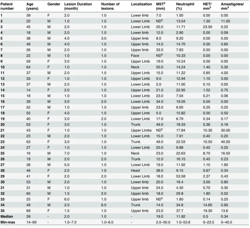
Table 1. Clinical and quantitative data of cutaneous lesions from 35 patients with American tegumentary leishmaniasis.
Patient number
Age (years)
Gender Lesion Duration (month)
Number of lesions
Localization MSTa
(mm)
Neutrophil (%)
NET/ mm2
Amastigotes/ mm2
1 39 F 2.0 1.0 Lower limb 7.0 1.00 0.00 0.00
2 20 M 1.0 1.0 Lower Limb NDb 13.54 1.30 11.00
3 45 M 2.0 1.0 Lower Limb 20.0 11.71 23.50 0.25
4 18 M 2.0 1.0 Lower limb 12.0 2.90 0.00 0.09
5 38 M 4.0 3.0 Upper limb 8.0 9.20 0.00 0.50
6 46 M 4.0 1.0 Upper limb 14.0 14.70 0.00 0.60
7 26 M 2.0 1.0 Upper limb 33.0 7.83 0.50 0.00
8 22 M 1.0 1.0 Face NDb 10.33 0.00 0.00
9 59 F 3.0 1.0 Upper Limb 19.0 10.24 0.00 0.00
10 64 F 1.0 1.0 Neck 50.0 14.24 1.40 5.30
11 37 M 2.0 1.0 Upper Limb 15.0 11.22 0.85 4.00
12 33 F 1.0 1.0 Upper Limb 9.0 12.94 1.10 3.00
13 37 M 2.0 1.0 Lower Limb 2.0 11.00 0.10 0.00
14 14 F 2.0 1.0 Upper Limb 21.0 22.95 1.52 0.75
15 18 M 1.0 1.0 Lower Limb 23.0 7.04 0.21 0.06
16 39 M 3.0 2.0 Lower Limb 34.0 19.05 0.00 0.00
17 52 M 1.0 1.0 Upper limb 23.0 6.60 0.25 0.25
18 55 F 4.0 1.0 Upper Limb 5.0 15.82 0.00 0.50
19 40 F 3.0 2.0 Lower Limb 17.0 6.79 0.34 0.17
20 63 F 3.0 1.0 Trunk 49.0 18.33 0.36 0.18
21 43 F 1.0 1.0 Upper Limbs NDb 17.84 10.30 30.00
22 23 M 2.0 1.0 Lower Limb 15.0 7.91 0.40 0.20
23 63 F 3.0 1.0 Trunk 49.0 22.03 10.50 40.00
24 27 F 1.0 1.0 Lower Limb 20.0 9.88 0.42 0.25
25 16 M 7.0 1.0 Neck 23.0 22.63 8.70 16.50
26 19 M 2.0 2.0 Trunk 12.0 16.15 0.45 0.23
27 36 M 5.0 1.0 Lower Limb 19.0 11.92 1.10 1.90
28 46 F 2.0 1.0 Head 38.0 9.15 0.67 0.34
29 41 F 2.0 2.0 Lower Limb 18.0 53.58 2.37 0.43
30 53 M 2.0 1.0 Lower limb 20.0 16.4 3.00 0.80
31 31 M 1.0 1.0 Upper limb 24.0 4.30 0.70 0.30
32 65 M 1.5 2.0 Upper limb 18.0 29.8 1.80 0.52
33 25 F 6.0 1.0 Upper limb NDb 1.80 0.14 0.20
34 49 M 2.0 6.0 Trunk 14.0 34.8 14.60 0.90
35 69 F 1.5 1.0 Upper limb 23.0 37.7 19.30 13.6
Median 39 - 2.0 1.0 - 19.0 11.92 0.5 0.34
Min-max 14–69 - 1.0–7.0 1.0–6.0 - 2.0–50.0 1.0–53.6 0–23.5 0–40.0
a
MST—Montenegro skin test. b
ND—not done.
Statistical analysis
The SPSS16 for Windows program (SPSS, Inc., Chicago, IL, USA) was used for statistical analy-sis. The results were analyzed using the nonparametric Mann-Whitney test and Spearman’s rank correlation coefficient for comparison between groups. The data are reported as the median and range. P-values0.05 were considered as significant.
Results
Clinical data (
Table 1
)
Lesion biopsies from 15 female and 20 male patients located on the lower limbs (n= 13), upper limbs (n= 14), face (n = 2), and trunk (n= 6), with lesion duration ranged from 1 to 7 months were studied. Clinical characteristics are shown inTable 1.
Neutrophils
Neutrophils were heterogeneously distributed in the granulomatous inflammatory infiltrate in all lesions analyzed and were also present in clusters, inside vessels or adhered to the endothe-lium. The percentage of neutrophils ranged from 1.0 to 53.6% (median: 11.9%) (Table 1,Fig 1A and 1B,S1–S7Figs).
Neutrophil extracellular traps (NET)
NET formation was observed in 27 (77.1%) of 35 lesions analyzed. In positive cases, the distri-bution of NETs/mm2ranged from 0.1 to 23.5 (median: 1.1) and the percentage of neutrophils
ranged from 1.8 to 53.6% (median: 12.9%). In patients in whom no NETs were identified, the percentage of neutrophils ranged from 1.0 to 19.1% (median: 10.3%), without significant differ-ences between the two groups. However, a correlation was observed between the number of NETs/tissue area and the percentage of neutrophils (r = 0.586; p = 0.0001), NETs and age (r = 0.368, p = 0.03), and NET and intensity of Montenegro skin test (MST) (r = 0.372; p = 0.039).
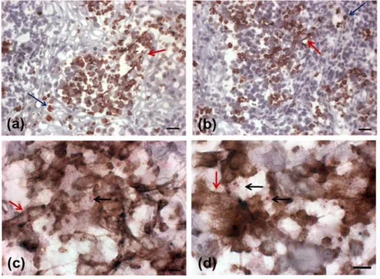
NETs were found throughout the lesion and varied in size according to the presence of neu-trophils (Fig 1A–1D). Two patterns of NET formation were observed: (1) large heterogeneous webs consisting of large-diameter fibers that could be observed at low magnification (100 a 400x) identified when the percentage of neutrophils were higher than 20% (Fig 1A and 1B) and, (2) smaller homogeneous networks consisting of small-diameter fibers that could be better observed at 1000x magnification in all lesions presenting NET formation (Fig 1C and 1D).Fig 2shows DNA and neutrophil elastase co-localization with typical features of NET formation (Fig 2D–2F), as well as neutrophil elastase positive cells presenting preserved nucleus (Fig 2A–
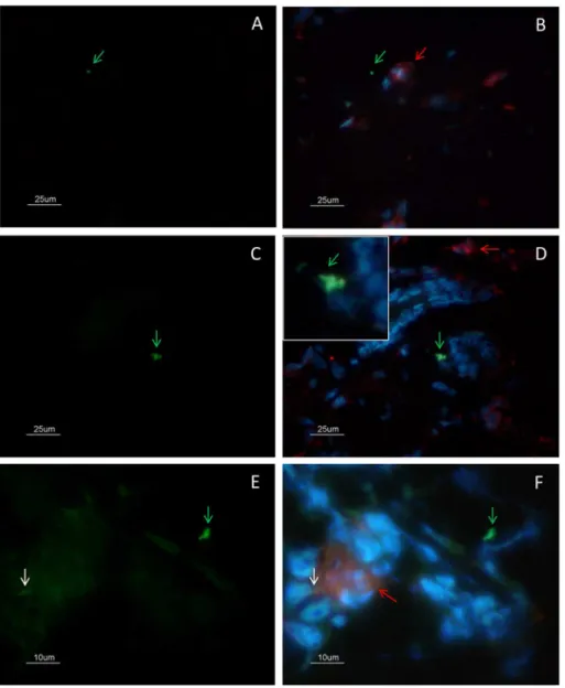
2C). SinceLeishmaniaamastigotes are typically found within lesion macrophages we evaluated the co-localization of macrophages and amastigotes. Amastigotes were observed outside cells (Fig 3A and 3B- green arrow) and inside CD68 negative cells (Fig 3C and 3D- zoom). We also observed amastigote antigens (white arrow) inside macrophages (Fig 3E and 3F- red arrow).
Since the presence of NETs was correlated with the number of neutrophils and since these cells might be involved in the control of parasite burden, we investigated the correlation between the presence of these structures and positivity for amastigotes and/or parasite antigens.
Amastigotes and NETs
Fig 1. Detection of NETs in active lesions.(A, B) Presence of NETs in an active lesion detected by neutrophil elastase immunostain(red arrows) in sections counterstained with Mayer`s hematoxylin (bar = 25μm). (C, D) Colocalization of neutrophils, NETs andLeishmaniaamastigotes. (neutrophil elastase
NETs: red arrows, neutrophils: blue arrows, amastigotes: black arrows) (bar = 10μm).
doi:10.1371/journal.pone.0133063.g001
Fig 2. Colocalization analysis of neutrophil elastase and DNA by immunofluorescence.(A) lesion stained for neutrophil elastase (red), and (B) DNA (blue). (C) merge: elastase and DNA showing a neutrophil elastase positive cell with morphologically preserved nucleus. (D) NET containing neutrophil elastase, (E) and DNA. (F) merge: elastase and DNA. All images were obtained from different regions of the same lesion with 45 days duration before diagnosis. Bar = 10μm.
presented NET formation with small diameters (0.10 and 0.50 NETs/mm2). Among the 29
lesions positive for amastigotes, 25 (86.2%) also presented NET scaffolds., In these patients, the number of amastigotes ranged from 0.06 to 40.0 / mm2(median: 0.5) and the number of NETs
ranged from 0.14 to 23.5 /mm2(median: 1.1). A correlation was observed between the number of amastigotes/mm2and the number of NETs/mm2(r = 0.710; p = 0.0001). An association
between the size of NETs and number of amastigotes could be observed (p = 0.02), since the lesions presenting larger NETs (n = 7;Table 1) showed the higher parasite burden (0.43 to 40.0 amastigotes/mm2-Median 0.90) than the lesions presenting the small NETs (0 to 30.0 amasti-gotes/mm2-Median 0.25). In the lesions of four patients who presented amastigotes in the
absence of NETs, 0.09, 0.6, 0.5 and 0.5 amastigotes/mm2were observed.
Fig 3. Colocalization analysis of CD68(+) macrophages and amastigotes by immunofluorescence. (A-B) Amastigotes (green arrow) were observed outside cells and close to macrophages (red arrow). (C-D) Amastigotes (green arrow) were observed inside CD68 negative cells (zoom). (E-F) Amastigote antigens inside macrophages (white arrow). (A-D) bar = 25μm; (E-F) bar = 10μm.
When the presence of amastigotes, neutrophils and NETs were compared, we noticed that all of them were in close proximity in various areas (Fig 1C and 1D,S8–S16Figs). A correlation was observed between the number of amastigotes/mm2and the percentage of neutrophils (r = 0.573; p = 0.0001). Amastigotes were often intermingled with NETs stained for elastase (Fig 1C and 1D). In addition, large clusters of NETs were noted close to sites containing a large number of parasites. In order to better demonstrate the correlation between the presence of NETs and amastigotes we performed a colocalization analysis by confocal microscopy.
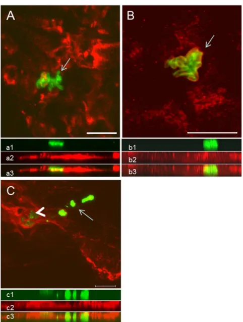
The confocal images reinforced our immunohistochemical findings since extensive areas occupied by NETs, evidenced by NE and histone staining, are in close contact with nests plenty of amastigotes (Fig 4). NETs were scattered throughout the tissue, forming webs of different shapes and diameters. Colocalization analysis of the parasites and NETs showed that most intact amastigotes were surrounded by NETs (Fig 4A–4C,S17andS18Figs,S1 Video). On the other hand, areas positive forLeishmaniaantigens in which no intact parasites could be detected were arranged in a heterogeneous fashion and were not directly associated with the presence of NETs (Fig 4C, arrow), despite the presence of a surrounding inflammatory infil-trate and small numbers of neutrophils (data not shown).
Discussion
and/or by IL-8 andLeishmaniachemotactic factor [34,35], followed by stimulation of NET release. Finally, NET degradation would occur and macrophages could remove cell and antigen remnants. Here, we observed NETs associated to granulomatous inflammatory infiltrate in close proximity with amastigotes. Recently, Braian et al [36] analyzed human macrophages and NETs induced byMycobacterium tuberculosis, and suggested that NETs can transfer dan-ger signals (as that provided by Hsp72) to adjacent macrophages stimulating them to produce cytokines such as IL-6, TNF-α, IL-1βand IL-10. It has been shown that NETs activate NLRP3 inflammasome [37]. Moreover, extracellular trap formation has also been demonstrated for
Fig 4. Colocalization analysis of NETs and amastigotes by confocal microscopy.(A) NETs containing neutrophil elastase (red) and amastigotes (green, arrow). (B) NETs containing histone (red) and amastigotes (green, arrow). (C) Area enclosing intact amastigotes (green—arrowhead) in intimate contact with NETs labeled with anti-neutrophil elastase (red). Area containing freeLeishmania spantigens (arrow). Below each image the respective side view (Z images) of (a1, b1, c1) amastigotes (green), (a2, c2) NET-elastase, (b2) NET-histones, merged sections showing the close contact between amastigotes and NETs (a3, b3, c3). Scale bar = 10μm.
other types of granulocytes, such as eosinophils and mast cells [38,39], an even for macro-phages [40,41]. However, as we have used specific markers for neutrophils, i.e. neutrophil elas-tase, colocalized with DNA, the events analyzed were due to the neutrophil extracellular traps.
Greater attention has been given over the last years to the function of neutrophils during subacute or chronic inflammatory processes. In the particular case of murine leishmaniasis, the results can vary. Lima et al [8] suggested the participation of neutrophils in the control of para-site burden since the absence of neutrophils resulted in the development of severe and dissemi-nated disease. In contrast, Tacchini-Cottier et al [12] showed that the absence of neutrophils had a protective effect associated with a reduced Th2 response and partial resolution of lesions in susceptible BALB/c mice infected withL.major. This apparent contradiction might be due to differences in the mouse models and parasites used since Novais et al [42], studying BALB/c mice infected withL.braziliensis, observed that the elimination of neutrophils increased para-site multiplication. However, the co-inoculation of neutrophils withL.braziliensishad the opposite effect suggesting cooperation between neutrophils and macrophages associated to the production of TNF-αand superoxides. It has been well established that macrophage activation is insufficient inL.majorinfected BALB/c mice due to the production of IL-4 and the smaller neutrophil elastase production [43]. Furthermore, Zandbergen et al [13] showed that neutro-phils may serve as vectors for the entry ofLeishmaniainto macrophages, a model called Trojan horse. Neutrophils enter a process of apoptosis and, together with the parasite, are phagocy-tosed by macrophages, which became inactivated [2,13]. However, in the present study we evaluated the presence of extracellular regions of NE and histone suggesting NETosis that is independent of both apoptosis and necrosis [16,33]. In this context, using cells from resistant mice (C57Bl/6) infected withL.major, Ribeiro-Gomes et al [44] observed that the interaction between dead neutrophils and macrophages promoted the death of the parasite through the secretion of TNF-α. Fadok et al [45] demonstrated that NE was able to activate human macro-phages and to induce the secretion of TNF-α. In addition, the inhibition of NE prevented the leishmanicidal activity of macrophages in a murine model, even in the presence of neutrophils, a fact suggesting the participation of NE in microbicidal activity [44]. Interestingly, during Gram-positive skin infection, Yipp et al [46] visualized live polymorphonuclear cellsin vivo
rapidly releasing NETs, which prevented systemic bacteria dissemination. NETosis occurred during crawling; thereby casting large areas of NETs associated with intact neutrophils that do not undergo lysis and contain phagocytosed pathogens retain the ability to multitask. In our study, active lesions of ATL also presented large areas of NETs composed NE and histones close to intact neutrophils distributed throughout the granulomatous infiltrate suggesting a probable cooperation between NETs, neutrophils and/or macrophages.
The duration of the lesions could play an important role in the relationship between neutro-phil function and parasites. In this respect, during the first moment of the infection, Leish-maniapromastigotes may use the invasion of apoptotic neutrophils as an escape mechanism to establish infection in cells of the mononuclear phagocyte system [2,13]. Once a local inflamma-tory reaction is initiated, the proinflammainflamma-tory environment would stimulate the degranulation and/or formation of NETs before the amastigote forms invade new cells, either neutrophils or macrophages. As a consequence, temporarily extracellular amastigotes would be destroyed, a process contributing to the reduction of local parasite burden. Our results show the presence of high numbers of neutrophils at the site of lesions and their intimate relationship with amasti-gotes, however it is important to mention that amastigotes could not be visualized inside neu-trophils. In addition, all lesions studied already presented an important and diffusely
distributed inflammatory process (data not shown and [31]).
highlighted. In the first, NETs were identified in peripheral blood neutrophils of patients with malaria and associated with both severe malaria in children and mechanisms of protection in adults [27]. Knowledge about this relationship with disease severity might be important for studies investigating protection against this infection. Similarly, herein we observed positive correlations between NET formation and age, neutrophil percentage and age, as well as, NET and number of lesions. The second article, anin vitrostudy of the interaction between neutro-phils andL.amazonensispromastigotes, demonstrated the trapping of these forms in the fiber networks [28]. In addition, the authors observed a toxic effect of histones released during the process of NET formation, reducing promastigote survival and observed extracellular regions of DNA and histone in human lesions suggesting NET activity duringin vivoinfection. In the third article, Bruns et al [22] showed that NETs are formedin vivoduring the immune
response againstAspergillus fumigatusand that their formation is a dynamic process. Although not playing a key role in the elimination of the fungus, the authors suggested that NETs exert a fungistatic effect, preventing dissemination of the fungus. In the same manner, NET could exert an additional parasiticidal effect, helping macrophages to clear parasites from the active lesions.
In conclusion, the present results suggest that 1) although neutrophils are classically identi-fied as cells of the innate immune response, they can be detected throughout the inflammatory reaction observed in established ATL lesions; 2) the presence of NETs is directly related to the presence of intact amastigotes, irrespective of lesion duration, suggesting that their formation is maintained by the stimulus derived from the parasite burden/parasite antigen in the extracel-lular environment; 3) the observation of areas containing only antigens not intermingled with NETs (elastase and histone) suggests that the involvement of these structures in the control of parasite burden is a dynamic process in which the formation of these networks is exhausted with the destruction of the parasites; 4) the presence of extracellular parasite antigens could facilitate the activation of macrophages to the microbicidal stage, thus activating the control of parasite burden.
Supporting Information
S1 Fig. Neutrophil elastase and NET formation (brown).Inflammatory infiltrate in skin lesions of American tegumentary leishmaniasis. Counterstain—Meyer`s hematoxilin; 400x magnification.
(PDF)
S2 Fig. Neutrophil elastase and NET formation (brown).Inflammatory infiltrate in skin lesions of American tegumentary leishmaniasis. Counterstain—Meyer`s hematoxilin; 400x magnification.
(PDF)
S3 Fig. Neutrophil elastase and NET formation (brown).Inflammatory infiltrate in skin lesions of American tegumentary leishmaniasis. Counterstain—Meyer`s hematoxilin; 400x magnification.
(PDF)
S4 Fig. Neutrophil elastase and NET formation (brown).Inflammatory infiltrate in skin lesions of American tegumentary leishmaniasis. Counterstain—Meyer`s hematoxilin; 400x magnification.
S5 Fig. Neutrophil elastase and NET formation (brown).Inflammatory infiltrate in skin lesions of American tegumentary leishmaniasis. Counterstain—Meyer`s hematoxilin; 200x magnification.
(PDF)
S6 Fig. Neutrophil elastase and NET formation (brown).Inflammatory infiltrate in skin lesions of American tegumentary leishmaniasis. Counterstain—Meyer`s hematoxilin; 200x magnification.
(PDF)
S7 Fig. Neutrophil elastase and NET formation (brown).Inflammatory infiltrate in skin lesions of American tegumentary leishmaniasis. Counterstain—Meyer`s hematoxilin; 200x magnification.
(PDF)
S8 Fig. Colocalization of neutrophil elastase (brown) and amastigotes (red).Inflammatory infiltrate in skin lesions of American tegumentary Leishmaniasis. counterstain—Meyer`s hematoxilin. 1000x magnification.
(PDF)
S9 Fig. Colocalization of neutrophil elastase (brown) and amastigotes (red).Inflammatory infiltrate in skin lesions of American tegumentary Leishmaniasis. counterstain—Meyer`s hematoxilin. 1000x magnification.
(PDF)
S10 Fig. Colocalization of neutrophil elastase (brown) and amastigotes (red).Inflammatory infiltrate in skin lesions of American tegumentary Leishmaniasis. counterstain—Meyer`s hematoxilin. 1000x magnification.
(PDF)
S11 Fig. Colocalization of neutrophil elastase (brown) and amastigotes (red).Inflammatory infiltrate in skin lesions of American tegumentary Leishmaniasis. counterstain—Meyer`s hematoxilin. 1000x magnification.
(PDF)
S12 Fig. Colocalization of neutrophil elastase (brown) and amastigotes (red).Inflammatory infiltrate in skin lesions of American tegumentary Leishmaniasis. counterstain—Meyer`s hematoxilin. 1000x magnification.
(PDF)
S13 Fig. Colocalization of neutrophil elastase (brown) and amastigotes (red).Inflammatory infiltrate in skin lesions of American tegumentary Leishmaniasis. counterstain—Meyer`s hematoxilin. 1000x magnification.
(PDF)
S14 Fig. Neutrophil elastase and NET formation (brown).Inflammatory infiltrate in skin lesions of American tegumentary Leishmaniasis. counterstain—Meyer`s hematoxilin. 1000x magnification.
(PDF)
S15 Fig. Colocalization of neutrophil elastase (brown) and amastigotes (red).Inflammatory infiltrate in skin lesions of American tegumentary Leishmaniasis. counterstain—Meyer`s hematoxilin. 1000x magnification.
S16 Fig. Colocalization of neutrophil elastase (brown) and amastigotes (red).Inflammatory infiltrate in skin lesions of American tegumentary Leishmaniasis. counterstain—Meyer`s hematoxilin. 1000x magnification.
(PDF)
S17 Fig. Colocalization analysis by confocal microscopy.(Figure A) Neutrophil elastase (red) and amastigotes (green); (Figure B) histone (red) and amastigotes (green); (Figure C) his-tone (red) and degraded amastigotes (green). Scale bar = 10um.
(PDF)
S18 Fig. Colocalization analysis by confocal microscopy.Neutrophil elastase (red) and amas-tigotes (green); Degraded amasamas-tigotes (arrow). Scale bar = 10um.
(PDF)
S1 Video. Colocalization analysis by confocal microscopy.Area containing intact amasti-gotes (green) surrounded by NETs that contain neutrophil elastase (black arrow). Area con-taining freeLeishmania spantigens (white arrow). Scale bar = 10μm.
(AVI)
S1 Supporting Information.
(PDF)
Acknowledgments
We thank the Program for Technological Development in Tools for Health—PDTIS-FIOCRUZ
for use of their confocal facilities.
Author Contributions
Conceived and designed the experiments: FNM MTCN EMS FCS. Performed the experiments: FNM MTCN COR MFM MCS ECFV MRL MIFP. Analyzed the data: FNM MTCN EMS FCS. Contributed reagents/materials/analysis tools: FCS EMS AOS FNM MRL MIFP. Wrote the paper: FNM MTCN EMS COR ECFV AOS FCS MRL MIFP.
References
1. Marzochi MC, Marzochi KB (1994) Tegumentary and visceral leishmaniases in Brazil: emerging anthro-pozoonosis and possibilities for their control. Cad Saúde Pública 10:359–75. PMID:15042226
2. Laskay T, van Zandbergen G, Solbach W (2008) Neutrophil granulocytes as host cells and transport vehicles for intracellular pathogens: apoptosis as infection-promoting factor. Immunobiology 213:183– 91. doi:10.1016/j.imbio.2007.11.010PMID:18406366
3. Bogdan C, Donhauser N, Döring R, Röllinghoff M, Diefenbach A, Rittiq M. (2000) Fibroblasts as host cells in latent leishmaniosis. J Exp Med 191:2121–30. PMID:10859337
4. Hespanhol RC, de Nazaré CSM, Meuser MB, de Nazareth S L, Meirelles M, Côrte-Real S. (2005) The expression of mannose receptors in skin fibroblast and their involvement in Leishmania (L.) amazonen-sis invasion. J Histochem Cytochem 53:35–44. PMID:15637336
5. Lugo-Yarbuh A, Valera M, Alarcón M, Moreno E, Premoli-Percoco G, Colasante C. (2003) [Detection of Leishmania (Viannia) braziliensis in vascular endothelium lesions of patients with localized cutaneous leishmaniasis]. Investig Clínica 44:61–76.
6. Quintella LP, Cuzzi T, Madeira M de F, Okamoto T, Schubach A de O (2009) Immunoperoxidase tech-nique using an anti-Leishmania (L.) chagasi hyperimmune serum in the diagnosis of culture-confirmed American tegumentary leishmaniasis. Rev Inst Med Trop São Paulo 51:83–6. PMID:19390736
8. Lima GM, Vallochi AL, Silva UR, Bevilacqua EM, Kiffer MM, Abrahamsohn IA. (1998) The role of poly-morphonuclear leukocytes in the resistance to cutaneous Leishmaniasis. Immunol Lett 64:145–51. PMID:9870666
9. Peters NC, Egen JG, Secundino N, Debrabant A, Kimblin N, Kamhavi S et al. (2008) In vivo imaging reveals an essential role for neutrophils in leishmaniasis transmitted by sand flies. Science 321:970–4. doi:10.1126/science.1159194PMID:18703742
10. Pompeu ML, Freitas LA, Santos ML, Khouri M, Barral-Netto M (1991) Granulocytes in the inflammatory process of BALB/c mice infected by Leishmania amazonensis. A quantitative approach. Acta Trop 48:185–93. PMID:1671620
11. Belkaid Y, Mendez S, Lira R, Kadambi N, Milon G, Sacks D. (2000) A natural model of Leishmania major infection reveals a prolonged“silent”phase of parasite amplification in the skin before the onset of lesion formation and immunity. J Immunol 165:969–77. PMID:10878373
12. Tacchini-Cottier F, Zweifel C, Belkaid Y, Mukankundiye C, Vasei M, Launois P et al. (2000) An immuno-modulatory function for neutrophils during the induction of a CD4+ Th2 response in BALB/c mice infected with Leishmania major. J Immunol 165:2628–36. PMID:10946291
13. Van Zandbergen G, Klinger M, Mueller A, Dannenberg S, Gebert A, Solbach W et al. (2004) Cutting edge: neutrophil granulocyte serves as a vector for Leishmania entry into macrophages. J Immunol 173:6521–5. PMID:15557140
14. Afonso L, Borges VM, Cruz H, Ribeiro-Gomes FL, DosReis GA, Dutra AN et al. (2008) Interactions with apoptotic but not with necrotic neutrophils increase parasite burden in human macrophages infected with Leishmania amazonensis. J Leukoc Biol 84:389–96. doi:10.1189/jlb.0108018PMID:18483206
15. Ribeiro-Gomes FL, Sacks D (2012) The influence of early neutrophil-Leishmania interactions on the host immune response to infection. Front Cell Infect Microbiol 2:59. doi:10.3389/fcimb.2012.00059
PMID:22919650
16. Abi Abdallah DS, Denkers EY (2012) Neutrophils cast extracellular traps in response to protozoan par-asites. Front Immunol 3:382. doi:10.3389/fimmu.2012.00382PMID:23248631
17. Brinkmann V, Reichard U, Goosmann C, Fauler B, Uhlemann Y, Weiss DS et al. (2004) Neutrophil extracellular traps kill bacteria. Science 303:1532–5. PMID:15001782
18. Urban CF, Reichard U, Brinkmann V, Zychlinsky A (2006) Neutrophil extracellular traps capture and kill Candida albicans yeast and hyphal forms. Cell Microbiol 8:668–76. PMID:16548892
19. Neeli I, Khan SN, Radic M (2008) Histone deimination as a response to inflammatory stimuli in neutro-phils. J Immunol 180:1895–902. PMID:18209087
20. Wang Y, Li M, Stadler S, Correll S, Li P, Hayama R et al. (2009) Histone hypercitrullination mediates chromatin decondensation and neutrophil extracellular trap formation. J Cell Biol 184:205–13. doi:10. 1083/jcb.200806072PMID:19153223
21. Ramos-Kichik V, Mondragón-Flores R, Mondragón-Castelán M, Gonzalez-Pozos S, Muñiz-Hernandez S, Rojas-Espinosa O et al. (2009) Neutrophil extracellular traps are induced by Mycobacterium tubercu-losis. Tuberc Edinb Scotl 89:29–37.
22. Bruns S, Kniemeyer O, Hasenberg M, Aimanianda V, Nietzsche S, Thywissen A et al. (2010) Produc-tion of extracellular traps against Aspergillus fumigatus in vitro and in infected lung tissue is dependent on invading neutrophils and influenced by hydrophobin RodA. Plos Pathog 6:e1000873. doi:10.1371/ journal.ppat.1000873PMID:20442864
23. Morgado FN, Schubach AO, Barros MBL, Conceição-Silva F (2011) The in situ inflammatory profile of lymphocutaneous and fixed forms of human sporotrichosis. Med Mycol 49:612–20. doi:10.3109/ 13693786.2011.552532PMID:21254963
24. Saitoh T, Komano J, Saitoh Y, Misawa T, Takahama M, Kozaki T et al. (2012) Neutrophil extracellular traps mediate a host defense response to human immunodeficiency virus-1. Cell Host Microbe 12:109–16. doi:10.1016/j.chom.2012.05.015PMID:22817992
25. Abi Abdallah DS, Lin C, Ball CJ, King MR, Duhamel GE, Denkers EY. (2012) Toxoplasma gondii trig-gers release of human and mouse neutrophil extracellular traps. Infect Immun 80:768–77. doi:10. 1128/IAI.05730-11PMID:22104111
26. Behrendt JH, Ruiz A, Zahner H, Taubert A, Hermosilla C (2010) Neutrophil extracellular trap formation as innate immune reactions against the apicomplexan parasite Eimeria bovis. Vet Immunol Immuno-pathol 133:1–8. doi:10.1016/j.vetimm.2009.06.012PMID:19625090
28. Guimarães-Costa AB, Nascimento MTC, Froment GS, Soares RPP, Morgado FN, Conceição-Silva F et al. (2009) Leishmania amazonensis promastigotes induce and are killed by neutrophil extracellular traps. Proc Natl Acad Sci USA 106:6748–53. doi:10.1073/pnas.0900226106PMID:19346483
29. Gabriel C, McMaster WR, Girard D, Descoteaux A (2010) Leishmania donovani promastigotes evade the antimicrobial activity of neutrophil extracellular traps. J Immunol 185:4319–27. doi:10.4049/ jimmunol.1000893PMID:20826753
30. Madeira MF, Schubach A, Schubach TMP, Pacheco RS, Oliveira FS, Pereira SA et al. (2006) Mixed infection with Leishmania (Viannia) braziliensis and Leishmania (Leishmania) chagasi in a naturally infected dog from Rio de Janeiro, Brazil. Trans R Soc Trop Med Hyg 100:442–5. PMID:16257024
31. Morgado FN, Schubach A, Rosalino CMV, Quintella LP, Santos G, Salgueiro M et al. (2008) Is the in situ inflammatory reaction an important tool to understand the cellular immune response in American tegumentary leishmaniasis? Br J Dermatol 158:50–8. PMID:17944980
32. Morgado FN, Schubach A, Vasconcellos E, Azeredo-Coutinho RB, Valete-Rosalino CM, Quintella LP et al. (2010) Signs of an in situ inflammatory reaction in scars of human American tegumentary leish-maniasis. Parasite Immunol 32:285–95. doi:10.1111/j.1365-3024.2009.01188.xPMID:20398229
33. Brinkmann V, Zychlinsky A (2012) Neutrophil extracellular traps: is immunity the second function of chromatin? J Cell Biol 198:773–83. doi:10.1083/jcb.201203170PMID:22945932
34. Van Zandbergen G, Hermann N, Laufs H, Solbach W, Laskay T (2002) Leishmania promastigotes release a granulocyte chemotactic factor and induce interleukin-8 release but inhibit gamma interferon-inducible protein 10 production by neutrophil granulocytes. Infect Immun 70:4177–84. PMID:
12117926
35. Menezes MJ, Costa DJ, Clarêncio J, Miranda JC, Barral A, Barral-Netto N et al. (2008) Immunomodula-tion of human monocytes following exposure to Lutzomyia intermedia saliva. Bmc Immunol 9:12. doi:
10.1186/1471-2172-9-12PMID:18402685
36. Braian C, Hogea V, Stendahl O (2013) Mycobacterium tuberculosis- Induced Neutrophil Extracellular Traps Activate Human Macrophages. J Innate Immun 5:591–602. doi:10.1159/000348676PMID:
23635526
37. Kahlenberg JM, Carmona-Rivera C, Smith CK, Kaplan MJ (2013) Neutrophil extracellular trap-associ-ated protein activation of the NLRP3 inflammasome is enhanced in lupus macrophages. J Immunol 190:1217–26. doi:10.4049/jimmunol.1202388PMID:23267025
38. Yousefi S, Gold JA, Andina N, Lee JJ, Kelly AM, Kozlowski E et al. (2008) Catapult-like release of mito-chondrial DNA by eosinophils contributes to antibacterial defense. Nat Med 14:949–53. doi:10.1038/ nm.1855PMID:18690244
39. Von Köckritz-Blickwede M, Goldmann O, Thulin P, Heinemann K, Norrby-Teglund A, Rohde M et al. (2008) Phagocytosis-independent antimicrobial activity of mast cells by means of extracellular trap for-mation. Blood 111:3070–80. doi:10.1182/blood-2007-07-104018PMID:18182576
40. Bartneck M, Keul HA, Zwadlo-Klarwasser G, Groll J (2010) Phagocytosis independent extracellular nanoparticle clearance by human immune cells. Nano Lett 10:59–63. doi:10.1021/nl902830xPMID:
19994869
41. Chow OA, von Köckritz-Blickwede M, Bright AT, Hensler ME, Zinkernagel AS, Coqen AL et al. (2010) Statins enhance formation of phagocyte extracellular traps. Cell Host Microbe 8:445–54. doi:10.1016/ j.chom.2010.10.005PMID:21075355
42. Novais FO, Santiago RC, Báfica A, Khouri R, Afonso L, Borges VM et al. (2009) Neutrophils and macro-phages cooperate in host resistance against Leishmania braziliensis infection. J Immunol 183:8088– 98. doi:10.4049/jimmunol.0803720PMID:19923470
43. Ribeiro-Gomes FL, Moniz-de-Souza MCA, Alexandre-Moreira MS, Dias WB, Lopes MF, Nunes MP et al. (2007) Neutrophils activate macrophages for intracellular killing of Leishmania major through recruitment of TLR4 by neutrophil elastase. J Immunol 179:3988–94. PMID:17785837
44. Ribeiro-Gomes FL, Otero AC, Gomes NA, Moniz-De-Souza MCA, Cysne-Finkelstein L, Arnholdt AC et al. (2004) Macrophage interactions with neutrophils regulate Leishmania major infection. J Immunol 172:4454–62. PMID:15034061
45. Fadok VA, Bratton DL, Guthrie L, Henson PM (2001) Differential effects of apoptotic versus lysed cells on macrophage production of cytokines: role of proteases. J Immunol 166:6847–54. PMID:11359844
46. Yipp BG, Petri B, Salina D, Jenne CN, Scott BNV, Zbytnuik LD et al. (2012) Infection-induced NETosis is a dynamic process involving neutrophil multitasking in vivo. Nat Med 18:1386–93. PMID:22922410
47. Guimarães-Costa AB, Nascimento MTC, Wardini AB, Pinto-da-Silva LH, Saraiva EM (2012) ETosis: A Microbicidal Mechanism beyond Cell Death. J Parasitol Res 2012:929743. doi:10.1155/2012/929743