S CIE
NT OR
U
M
ACTA
Acta Sci. Pol., Technol. Aliment. 9(1) 2010, 45-59
ISSN 1644-0730 (print) ISSN 1889-9594 (online)
© Copyright by Wydawnictwo Uniwersytetu Przyrodniczego w Poznaniu
BIOTRANSFORMATION OF FERULIC ACID
BY LACTOBACILLUS ACIDOPHILUS K1
AND SELECTED BIFIDOBACTERIUM STRAINS
Dominik Szwajgier, Anna Jakubczyk
University of Life Sciences in LublinBackground. Lactic acid bacteria (LAB) were pointed out to produce ferulic acid es-terase. Except the release of phenolic acids from esterified forms, it was postulated that the biotransformations of these compounds can occur during the bacterial growth. In the presented work, the biotransformation of ferulic acid by Lactobacillus acidophilus K1 and three Bifidibacterium strains (B. animalis Bi30, B. catenulatum KD 14 and B. longum KN 29) was studied.
Material and methods. The microorganisms were grown in media containing methyl es-ters of phenolic acids as carbon sources. The feruloyl esterase activity as well as the con-tents of phenolic acids in supernatants were estimated using HPLC-DAD.
Results. The enzyme activity was evaluated using methyl ferulate exclusively, but p- -coumaric acid and another chromatographic peak (probably caffeic acid, but its identity was not positively confirmed by the DAD analysis) were present in reaction mixtures con-taining the supernatants of Lactobacillus acidophilus K1 cultivars with methyl p-cou-marate or methyl syringate. Both peaks of p-coumaric acid and another phenolic com-pound were also present in the solutions containing the supernatants of B. catenulatum and B. longum grown in the presence of methyl vanillate and the supernatants of B. ani-malis Bi30 grown using methyl p-coumarate, methyl syringate or methyl vanillate. Conclusions. The results suggest a distinct ability of the studied LAB strains to transform free ferulic acid yielding p-coumaric acid and probably caffeic acid although no mecha-nism involved in this transformation was proposed and closer characterised in the frames of this work.
INTRODUCTION
In the previous decades, a considerable number of ferulic acid esterases (FAEs) of fungal origin were well characterised but the knowledge of the enzymes produced by lactic acid bacteria (LAB) is rather limited [Donaghy et al. 1998, Vardakou et al. 2007, 2008, Nsereko et al. 2008, Yuan et al. 2005, Wang et al. 2004]. Esterases (EC 3.1.1.x) is the class of hydrolytic enzymes broadly distributed in the plant and animal kingdom. Ferulic acid esterases produced by LAB present in human gastrointestinal tract can be a very interesting object of nutritional studies. Dietary fiber is a rich source of strong antioxidants- phenolic acids esterified to polymeric non-starch polysaccharides, with ferulic or p-coumaric acids known to be very strong antioxidants in vitro [Gerhäuser 2005, Maillard and Berset 1995, Maillard et al. 1996, Nardini et al. 1995] or in vivo [Tanaka et al. 1993, Itagaki et al. 2009, Joshi et al. 2006, Saija et al. 2000, Young et al. 2008]. Prior absorption, phenolic acids esters are deesterified from dietary fiber in the gastrointestinal tract due to the action of bacterial FAEs, also originating from LAB [Andreason et al. 2001, Couteau et al. 2001] followed by the transformations in the liver [Scalbert and Williamson 2000, Olthof et al. 2003, Plumb et al. 1999, Rondini et al. 2002, Hollman and Katan 1998, Nardini et al. 1997, Ghiselli et al. 2000]. A considerable number of works reveal that free phenolic acids, including ferulic acid, are very easily absorbed into blood plasma [Nardini et al. 2002, 2006] and excreted in urine [Choud-hury et al. 1999, Bourne and Rice-Evans 1998].
MATERIAL AND METHODS
Strains and reagents
Bacterial strains were isolated from gastrointestinal tract either from infants (B. lon-gum KN29) or adults (B. catenulatum KD14, B. bifidum Bi30, L. acidophilus K1) by Prof. dr hab. Maria Bielecka from the Department of Food Microbiology, Institute of Animal Reproduction and Food Research of Polish Academy of Sciences in Olsztyn, Poland [Bielecka et al. 2002]. Methyl ferulate, methyl p-coumarate, methyl syringate and methyl vanillate were obtained from Apin Chemicals, Oxon, UK. Garche’s and MRS broths were purchased from BTL Ltd. Zakład Enzymów i Peptonów, Poland. All other reagents were from POCh Gliwice, Poland. Chromatography reagents (POCh Gliwice, Poland) were of HPLC grade.
Culture conditions
Bifidobacterium strains were grown using the slightly modified method of
Bie-drzycka et al. [2003]. The bacteria were thermostated in Garche’s medium (10 cm3)
in test tubes at 37°C in anaerobic conditions (0.5 cm3 of 15% (w/v) NaHCO3 solution
and 0.5 cm3 20% (w/v) pirogallol solution injected into cotton stoppers followed by the
aseptical closing of the tubes using rubber stoppers). Every 24 hours, cultures were
repeatedly inoculated in a new broth using 5% (v/v) of inoculum. Lactobacillus
aci-dophilus K1 was grown using MRS broth (10 cm3) at 37°C in 30 cm3 test tubes and then
every 24 hours it was repeatedly inoculated using new MRS broths and 3% (v/v) of inoculum. These bacterial cultures were used for subsequent studies as described below.
Method of cultivation for FAE
All bacterial strains were cultivated at 37°C in tubes with cotton and rubber
stop-pers, in anaerobic conditions, as described above, in 10 cm3 of minimal growth medium
composed of (g in dm3): peptone – 2.0, yeast extract – 2.0, L-cysteine·HCl – 0.5, NaCl –
0.1, NaHCO3 – 2.0, K2HPO4 – 0.04, KH2PO4 – 0.04, MgSO4·7H2O – 0.01, CaCl2·6H2O
– 0.01, Tween 80 – 2 cm3. As a carbon source, 0.25% (w/v) of methyl syringate, methyl
FAE activity determination
The ability of FAE to hydrolyze methyl ferulate was used and the free ferulic acid content was determined using HPLC with Diode Array Detection. Methyl ferulate was
dissolved in minimal volume of 96.0% ethanol (v/v, typically 0.05 cm3) followed by the
dilution using Tris-HCl buffer (100 mM × dm-3, pH 6.5) until 6 mM × dm-3
concentra-tion was obtained. 0.5 cm3 of the studied sample was added to 0.1 cm3 of substrate
solu-tion and the samples were incubated at 37°C for 5 hours. The enzyme was then inacti-vated in boiling water (5 min) followed by the cooling and centrifugation (7000 × g, 30 min, 6°C, centrifuge MPW-365, Mechanika Precyzyjna, Warsaw, Poland). Free phe-nolic acids and methyl ferulate in studied and double blank samples (lacking substrate or supernatant solution) were separated and analysed simultaneously as described be-low. Both blank samples were subtracted from corresponding studied samples. FAE
activity was expressed in units (1 unit is equal to 1 nM of ferulic acid released in 1 cm3
of reaction medium after 1 min of incubation). Analyses were duplicated and mean values with standard deviations were calculated.
HPLC-DAD identification of phenolic acids and methyl ferulate
The HPLC system consisted of two Gilson 306 Separation Module piston pumps,
Gilson PhotoDiode Array Detector 170, Gilson loop (0.02 cm3), manometric module
Gilson 805, dynamic mixer 811C. Waters Symmetry C18 column (USA, 250 mm, 4.6 mm
i.d., 5 m), and Waters Symmetry C18 pre-column (5 m, 8 × 20 mm) were used for
separations. The method of Kim et al. [2006] was used. Eluents used were: A-1% (w/v) acetic acid solution in DDI water. Eluent B- 50% HPLC-grade acetonitrile in DDI wa-ter. Signals were monitored at 320 nm, 280 nm, 260 nm and 360 nm. The program ap-plied was as follows: START 92% A, 8% B 0-10 min; 70% A, 30% B 10-40 min; 60%
A, 40% B 40-55 min; 92% A, 8% B 55-70 min. The effluent flow was 0.8 cm3 × min-1
(17 MPa). Phenolic acids concentrations were calculated using the calibration curves plotted using the series of HPLC grade phenolic acids standards individually injected into HPLC-DAD system. Diode Array spectra of studied chromatographic peaks were obtained and compared with spectra of phenolic acids standards in the database (Gilson Unipoint ver. 3.01).
RESULTS AND DISCUSSION
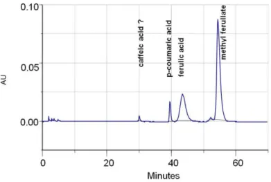
Methyl ferulate concentration in the enzyme reaction mixture was significantly higher than the concentrations of the compounds originating from the studied supernatants. If this is the case, it can be concluded that both p-coumaric acid peak and unidentified peak originate from methyl ferulate. The unidentified peak can be a caffeic acid deriva-tive as well as other phenolic acid but no further identification was performed. What is obvious, quantitative analysis of this compound was also omitted in the results.
Fig. 1. An example chromatogram obtained after the separation of phenolic acids and methyl ferulate during ferulic acid esterase activity determination
Free p-coumaric acid was present in the reaction mixtures containing the post-culti-vation supernatants of Lactobacillus acidophilus K1 after 42 hours of cultipost-culti-vation fol-lowed by the continuous increase in the phenolic acid concentration until 60 hours (Fig. 2). As it can be seen, free ferulic acid was released from methyl ferulate as a result of FAE activity in the reaction mixtures containing post-cultivation supernatants except the last hours of bacterial growth (84 hours of cultivation). It is interesting that p-cou-maric acid concentrations were higher than corresponding ferulic acid contents in post-cultivation supernatants and that after 84 hours of post-cultivation, no free ferulic acid was detected in the solution.
Fig. 2. Phenolic acids concentrations in reaction mixtures ob-tained during ferulic acid esterase activity determina-tion (methyl p-coumarate was the carbon source for L. acidophilus K1 growth)
Fig. 3. Phenolic acids concentrations in reaction mixtures ob-tained during ferulic acid esterase activity determination (methyl p-coumarate was the carbon source for B. anima-lis Bi30 growth)
0 2 4 6 8 10
48 60 84
Cultivation time, h
Phenolic a
c
id
co
nte
n
t, nM·
c
m
-3
p-coumaric acid
ferulic acid 0
2 4 6 8 10
36 42 48 60 84
Cultivation time, h
Phenolic a
c
id
co
nte
n
t, nM·
c
m
-3
p-coumaric acid
Fig. 4. Phenolic acids concentrations in reaction mixtures ob-tained during ferulic acid esterase activity determina-tion (methyl syringate was the carbon source for L. acidophilus K1 growth)
P-coumaric acid was also present when the reaction mixture was composed of methyl ferulate solution (substrate) and the supernatant obtained after the cultivation of L. acidophilus K1 (Fig. 4) or B. animalis Bi30 (Fig. 5) using methyl syringate as a carbon source.
Fig. 5. P-coumaric acid concentrations in reaction mixtures obtained during ferulic acid esterase activity deter-mination (methyl syringate was the carbon source for B. animalis Bi30 growth)
0 0.06 0.12 0.18
36 42 48 60 84
Cultivation time, h
P-coumari
c
acid
co
nten
t, nM·cm
-3
0 0.2 0.4 0.6 0.8
36 42 48 60 84
Cultivation time, h
Phenolic a
c
id
co
nte
n
t, nM·
c
m
-3
p-coumaric acid
In the case of L. acidophilus K1 (Fig. 4), p-coumaric acid was present in the reaction medium during FAE activity determination beginning from the 36. hour of cultivation. The content of the acid significantly increased after 42. and 84. hour of cultivation. It must be pointed out that ferulic acid was released from methyl ferulate during enzyme activity determination. Nevertheless, it was not present in the reaction mixtures contain-ing post-cultivation supernatants originatcontain-ing from the later stages of bacterial growth (42-84 hours).
Similarly, no free ferulic acid was detected in the reaction mixtures containing post-cultivation supernatants originating from all stages of B. animalis Bi30 growth (36-84 hours) when methyl syringate was the only carbon source for the bacterial growth (Fig. 5). After the initial increase in the content of p-coumaric acid in the reaction mix-tures, the decrease after 84 hours of bacterial growth was seen what could suggest the corresponding changes in FAE activity or the loss of p-coumaric acid due to the bacte-rial decarboxylation of this acid yielding 4-ethylguaiacol. Anyway, this observation was not further elaborated within the frames of this study and needs a detailed explanation.
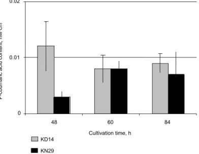
Other two LAB strains- B. catenulatum KD14 and B. longum KN29 exhibited the limited ability to transform ferulic acid into p-coumaric acid when cultivated with the use of methyl vanillate (Fig. 6). No ferulic acid originating from the hydrolysis of methyl ferulate was detected and the concentrations of p-coumaric acid in the case of both bacterial strains were similar especially at the later stages of cultivations.
Fig. 6. P-coumaric acid concentrations in reaction mixtures obtained during ferulic acid esterase activity determination (methyl va-nillate was the carbon source for B. catenulatum KD14 and B. longum KN29 growth)
0 0.01 0.02
48 60 84
Cultivation time, h
P-coumari
c
acid
co
nten
t, nM·cm
-3
KD14
DISCUSSION
It is evident that lactic acid bacteria are able to transform the components of the die-tary fiber fractions to the limited extent due to the production of some secondary en-zymes like FAE [Wang et al. 2004]. Donaghy et al. [1998] detected FAE activities in 7 strains of lactic acid bacteria: 1 Lactobacillus fermentum and 6 Bacillus subtilis strains, but as many as 80 Bacillus-type strains and 50 gram positive bacteria (Enterococcus,
Lactobacillus, Lactococcus, Leuconostoc, Pediococcus and Propionibacterium) were
studied in the frames of this work. L. fermentum NCF 1751 released free ferulic acid from methyl ferulate, methyl p-coumarate and a natural feruloylated oligomer-2-O-[5- -O-(trans-ferulic acid)--L-ara-f]-D-xyl-p. In 2008, Nsereko et al. [2008] examined approx. 10 000 LAB strains for the ability to produce FAE and some FAE-producing microorganisms were selected for the further studies, including: Lactobacillus buchneri,
L. crispatus, L. reuteri, L. brevis and one not identified Lactobacillus strain. Live
L. fermentum 11976 cells were immobilized in alginate-poly(L-lysine)-alginate gel and the FAE activity was detected in the supernatant solution. The alginate microcapsules were used as the source for the enzyme in the in vitro experiments using the model of the gastrointestinal tract [Bhathena et al. 2008].
Some authors successfully used wheat bran, a cheap and valuable by-product, as natural carbon source for the LAB growth [Wang et al. 2005]. FAE activity was also
detected in batch cultures of faecal microorganisms with water-unextractable
feruloy-lated arabinoxylans from de-starched wheat flour [Vardakou et al. 2007] but no release or transformations of phenolic acids were studied in the frames of this work. Lactobacil-lus and Bifidobacterium were repeatedly pointed out as FAE producers in mixed batch cultures of faecal microorganisms containing water-unextractable (untreated and pre-treated with fungal xylanase) feruloylated arabinoxylans from de-starched wheat flour [Vardakou et al. 2008]. The both citied works of Vardakou et al. focused on the ability of LAB strains to grow in the medium containing non-starch polysaccharides from wheat bran. The detailed analysis of the decomposition of polysaccharides was not performed and the phenolic acids concentrations in the growth media were not determined.
The analysis of the scientific databases can give a clear assumption, that the litera-ture concerning the biotransformation of phenolic acids by lactic acid bacteria is incom-plete. Phenolic acids side chains undergo biotransformations but the phenol ring re-mains unchanged what causes the modifications of biological activities of these pheno-lic compounds. Chlorogenic acid subjected to the action of the consortium of human faecal bacteria underwent epimerization to the mixture of 3-, 4- and 5-caffeoyl quinic acid followed by the hydrolysis yielding caffeic acid, then after 8 hours of incubation caffeic acid was reduced to dihydrocaffeic acid. After another 24 hours, dihydrocaffeic acid was dehydroxylated yielding 3-(3,4-dihydroxyphenyl)-priopionic acid [Rechner et al. 2004]. Taking under consideration individual bacterial strains, there is a considerable number of works concerning Lactobacillus strains. In the presented work it was shown
that Lactobacillus acidophilus K1 was able to effectively release free ferulic acid but
the post-cultivations supernatants contained significant concentrations of p-coumaric acid. Cavin et al. [1997] purified and characterised a p-coumaric acid decarboxylase
from Lactobacillus plantarum. The enzyme activity was 60-fold higher towards p-
de-termined the ability of Lactobacillus plantarum CECT 748T strain to metabolize 19 food phenolic acids. Only p-coumaric, m-coumaric, ferulic and caffeic acids were metabo-lized. Caffeic and p-coumaric acids were transformed into their corresponding ethyl and vinyl derivatives, ferulic acid was transformed into 4-vinylguaiacol, and 3-(3-OH- -phenyl) propionic acid was produced from m-coumaric acid. Gallic and protocatechuic acid were decarboxylated yielding pyrogallol and catechol, respectively. The enzymes involved in these transformations were inducible as verified in phenolic acids free bac-terial cultures. On the other hand, the ability to metabolize the phenolic acids is strain-specific, because in another work [Landete et al. 2008] it was showed that only proto-catechuic acid was metabolized to catechol (via decarboxylation) during green olives fermentation by a number of different Lactobacillus plantarum strains, including CECT
748T strain. Alberto et al. [2001] showed that (+)-catechin and gallic acid were utilized
from the very beginning growth of Lactobacillus hilgardi in the experimental FT80 medium containing tomato juice and phenolic standard. The bacterial cell number was
higher in the medium containing gallic acid up to 100 mg·dm-3 compared to the cell
number in control medium what suggested the use of this phenolic acid as the carbon source. When MRS broth was used for bacterial growth in the presence of gallic acid, no difference between studied samples and controls (without gallic acid) was seen. Unfortunatelly, the authors did not proceed with the closer characterisation of the gallic acid biotransformation by the studied Lactobacillus hilgardii strain.
Different LAB wild strains are able to modify different hydroxycinnamic acids and their derivatives during wine fermentations [Hernandez et al. 2007]. Phenolic acids like p-coumaric or ferulic acids released from their correspnding esters (mainly with tartaric acid) were further decarboxylated to corresponding 4-vinyl derivatives and reduced to 4-ethyl derivatives by the yeast [Chatonnet et al. 1995, Dugelay et al. 1993] or LAB species like Lactobacillus brevis, L. plantarum, Pediococcus [Cavin et al. 1993]. LAB
strains from wine (Oenococcus oeni, Lactobacillus brevis, L. hilgardii, L. plantarum,
Pediococcus damnosus) were also able to transform ferulic acid to vanillin.
Lactobacil-lus sp. and Pediococcus very effectively degraded ferulic acid to 4-vinylguaiacol, and
only L. plantarum was the only bacteria that was able to reduce 4-vinylguaiacol to 4-
Lactobacillus brevis, L. collinoides, and L. plantarum. As for Pediococcus strains, most of these microorganisms transformed p-coumaric acid to 4-vinylphenol but not to 4- -ethylphenol. The results concerning Oenococcus oeni strains and Leuconostoc mesente-roides were in oposition to some resultes citied above because these lactic acid bacteria did not produce any p-coumaric acid derivatives in the frames of this study [Couto et al. 2006]. In general, studied bacteria were predominantly able to transform ferulic acid and not p-coumaric acid to volatile phenols. In another work [Chatonnet et al. 1997] it was showed that only Lactobacillus plantarum was able to form considerable quanti-ties of 4-ethylphenol and no 4-ethylphenol in medium containing Pediococcus damno-sus or Leuconostoc oenos.
In herein presented work, methyl ferulate was used as the substrate for ferulic acid esterase activity and p-coumaric appeared in the post-cultivation supernatant solution. Within the frames of this work, caffeic acid was not identified although the retention time of unidentified chromatographic peak was in agreement with the retention time of caffeic acid standard. Other authors [Micard et al. 2002] detected the O-demethylation of ferulic acid by the anaerobic bacteria Clostridium methoxybenzovorans SR3 and Enterobacter cloacae DG6 yielding caffeic acid. The enzymes originating from both microorganisms were intracellular and both were unable to demethylate ferulic acid in the ester form in feruloyl-arabinoxylan. Other study also reported the ability of Clostrid-ium methoxybenzovorans to o-demethylate vanillate, isovanillate, vanillin, anisate, ferulate and veratrate to their corresponding hydroxylated derivatives and to ferment the side chains to acetate and butyrate [Mechichi et al. 2005]. Veratrate was initially o- -demethylated to vanillate and then to protocatechuate together with the production of acetate and butyrate from the side chains. Other human intestinal microorganism – Pep-tostreptococcus productus was also able to demethylate ferulic acid in the frames of the study involving the conversion of dietary lignan seicosolariciresinol in the system of synthetic stomach or intestinal juice [Clavel et al. 2006]. The results presented in the citied works prove that the problem of the biotransformation of phenolic acids and their derivatives by LAB strains is very interesting. In this context, the extended studies should be continued in this field, especially concerning probiotic strains belonging to the group of LAB.
SUMMARY
1. The results obtained within the frames of this study prove the ability of studied bacterial strains to transform ferulic acid methyl ester.
2. The supernatants of Lactobacillus acidophilus K1 contained p-coumaric acid and probably caffeic acid but the presence of the latter phenolic acid was not confirmed by HPLC-DAD spectrum analysis.
3. Similarly, supernatants of B. catenulatum and B. longum obtained using methyl vanillate and the supernatants of B. animalis Bi30 obtained using methyl p-coumarate, methyl syringate or methyl vanillate contained both free p-coumaric and caffeic acid as confirmed by HPLC-UV analysis.
REFERENCES
Alberto M.R., Farias M.E., Manca De Nadra M.C., 2001. Effect of gallic acid and catechin on Lactobacillus hilgardii 5w growth and metabolism of organic compounds. J. Agric. Food Chem. 49, 4359-4363.
Andreason M.F., Kroon P., Williamson G., Garcia-Conesa M.T., 2001. Esterase activity able to hydrolyze dietary antioxidant hydroxycinnamates is distributed along the intestine of mam-mals. J. Agric. Food Chem. 49, 5679-5684.
Barthelmebs L., Divies C., Cavin J.F., 2001. Molecular characterization of the phenolic acid metabolism in the lactic acid bacteria Lactobacillus plantarum. Lait. 81, 161-171.
Bhathena J., Kulamarva A., Martoni C., Urbanska A.M., Prakash S., 2008. Preparation and in vitro analysis of microencapsulated live Lactobacillus fermentum 11976 for augmentation of feruloyl esterase in the gastrointestinal tract. Biotechnol. Appl. Biochem. 50(1), 1-9.
Biedrzycka E., Kielecka M., Wróblewska B., Jędrychowski L., Duńczyk Z., Heros C.M., 2003. Immunostimulative activity of probiotic Bifidobacterium strains determined In vivo using ELISA method. Pol. J. Food Nutr. Sci. 12(53), 20-23.
Bielecka M., Biedrzycka E., Majkowska A., 2002. Selection of probiotics and prebiotics for synbiotics and confirmation of their in vivo effectiveness. Food Res. Int. 35, 125-131. Bloem A., Bertrand A., Lonvaud-Funel A., De Revel G., 2007. Vanillin production from simple
phenols by wine-associated lactic acid bacteria. Lett. Appl. Microbiol. 44, 62-67.
Bourne L.C., Rice-Evans S.C., 1998. Bioavailability of ferulic acid. Biochem. Biophys. Res. Commun. 18, 253.
Cavin J.F., Andioc V., Etievant P.X., Divies C., 1993. Ability of wine LAB to metabolize phenol carboxylic acids. Am. J. Enol. Vitic. 44, 76-80.
Cavin J.-F., Barthelmebs L., Guzzo J., Van Beeumen J., Samyn B., Travers J.F., Divies C., 1997. Purification and characterization of an inducible p-coumaric acid decarboxylase from Lacto-bacillus plantarum. FEMS Microbiol. Lett. 147, 291-295.
Chatonnet P., Dobourdieu D., Boidron J., 1995. The influence of Brettanomyces/Dekkera sp. yeast and LAB on the ethylphenol content of red wine. Am. J. Enol. Vitic. 46, 63-68. Chatonnet P., Viala C., Dubourdieu D., 1997. Influence of polyphenolic components of red wines
on the microbial synthesis of volatile phenols. Am. J. Enol. Vitic. 48(4), 443-448.
Choudhury R., Srai K., Debnam E., Rice-Evans C.A., 1999. Urinary excretion of hydroxycinna-mates and flavonoids after oral and intravenous administration. Free Rad. Biol. Med. 27, 278- -286.
Clavel T., Borrmann D., Braune A., Dore J., Blaut M., 2006. Occurence and activity of human intestinal bacteria involved in the conversion of dietary lignans. Anaerobe (Food Microbiol.) 12, 140-147.
Clifford M.N., Copeland E.L., Bloxsidge J.P., Mitchell L.A., 2000. Hippuric acid as a major excretion product associated with black tea consumption. Xenobiotica 50, 317-326.
Couteau D., Mccartney A.L., Gibson, G.R., Williamson G., Faulds CB., 2001. Isolation and characterization of human colonic bacteria able to hydrolase chlorogenic acid. J. Appl. Mi-crobiol. 90, 873-881.
Couto J.A., Campos F.M., Figueiredo A.R., Hogg T.A., 2006. Ability of lactic acid bacteria to produce volatile phenols. Am. J. Enol. Vitic. 57(2), 166-171.
De Las Rivas B., Rodriguez H., Curiel J.A., Landete J.M., Munoz R., 2009. Molecular screening of wine lactic acid bacteria degrading hydroxycinnamic acids. J. Agric. Food Chem. 57, 490- -494.
Deprez S., Brezillon C., Raport S., Philippe C., Mila I., Lapierre C., Scalbert A., 2000. Polymeric proanthocyanidins are catabolized by human colonic microflora into low-molecular-weight phenolic acids. J. Nutr. 130, 2733-2738.
Dugelay I., Gunata Z., Sapis J.C., Baumes R., Bayonove C., 1993. Role of cinnamoyl esterase activities from enzyme preparations on the formation of volatile phenols during winemaking. J. Agric. Food Chem. 41, 2092-2096.
Gerhäuser C., 2005. Beer constituents as potential cancer chemopreventive agents. Eur. J. Cancer. 41, 1941-1954.
Ghiselli A., Natella F., Guidi A., Montanari L., Fantozzi P. Scaccini C., 2000. Beer increases plasma antioxidant capacity in humans. J. Nutr. Biochem. 11, 76-80.
Gross M., Pfeiffer M., Martini M., Campbell D., Slavin J., Potter J., 1996. The quantitation of metabolites of quercetin flavonols in human urine. Cancer Epidemiol. Biomarkers Prev. 5, 711-720.
Hernandez T., Estrella I., Perez-Gordo M., Alergia E.G., Tenorio C., Ruiz-Larrrea F., Moreno-Arribas, M.V., 2007. Contribution of malolactic fermentation by Oenococcus oeni and Lacto-bacillus plantarum to the changes in the anthocyanin polyphenolic composition of red wine. J. Agric. Food Chem. 55, 5260-5266.
Hollman P.C., Katan M.B., 1998. Bioavailability and health effects of dietary flavonols in man. Arch. Toxicol. 20, 237-248.
Itagaki S., Kurokawa T., Nakata C., Saito Y., Oikawa S., Kobayashi M., Hirano T., Iseki K., 2009. In vitro and in vivo antioxidant properties of ferulic acid. A comparative study with other natural oxidation inhibitors. Food Chem. 114(2), 466-471.
Joshi G., Perluigi M., Sultana R., Agrippino R., Calabrese V., Butterfield D.A., 2006. In vivo protection of synaptosomes by ferulic acid ethyl ester (FAEE) from oxidative stress mediated by 2,2-azobis(2-amidino-propane)dihydrochloride (AAPH) or Fe2+/H2O2: Insight into mecha-nisms of neuroprotection and relevance to oxidative stress-related neurodegenerative disor-ders. Neurochem. Int. 48(4), 318-327.
Kanski J., Aksenova M., Stoyanova A., Butterfield D.A., 2002. Ferulic acid antioxidant protec-tion against hydroxyl and peroxyl radical oxidaprotec-tion in synaptosomal and neuronal cell culture systems in vitro: structure-activity studies. J. Nutr. Biochem. 13, 273-281.
Kim K.-H., Tsao R., Yang R., Cui S.W., 2006. Phenolic acid profiles and antioxidant activities of wheat bran extracts and the effect of hydrolysis conditions. Food Chem. 95, 466-473. Landete J.M., Curiel J.A., Rodriguez H., De La Rivas B., Munoz R., 2008. Study of the inhibitory
activity of phenolic compounds found in olive products and their degradation by Lactobacil-lus plantarum strains. Food Chem. 107, 320-326.
Maillard M.-N., Berset C., 1995. Evolution of antioxidant activity during kilning: role of insolu-ble bound phenolic acids of barley and malt. J. Agric. Food Chem. 43, 1789-1793.
Maillard M.N., Soum M.H., Boivin P., Berset C., 1996. Antioxidant activity of barley and malt: relationship with phenolic content. Lebensm.-Wiss. Technol. 29, 238-244.
Mechichi T., Patel B.K.C., Sayadi S., 2005. Anaerobic degradation of methoxylated aromatic compounds by Clostridium methoxybenzovorans and a nitrate-reducing bacterium Thauera sp. Strain Cin3,4. Int. Biodeter. Biodegr. 56, 224-230.
Micard V., Landazuri T., Surget A., Moukha S., Labat M., Rouau X., 2002. Demethylation of ferulic acid and feruloyl-arabinoxylan by microbial cell extracts. Lebensm.-Wiss. Technol. 35, 272-276.
Nardini M., Cirillo E., Natella F., Scaccini C., 2002. Absorption of phenolic acids in humans after coffee consumption. J. Agric. Food Chem. 50, 5735-5741.
Nardini M., D'aquino M., Tomassi G., Gentili V., Di Felice M., Scaccini C., 1995. Inhibition of human low-density lipoprotein oxidation by caffeic acid and other hydrocinnamic acid deriva-tives. Free Rad. Biol. Med. 19, 541-552.
Nardini M., Natella F., Gentili V., Di Felice M., Scaccini C., 1997. Effect of caffeic acid dietary supplementation on the antioxidant defense system in rat: an in vivo study. Arch. Biochem. Biophys. 342(1), 157-160.
Nsereko V., Smiley B.K., Rutherford W.M., Spielbauer A., Forrester K.J., Hettinger G.H., Har-man E.K., HarHar-man B.R., 2008. Influence of inoculating forage with lactic acid bacterial strains that produce ferulate esterase on ensilage and ruminal degradation of fiber. Anim. Feed Sci. Tech. 145, 122-135.
Olthof M.R., Hollman P.C., Bujisman M.N., Van Amelsvoort J.M., Katan M.B., 2003. Chloro-genic acid, quercetin-3-rutinoside and black tea phenols are extensively metabolised in hu-mans. J. Nutr. 133, 1806-1814.
Plumb G.W., Garcia-Conesa M.T., Kroon P.A., Rhodes M., Ridley S., Williamson G., 1999. Metabolism of chlorogenic acid by human plasma, liver, intestine and gut microflora. J. Sci. Food Agric. 79, 390-392.
Rechner A.R., Smith M.A., Kuhnle G., Gibson G.R., Debnam E.S., Srai S.K.S., Moore K.P., Rice-Evans C.A., 2004. Colonic metabolism of dietary polyphenols: influence of structure on microbial fermentation products. Free Rad. Biol. Med. 36, 212-225.
Rechner A.R., Pannala A.S., Rice-Evans C.A., 2001 a. Caffeic acid derivatives in artichoke ex-tract are metabolized to phenolic acids in vivo. Free Rad. Res. 35, 195-202.
Rechner A.R., Spencer J.P.E., Kuhnle G., Hahn U., Rice-Evans C.A., 2001 b. Novel biomarkers of the bioavailability and metabolism of caffeic acid derivatives in humans. Free Rad. Biol. Med. 30, 1213-1222.
Rodriguez H., Landete J.M., De La Rivas B., Munoz R., 2008. Metabolism of food phenolic acids by Lactobacillus plantarum CECT 748T. Food Chem. 107, 1393-1398.
Rondini L., Peyrat-Maillard M.N., Marsset-Baglieri A., Berset C., 2002. Sulfated ferulic acid is the main in vivo metabolite found after short-term ingestion of free ferulic acid in rats. J. Ag-ric. Food Chem. 50, 3037-3041.
Saija A., Tomaino A., Trombetta D., De Pasquale A., Uccella N., Barbuzzi T., Paolino D., Bonina F., 2000. In vitro and in vivo evaluation of caffeic and ferulic acids as topical photoprotective agents. Int. J. Pharm. 199(1), 39-47.
Scalbert A., Williamson G., 2000. Dietary intake and bioavailability of polyphenols. J. Nutr. 130, 2073S-2085S.
Tanaka T., Kojima T., Kawamori T., Wang A., Suzui M., Okamoto K., Mori H., 1993. Inhibition of 4-nitroquinoline-1-oxide-induced rat tongue carcinogenesis by the naturally occuring plant phenolics caffeic, ellagic, chlorogenic and ferulic acids. In: Chemopreventions by plant phe-nolics. Oxford Univ. Press London, 1321-1325.
Vardakou M., Palop C.N., Christakopoulos P., Faulds C.B., Gasson M.A., Narbad A., 2008. Evaluation of the prebiotic properties of wheat arabinoxylan fractions and induction of hy-drolase activity in gut microflora. Int. J. Food Microbiol. 123, 166-170.
Vardakou M., Palop C.N., Gasson M.A., Narbad A., Christakopoulos P., 2007. In vitro three-stage continuous fermentation of wheat arabinoxylan fractions and induction of hydrolase ac-tivity by the gut microflora. Int. J. Biol Macromol. 41, 584-589.
Wang X., Geng X., Egashira Y., Sanada H., 2004. Purification and characterization of a feruloyl esterase from the intestinal bacterium Lactobacillus acidophilus. Appl. Environ. Microb. 70, 2367-2372.
Wang X., Geng X., Egashira Y., Sanada H., 2005. Release of ferulic acid from wheat bran by an inducible FAE from an intestinal bacterium Lactobacillus acidophilus. Food Sci. Technol. Res. 11, 241-247.
Yan J.-J., Cho J.-Y., Kim H.-S., Kim K.-L., Jung J.-S., Huh S.-O., Suh H.-W., Kim Y.-H., Song D.-K., 2001. Protection against β-amyloid peptide tixicity in vivo with long-term administra-tion of ferulic acid. Brit. J. Pharmacol. 133, 89-96.
Young J., Wahle K.W.J., Boyle S.P., 2008. Cytoprotective effects of phenolic antioxidants and essential fatty acids in human blood monocyte and neuroblastoma cell lines: Surrogates for neurological damage in vivo. Prostag. Leukotr. Ess. 78, 145-59.
BIOTRANSFORMACJA KWASU FERULOWEGO
PRZEZ BAKTERIE LACTOBACILLUS ACIDOPHILUS K1
ORAZ WYBRANE BAKTERIE Z RODZAJU BIFIDOBACTERIUM
Wprowadzenie. Bakterie kwasu mlekowego były w przeszło ci wskazywane jako
produ-cent esterazy kwasu ferulowego. Poza uwalnianiem kwasów fenolowych z form estro-wych, wskazywano na mo liwoć biotransformacji kwasów fenolowych w czasie wzrostu bakterii. W pracy badano zdolnoć bakterii Lactobacillus acidophilus K1 i trzech szcze-pów bakterii z rodzaju Bifidibacterium (B. animalis Bi30, B. catenulatum KD 14 i B. lon-gum KN 29) do biotransformacji kwasu ferulowego.
Materiał i metody. Drobnoustroje hodowano w po ywkach zawierających metylowe
es-try wybranych kwasów fenolowych jako jedyne ródło węgla. Aktywno ci esterazy kwa-su ferulowego oraz zawarto ci kwasów fenolowych w kwa-supernatantach okre lano za pomo-cą techniki HPLC z detekcją DAD.
Wyniki. Aktywnoć enzymatyczna była oznaczana wyłącznie z u yciem ferulanu metylu,
ale w supernatantach pohodowlanych wszystkich bakterii stwierdzono obecnoć piku chromatograficznego kwasu p-kumarowego. Ponadto zarejestrowano dodatkowo jeden
niezidentyfikowany pik (prawdopodobnie kwasu kawowego, jednak obecnoć tego
związku nie została potwierdzona poprzez analizę widmową DAD) w próbach zawierają -cych supernatant uzyskany po hodowlach bakterii Lactobacillus acidophilus K1 z u y-ciem p-kumaranu metylu lub syringanu metylu. Obecnoć obu pików (kwasu p-kumaro-wego oraz niezidentyfikowany) stwierdzono równie w obrazie chromatograficznym w czasie analizy supernatantów uzyskanych po hodowlach B. catenulatum i B. longum na wanilianie metylu i B. animalis Bi30 z u yciem p-kumaranu metylu, syringanu metylu lub wanilianu metylu jako ródła węgla.
Wnioski. Powy sze wyniki wskazują na zdolno ć badanych bakterii mlekowych do
prze-kształcania kwasu ferulowego do kwasu p-kumarowego i prawdopodobnie kwasu kawo-wego, ale w pracy nie podjęto próby bli szego scharakteryzowania mechanizmów enzy-matycznych biorących udział w omawianych transformacjach.
Słowa kluczowe: kwas ferulowy, demetylacja, Bifidobacterium, Lactobacillus, probiotyk, przeciwutleniacz
Accepted for print – Zaakceptowano do druku: 20.10.2009