O
RIGINALA
RTICLE Revista Brasileira de FisioterapiaStrength of pelvic floor muscles and sexual
function during pregnancy
Força dos músculos do assoalho pélvico e função sexual em gestantes
Joseli Franceschet, Cinara Sacomori, Fernando L. Cardoso
Abstract
Background: Sexual well-being depends on pelvic floor muscles (PFMs) that are strong enough to maintain their function. During pregnancy, both the sexual function and the strength of the PFMs may be altered. Objectives: to compare the degree of PFM strength and the sexual function of pregnant women in the second and the third trimesters. Methods:a descriptive, causal-comparative study was carried out with 37 pregnant women in Florianópolis (18 in the second trimester and 19 in the third trimester) with a mean age of 25.22 years (±5.7 years). The instruments used were the Female Sexual Function Index (FSFI) Questionnaire and the Manual Test of Pelvic Floor Muscle Strength, using the modified Oxford scale to grade strength. The data were analyzed using descriptive and inferential statistics (independent t test, the Mann-Whitney U test, Spearman’s correlation) with a significance level of 0.05. Results:
There was no significant difference between the mean rank values of PFM strength of pregnant women in the second and third trimester (U=150.5; p=0.512). However, the sexual function of the pregnant women in the second trimester of pregnancy was better than that of the women in the third trimester (U=104; p=0.042). In addition, PFM strength had statistically significant correlations with age (ρ=0.320, p=0.041) and with FSFI score (ρ=0.540, p<0.001). Conclusions: Sexual function decreased significantly from the second to the third trimester while PFM strength did not differ between trimesters.
Key words: sexual function; pelvic floor; pregnancy.
Resumo
Contextualização: O bem-estar sexual depende de músculos do assoalho pélvico (MAP) fortes o suficiente para manter a sua função.
Durante a gestação, tanto a função sexual como a força dos MAP podem modificar-se. Objetivos: Comparar o grau de força dos MAP e a função sexual em gestantes do segundo e terceiro trimestres. Métodos:Pesquisa descritiva causal comparativa realizada com 37 gestantes de Florianópolis (18 do segundo e 19 do terceiro trimestre), com média de idade de 25,22 anos (±5,7 anos). Os instrumentos utilizados foram o Questionário Female Sexual Function Index (FSFI) e o Teste Manual da Musculatura do Assoalho Pélvico, utilizando a escala de Oxford modificada para graduação da força. Os dados foram analisados por meio de estatística descritiva e inferencial (teste t independente, teste U de Mann Whitney, correlação de Spearman), nível de significância de 0,05. Resultados: Não houve diferença significativa entre a média dos valores dos postos do grau de contração dos MAP de gestantes do segundo e do terceiro trimestre (U=150,5; p=0,512). Todavia, a função sexual das gestantes do segundo trimestre de gestação foi melhor que as do terceiro (U=104; p=0,042), e o grau de contração dos MAP apresentou correlações estatisticamente significativas com a idade (ρ=0,320, p=0,041) e com o escore do FSFI (ρ=0,540, p<0,001).Conclusões:A função sexual diminuiu significativamente do segundo para o terceiro trimestre, enquanto que a força dos MAP não apresentou diferença entre os trimestres.
Palavras-Chave: função sexual; assoalho pélvico; gestação.
Received: 14/07/2008 – Revised: 18/12/2008 – Accepted: 20/03/2009
Center of Health Sciences and Sports, Universidade do Estado de Santa Catarina (UDESC), Florianópolis (SC), Brazil
Correspondence to: Cinara Sacomori, Rua Paula Ramos, 1.223, Coqueiros, CEP 88080-401, Florianópolis (SC), Brasil, e-mail: [email protected]
Introduction
he pelvic loor constitutes the inferior portion of the abdomino-pelvic cavity1, and its strength refers to the degree
of maximum voluntary contraction, with the recruitment of the greatest possible number of ibers2,3. he events which
oc-cur during a woman’s lifetime, such as pregnancy, childbirth, weight gain, menopause and aging, afect the strength of the pelvic loor muscles (PFMs) and other structures which provide support to the pelvic organs4. he pelvic loor is the only
trans-verse muscle group of the human body that supports load, and it is responsible for several functions such as: support of the abdominal and pelvic organs5-7, maintenance of urinary and
fe-cal continence1,3,8, aid in increasing intra-abdominal pressure,
in breathing and in stabilizing the body1,6. hese muscles also
allow sexual intercourse and childbirth; their involuntary con-tractions are the main characteristics of orgasm6,7,9 and, when
these muscles are weak, they can cause vaginal hypoesthesia and anorgasmia7. herefore, PFMs can interfere negatively in
female sexual function10,11.
he importance of sexual health in quality of life has been increasingly recognized in recent years12,13. hus, sexual
dys-function can have harmful efects on women’s self-esteem and relationships. Studies have shown a signiicant correla-tion between sexual dysfunccorrela-tion and feelings of physical and emotional dissatisfaction, as well as a reduction in the general well-being of these women13-15. Pregnancy is a time of
physi-cal and psychologiphysi-cal changes which, combined with cultural, social, religious and emotional inluences, can have an impact on sexual activity and behavior13,15,16. As pregnancy progresses,
women usually have a decrease in sexual desire, frequency and satisfaction17. However, the relationship between pregnancy,
pelvic loor muscles strength and female sexual function is still unclear. hus, the present study aims to compare the strength
of PFMs and the sexual function of pregnant women in the sec-ond and third trimester.
Methods
Participants
his is a descriptive, causal-comparative and non-probabil-istic study. he participants of this study were pregnant women in the second and third trimester who received care at health centers in Florianópolis between April 23rd and May 09th, 2008.
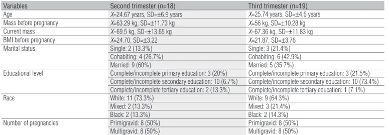
he exclusion criteria were: clinical complications (preeclamp-sia, risk of premature labor and urinary infection) which could interfere in the evaluation; obesity (pre-pregnancy BMI>30) and previous PFM contraction exercise. he study was ap-proved by Human Research Ethics Committee of Universidade do Estado de Santa Catarina, protocol 149/2007. Table 1 shows the characteristics of pregnant women in the second and third trimesters.
Procedures
he procedure for data collection consisted of: a) infor-mation to the participants about the study and request for informed consent; b) anamnesis of prior knowledge and performance of PFM contraction exercises; c) veriication of PFM strength through digital assessment; d) application of the Female Sexual Function Index (FSFI) questionnaire and, inally, e) sociodemographic data collection based on a semi-structured questionnaire. he entire data collection proce-dure was conducted in a private room of the health center and a single individual carried out the PFM evaluation for greater reliability.
Table 1. Participant’s characteristics.
Variables Second trimester (n=18) Third trimester (n=19)
Age X=24.67 years, SD=±6.9 years X=25.74 years, SD=±4.6 years
Mass before pregnancy X=63.29 kg, SD=±11,73 kg X=56 kg, SD=±10.28 kg
Current mass X=69.5 kg, SD=±13.65 kg X=67.36 kg, SD=±11.83 kg
BMI before pregnancy X=24.70, SD=±3.22 X=21.87, SD=±3.76
Marital status Single: 2 (13.3%) Single: 3 (21.4%)
Cohabiting: 4 (26.7%) Cohabiting: 6 (42.9%)
Married: 9 (60%) Married: 5 (35.7%)
Educational level Complete/incomplete primary education: 3 (20%) Complete/incomplete primary education: 3 (21.5%) Complete/incomplete secondary education: 10 (6.7%) Complete/incomplete secondary education: 10 (73.4%) Complete/incomplete tertiary education: 2 (13.3%) Complete/incomplete tertiary education: 1 (7.1%)
Race White: 11 (73.3%) White: 9 (64.3%)
Mixed: 2 (13.3%) Mixed: 3 (21.4%)
Black: 2 (13.3%) Black: 2 (14.3%)
Number of pregnancies Primigravid: 8 (50%) Primigravid: 8 (50%)
Multigravid: 8 (50%) Multigravid: 8 (50%)
Evaluation instruments
To measure PFM strength, the participant was in the supine position, with knees bent and covered with a sheet; the digital assessment was performed with lubricating gel and sterile gloves, and the patient was irst instructed on how to perform the contraction appropriately and, then, requested to carry out a maximum PFM contraction. he test was carried out by a single physical therapist in order to minimize possible measurement errors, and the Modiied Oxford Grading Scale by Laycock18 (Appendix 1) was used
to grade muscle strength. he participants’ sexual function was evaluated through the FSFI questionnaire, developed and validated in the United States of America14, translated
into Portuguese language19 and later also applied to Brazilian
pregnant women13. he questionnaire contains 19 multiple
choice questions grouped into six domains: desire, arousal, lubrication, orgasm, satisfaction and pain. A value of 0 to 5 is attributed to each answer. A mathematical calculation is then performed to obtain a inal index, the FSFI score. he scores range from 2 to 36, and the lower the score, the worse the sexual function.
Statistical analysis
he data were analyzed using the statistical program SPSS, version 13.0, through descriptive statistics ( frequency, mean and standard deviation) and inferential statistics (independent t test, Mann-Whitney U test and Spearman’s correlation). he
conidence interval used was of 95% and p values lower than 0.05 were considered signiicant. he independent variable was the gestational trimester (second or third) and the dependent variables were: FSFI score and PFM contraction strength. Ac-cording to the Shapiro-Wilk normality test, none of the de-pendent variables followed normal distribution (p=0.001 and p=0.013, respectively). For the control variable (age), the data were normal (p=0.119).
Results
PFM strength
For the variable PFM strength, the median and the mode were 3. Figure 1 shows this distribution in the studied tri-mesters. Most of the women in the third trimester had me-dium strength, and most of those in the second trimester had medium to high strength. Additionally, the mean rank of PFM strength for second-trimester women was 20.14 (sum of
ranks=362.5) compared to 17.92 for third-trimester women (sum of ranks=340.5). However, the diference between the means was not statistically signiicant (Mann-Whitney U test=150.5; p=0.512).
Sexual function
he FSFI mean score was 21.73 (±8.02). he Mann-Whitney U test was used to compare the FSFI mean score of the second-trimester women (n=18) to that of the third-second-trimester women (n=19). he mean ranks for the second-trimester women was 22.72 (sum of ranks=409.00) compared to 15.47 (sum of ranks=294.00) for the third-trimester women. he test also showed a statistically signiicant diference between second and third-trimester women (Mann-Whitney U test=104.00; p=0.042).
To determine whether this diference could be credited to the age diference between the participants, the mean age of the second and third-trimester women was compared. he mean age of the second-trimester women was 24.67 yrs (±6.90 yrs) and the mean age of the third-trimester women was 25.85 yrs (±4.50 yrs). Although the second-trimester women had a lower mean age compared to third-trimester women, that diference was not signiicant (t=-0.619; df=28.72; p=0.541).
PFM strength and sexual function
PFM strength had statistically signiicant correlations with some controlled variables of the present study: age, FSFI score and with 15 of the 19 variables of the FSFI (Table 2).
Figure 1. Pelvic Floor Muscle (PFM) strength according to gestational trimester.
5 4 3 2 1 0
PFM strength according to the Modified Oxford Grading Scale 8
6
4
2
0 N
Third Trimester Second Trimester
Discussion
PFM strength
No studies were found comparing PFM strength between gestational periods. However, asymptomatic non-pregnant Austrian women with a mean age of 41.2±14.6 yrs old were evaluated in relation to PFM strength according to the Modi-ied Oxford Grading Scale by Laycock18. he authors reported
that: 2.6% showed grade 0 muscle contraction; 12.5% showed grade 1 muscle contraction; 29.7%, grade 2 muscle contraction; 31.2%, grade 3 muscle contraction; 18.4%, grade 4 muscle con-traction and 5.5%, grade 5 muscle concon-traction20. Although the
non-pregnant women of this study had a higher mean age than the participants of the present study, the results were similar.
Digital assessment is one of the most widely used meth-ods of evaluating pelvic loor strength because it is simple and does not require expensive equipment2, despite the fact that
evidence does not ensure its intra-rater reliability21. According
to other authors, this is the most sensitive method of assess-ing PFM strength and tonus3. Although not the most reliable
method, it is the most accessible and afordable and, in the present study, a single physical therapist carried out the assess-ment to minimize possible measureassess-ment errors.
Only one participant of the present study (2.7%) had heard about the PFM contraction exercise needed to measure the strength of these muscles through digital assessment. In
contrast, 55.3% of pregnant women in England received some instruction about pelvic loor exercise22; in the United States,
most of the asymptomatic and non-pregnant women had al-ready heard about this exercise, but had not been instructed on how to perform it23. Another study reported that only 10%
of pregnant/postpartum women who received instructions on the correct contraction of these muscles were instructed dur-ing the pelvic exam24.
When asymptomatic women were instructed on how to perform this exercise, 68% were capable of performing an ap-propriate PFM contraction for up to 3 seconds23, and a high
percentage (15.2%) of the women was not able to voluntarily contract the PFMs20. More than 30% of the women were not
able to perform an appropriate PFM contraction during the irst assessment2. his could jeopardize the results in relation
to PFM strength grading, even though the participants were in-structed on how to perform the exercise. hus, there is poten-tial for improvement of the awareness of pelvic loor exercises and their beneits for pregnant women in Brazil.
Sexual function
A signiicant decrease in sexual function was observed from the second to the third trimester, and this diference could not be credited to the age diference between groups. Similarly, a study that evaluated sexual function at every gestational trimester (participants’ age=25.5±4.5 yrs) found
Table 2. Correlations with pelvic floor muscle strength.
Variables Spearman’s ρ P
Participant’s age 0.320 0.041*
FSFI score 0.540 0.000*
Frequency of desire** 0.402 0.010*
Level of desire** 0.261 0.104
Frequency of arousal** 0.264 0.099
Level of arousal** 0.298 0.062
Confidence of arousal** 0.327 0.039*
Satisfaction with arousal** 0.410 0.009*
Frequency of lubrication ** 0.491 0.001*
Difficulty of lubrication ** 0.516 0.001*
Frequency of maintaining lubrication ** 0.493 0.001* Difficulty in maintaining lubrication ** 0.413 0.008*
Frequency of orgasm ** 0.465 0.003*
Difficulty to obtain orgasm ** 0.446 0.004*
Satisfaction with orgasm ** 0.285 0.075
Satisfaction with amount of closeness with partner ** 0.393 0.012* Satisfaction with sexual relationship** 0.433 0.005* Satisfaction with overall sex life ** 0.429 0.006* Frequency of pain during vaginal penetration ** 0.448 0.004* Frequency of pain following vaginal penetration ** 0.366 0.020* Level of pain during or following vaginal penetration ** 0.443 0.004*
a signiicant decline in the scores in all the FSFI domains during gestation, with a signiicant decrease in the third tri-mester compared to the other periods25. Another study found
no signiicant diference between the FSFI total scores of the irst and third gestational trimesters in the participants of both groups26.
he analysis of each FSFI domain showed a decrease in sexual desire, arousal, vaginal lubrication, orgasm and sexual satisfaction from the second to the third trimester of gestation, as well as greater discomfort or pain related to the sexual activ-ity. Several studies have reported similar results and attributed them to a series of factors such as: body changes that afect self-esteem, discomfort, fear of harming the baby and physi-cal symptoms as nauseas, sleepiness and fatigue16,26-33. Some
authors stipulated a total FSFI score of 26.5 as the cut-of score between women with and without sexual dysfunction34. he
mean value of the total FSFI score obtained in the present study was of 21.73; therefore, based on the reference score34,
the pregnant women of the present study had a low mean FSFI score, which may not represent a dysfunction, but an adapta-tion due to the pregnancy.
PFM strength and sexual function
here was a signiicant correlation between the grade of PFM strength and the sexual function score in pregnant women (p=0.540 and p<0.001). Meanwhile, Baytur et al.9 did
not ind a correlation between PFM strength (using the perin-eometer) and sexual function (through the FSFI questionnaire)
postpartum. However, a PFM training program with non-pregnant women found that those with weak PFMs and who received training had positive results in sexual function, also veriied through the FSFI35. his demonstrates that stronger
muscles are linked to better sexual function.
Four of the 19 FSFI variables (“level of desire”, “frequency of arousal”, “level of arousal” and “satisfaction with or-gasm”) did not correlate with the grade of PFM strength. his result is attributed to the fact that the abovementioned vari-ables of sexual function are more psychological than physical.
Final considerations
he pregnant women in the second trimester demon-strated better sexual function than those in the third trimester. However, the grade of PFM strength was similar. Furthermore, higher grades of PFMs strength were correlated with better sexual function.
he present study compared only the second and the third gestational trimesters. It did not assess the irst trimester and it did not include a control group, which would have been the ideal model. It is suggested that a new study be carried out comparing all the periods (before gestation, irst, second and third trimesters) and controlling other factors, such as the physical symptoms resulting from gestation.
he relevance of this study goes beyond the research. Dur-ing the data collection, it was possible to instruct the pregnant women about the importance of PFM exercises. hus, the study also became an educational intervention.
1. Thompson JA, O´Sullivan PB, Briffa NK, Neumann P. Differences in muscle activation patterns during pelvic floor muscle contraction and valsalva manouevre. Neurourol Urodyn. 2006;25(2):148-55.
2. Bo K, Sherburn M. Evaluation of female pelvic-floor muscle function and strength. Phys Ther. 2005;85(3):269-82.
3. Rosenbaum TY. Pelvic floor involvement in male and female sexual dysfunction and the role of pelvic floor rehabilitation in treatment: a literature review. J Sex Med. 2007;4(1):4-13.
4. Phillips C, Monga A. Childbirth and the pelvic floor: ‘‘the gynaecological conse-quences’’. Reviews in Gynaecological and Parinatal Practice. 2005;5(1):15-22.
5. Nagib ABL, Guirro ECO, Palauro VA, Guirro RRJ. Avaliação da sinergia da musculatura abdomino-pélvica em nulíparas com eletromiografia e biofeedback perineal. Rev Bras Ginecol Obstret. 2005;27(4):210-5.
6. Sapsford R. Rehabilitation of pelvic floor muscles utilizing trunk stabilization. Man Ther. 2004;9(1):3-12.
7. Azar M, Noohi S, Radfar S, Radfar MH. Sexual function in women after surgery for pelvic organ prolapse. Int Urogynecol J Pelvic Floor Disfunct. 2008;19(1):53-7.
8. Bharucha AE. Pelvic floor: anatomy and function. Neurogastroenterol Motil. 2006;18(7):507-19.
9. Baytur YB, Deveci A, Uyar Y, Ozcakir HT, Kizilkaya S, Caglar H. Mode of delivery and pelvic floor muscle strength and sexual function after childbirth. Int J Gynaecol Obstet. 2005;88(3):276-80.
10. Özel B, White T, Urwitz-Lane R, Minaglia S. The impact of pelvic organ prolapse on sexual function in women with urinary incontinence. Int Urogynecol J Pelvic Floor Dysfunct. 2006;17(1):14-7.
11. Morokoff P. Determinantes of female orgasm. In: Lopiccolo J, Lopiccolo L (editors). Handbook of sex therapy. New York: Plenum Press; 1978. p. 147-65.
12. Edwards WM, Coleman E. Defining sexual health: a descriptive overview. Arch Sex Behav. 2004;33(3):189-95.
387
13. Leite APL, Moura EA, Campos AAS, Mattar R, Souza E, Camano L. Validação do índice da função sexual feminina em grávidas brasileiras. Rev Bras Ginecol Obstet. 2007;29(8):396-401.
14. Rosen R, Brown C, Heiman J, Leiblum S, Meston C, Shabsigh R, et al. The female sexual function index (FSFI): a multidimensional self-report instrument for the assessment of female sexual function. J Sex Marital Ther. 2000;26(2):191-208.
15. von Sydow K. Sexuality during pregnancy and after childbirth: a metacontent analysis of 59 studies. J Psychosom Res. 1999;47(1):27-49.
16. Fok WY, Chan LY, Yuen PM. Sexual behavior and activity in Chinese pregnant women. Acta Obstet Gynecol Scand. 2005;84(10):934-8.
17. Brown HL, McDaniel ML. A review of the implications and impact of pregnancy on sexual function. Curr Sexual Health Reports. 2008;5(1):51-5.
18. Laycock J. Female pelvic floor assessment: the Laycock ring of continence. J Natl Women Health Group Aust Physiother Assoc. 1994;40-51.
19. Hentschel H, Alberton DL, Capp E, Goldim JR, Passos EP. Validação do female sexual function index (FSFI) para uso em língua portuguesa. Rev HCPA. 2007;27(1):10-4.
20. Talasz H, Himmer-Perschak G, Marth E, Fischer-Colbrie J, Hoefner E, Lechleitner M. Evaluation of pelvic floor muscle function in a random group of adult women in Austria. Int Urogynecol J Pelvic Floor Dysfunct. 2008;19(1):131-5.
21. Sherburn M, Murphy CA, Carroll S, Allen TJ, Galea MP. Investigation of transabdominal real-time ultrasound to visualise the muscles of the pelvic floor. Aust J Physiother. 2005;51(3):167-70.
22. Mason L, Glenn S, Walton I, Hughes C. The instruction in pelvic floor exercises provided towomen during pregnancy or following delivery. Midwifery. 2001;17(1):55-64.
23. Moen M, Noone M, Vassallo B, Lopata R, Nash M, Sum B, et al. Knowledge and performance of pelvic muscle exercises in woman. J Pelvic Med Surg. 2007;13(3):113-7.
24. Fine P, Burgio K, Borello-France D, Richter H, Whitehead W, Weber A, et al. Teaching and practicing of pelvic floor muscle exercises in primiparous women during pregnancy and the postpartum period. Am J Obstet Gynecol. 2007;197(1):107e1-5.
25. Aslan G, Aslan D, Kizilyar A, Ispahi C, Esen A.A prospective analysis of sexual functions during pregnancy. Int J Impot Res. 2005;17(2): 154-7.
26. Sproul K, Deugarte CM, Yamini E, DeCherney A, Berman J. Female sexual function during pregnancy. Fertil Steril. 2004;82 Suppl 2: S339.
27. DeJudicibus MA, McCabe MP. Psychological factors and the sexuality of pregnant and postpartum women. J Sex Res. 2002;39(2):94-103.
28. Sipinski A, Kazimierczak M, Buchacz P, Sipinska K. Sexual behavior of pregnant women. Wiad Lek.2004;57 Suppl 1:281-4.
29. Masters WH, Johnson VE. A resposta sexual humana. São Paulo: Roca; 1984.
30. Gökyildiz Ş, Beji N. The Effects of pregnancy on sexual life. J Sex Marital
Ther. 2005;31(3):201-15.
31. Kitzinger S. A mulher e o sexo. Rio de Janeiro: Interamericana; 1985.
32. Lazar MCS. Práticas sexuais de mulheres no ciclo gravídico-puerperal [tese]. Campinas (SP): UNICAMP; 2002.
33. Uwpusitanon W, Choobun T. Sexuality and sexual activity in pregnancy. J Med Assoc Thai. 2004;87 Suppl 3:S45-9.
34. Wiegel M, Meston C, Rosen R. The female sexual function index (FSFI): cross-validation and development of clinical cut-off scores. J Sex Marital Ther. 2005;31(1):1-20.
Appendix 1
The modified Oxford Grading Scale
0) No discernible contraction.
1) Flicker contraction.
2) Weak contraction, felt as slight pressure on the examining finger.
3) Moderate contraction, distinct pressure on the examining finger, and palpable upward and forward movement.
4) Good contraction, elevation possible against slight resistance, the fingers are pressed against each other in the direction of pubic symphysis.
5) Very strong muscle strength, with suction-type effect on the examining finger, these are pressed against each other in the direction of pubic symphysis.
Source: Bo and Sherburn2, Laycock18.