O
RIGINALA
RTICLE Revista Brasileira de FisioterapiaProposal of non-invasive experimental model
to induce scoliosis in rats
Proposta de modelo experimental não-invasivo para indução de escoliose
em ratos
Carlos A. Silva1, Rinaldo R. J. Guirro2, Gabriel B. Delfino3, Eder J. Arruda3
Abstract
Background: In the literature, there are several experimental models that induce scoliosis in rats; however, they make use of drugs or invasive interventions to generate a scoliotic curve. Objectives:To design and apply a non-invasive immobilization model to induce scoliosis in rats. Methods: Four-week old male Wistar rats (85±3.3 g) were divided into two groups: control (CG) and scoliosis (SG). The animals in the SG were immobilized by two vests (scapular and pelvic) made from polyvinyl chloride (PVC) and externally attached to each other by a retainer that regulated the scoliosis angle for twelve weeks with left convexity. After immobilization, the abdominal, intercostal, paravertebral, and pectoral muscles were collected for chemical and metabolic analyses. Radiographic reports were performed every 30 days over a 16-week period. Results: The model was effective in the induction of scoliosis, even 30 days after immobilization, with a stable angle of 28±5º. The chemical and metabolic analyses showed a decrease (p<0.05) in the glycogenic reserves and in the relationship between DNA and total protein reserves of all the muscles analyzed in the scoliosis group, being lower (p<0.05) in the convex side. The values for the Homeostatic Model Assessment of Insulin Resistance indicated a resistance condition to insulin (p<0.05) in the scoliosis group (0.66±0.03), when compared to the control group (0.81±0.02). Conclusions: The scoliosis curvature remained stable 30 days after immobilization. The chemical and metabolic analyses suggest changes in muscular homeostasis during the induced scoliosis process.
Keywords: scoliosis; musculoskeletal; non-invasive model.
Resumo
Contextualização: Encontram-se na literatura diversos modelos experimentais de indução de escoliose em ratos, porém evidencia-se o uso de drogas ou intervenções invasivas para a geração da curvatura escoliótica. Objetivos: Projetar e aplicar um modelo de imobilização não-invasiva para a indução de escoliose em ratos. Métodos: Ratos Wistar machos com idade inicial de quatro semanas (85±3,3 g) foram divididos nos grupos controle (GC) e escoliose (GE). Os animais do GE foram imobilizados por dois cintos (escapular e pélvico) de policloreto de vinila (PVC), interligados externamente por um limitador que regulava o ângulo da escoliose durante 12 semanas, com convexidade à esquerda. Após a imobilização, os músculos abdominais, intercostais, paravertebrais e peitorais bilateralmente foram coletados para as análises químio-metabólicas. Os registros radiológicos foram realizados a cada 30 dias, num total de 16 semanas.
Resultados: O modelo foi eficiente e eficaz na indução da escoliose, mesmo após 30 dias da desmobilização, mantendo um ângulo
estável de 28±5 graus. Quanto às análises químio-metabólicas, observou-se diminuição (p<0,05) nas reservas glicogênicas e na relação proteína total/DNA de todos os músculos analisados do GE, sendo menores (p<0,05) no lado da convexidade. Os valores do HOMA-IR indicaram um quadro de resistência à insulina (p<0,05) no GE (0,66±0,03) quando comparado ao GC (0,81±0,02). Conclusões: A curvatura escoliótica manteve-se estável após 30 dias da desmobilização, e as alterações químio-metabólicas sugeriram a ocorrência de modificações na homeostasia muscular durante o processo indutor da escoliose.
Palavras-chave: escoliose; musculoesquelético; modelo não-invasivo.
Received: 12/13/2011 – Revised: 12/19/2011 – Accepted: 12/21/2011
1 Master’s Degree Program in Physical Therapy, Universidade Metodista de Piracicaba (UNIMEP), Piracicaba, SP, Brazil
2 Department of Biomechanics, Medicine, and Rehabilitation of the Locomotor System, Faculdade de Medicina de Ribeirão Preto, Universidade de São Paulo (USP), Ribeirão Preto, SP, Brazil 3 Undergraduate Program in Physical Therapy, UNIMEP, Piracicaba, SP, Brazil
Correspondence to: Rinaldo Roberto de Jesus Guirro, Faculdade de Medicina de Ribeiro Preto - USP, Departamento de Biomecânica, Medicina e Reabilitação do Aparelho Locomotor, Av. Bandeirantes, 3900, Monte Alegre, CEP 14049-900, Ribeirão Preto, SP, Brazil, e-mail: [email protected]
Introduction
Anatomic alterations of the spine have been investigated in clinical and experimental studies not only to create new forms of treatment, but also to elucidate the etiology and the efects that these alterations can have on the body. In this context, researchers have been developing experimental models to pro-mote scoliosis in animals.
For several decades, these models have been developed and many are currently in use, but they are all invasive. Some of them are: the administration of intraperitoneal
beta-aminopropionitrile to alter the vertebral ligaments1;
altera-tions to the intervertebral cartilage of rats2; osteolathyrism
with carbazide3; mechanical separation of the vertebrae
of rats to limit movement4; suture of musculature next to
vertebrae to limit movement5; increase in flexibility after
chymopapain injection6; growth modulation after fusion
of vertebrae7; model to correct scoliosis with transfixation
pins in the spine8; transfixation pins to maintain spinal
po-sition9; maintenance system applied to the intervertebral
discs interfering in the scoliotic curve10, and more recently,
an experimental model to induce scoliosis based on the use
of steel wires11. Obsolescence was observed along the years
regarding the development of new methodologies to induce experimental scoliosis in animals. In more recent articles, old and obsolete induction methods have been reported.
It has been demonstrated that scoliosis is not exclusive to
bi-pedalism12.In this context, our research group strived to develop a
methodology that would be diferent from the existing ones. Some aspects were considered: time, risk of losing the animals, simple planning, and especially no surgery so that the surrounding tis-sue structures in the spine would not be afected and, therefore, not interfere with the parameters that could be analyzed for the development and treatment of scoliosis. hus, the main objective of this study was to design and apply a non-invasive method to induce scoliosis in rats.
Methods
Animals
Twenty-four male Wistar rats were divided into two groups: control (CG, n=8) and scoliosis (SG, n=16), with
initial weight of 85±3.3 g. The SG was subdivided into two
other groups: scoliosis x-ray and chemical-metabolic. The
animals were given ordinary feed as well as water ad libitum
and submitted to bioterium facilities, with a 12-hour light-dark cycle. This study was approved by the Ethics Commit-tee for Animal Experimentation at Universidade Federal de
São Carlos (UFSCar), São Carlos, SP, Brazil, under protocol number 024/2006.
Orthosis design
The orthosis comprised two vests made from polyvinyl chloride (PVC), 0.50mm thick, in nine different sizes to allow growth. The sequence and measures are shown in Figure 1. A scoliosis angle of 40º was delimited by the fixa-tion of two plastic holders, each one fixed to one of the vests connected by a metallic wire. To avoid cutaneous lesions, the edges of the vest were covered with rubber adhesive and synthetic resin (Figure 2). The vests were made to protect against any cutaneous lesions as well as to allow breathing and moving.
Immobilization
he immobilization period began after the weaning stage (28 days of age) and lasted 12 weeks. he PVC vest was adjusted to develop left convexity scoliosis. hey were changed every 7 days and maintained for a period of 12 weeks. Afterwards, the animals were followed up for another 4 weeks (immobilization-free period).
Radiological analysis
The animals were x-rayed after 24h of immobilization and every 4 weeks. The digital imaging was analyzed with the software ALCimagem to measure the scoliosis angle. The following parameters were used: Posterior-Anterior in-cidence (PA), 50 kVp, 6-pulse exposure time, focal distance/
chassis 75 cm, and film for 20.2x25.3 cm x-ray (Kodak®
). The animals were previously anesthetized with sodium pento-barbital (40 mg/Kg, i.p.).
Sampling
he animals were anesthetized with sodium pentobarbital (40 mg/Kg, i.p.) to collect abdominal, intercostal, paravertebral, and pectoral muscles bilaterally.
Determining the muscular glycogen
The muscle samples were digested in KOH 30% hot, and the glycogen was precipitated from the passage of hot ethanol. The sample was centrifuged at 3000 rpm, and the glycogen was subjected to acid hydrolysis in the
pres-ence of phenol13. Values were expressed in mg/100 mg wet
weight.
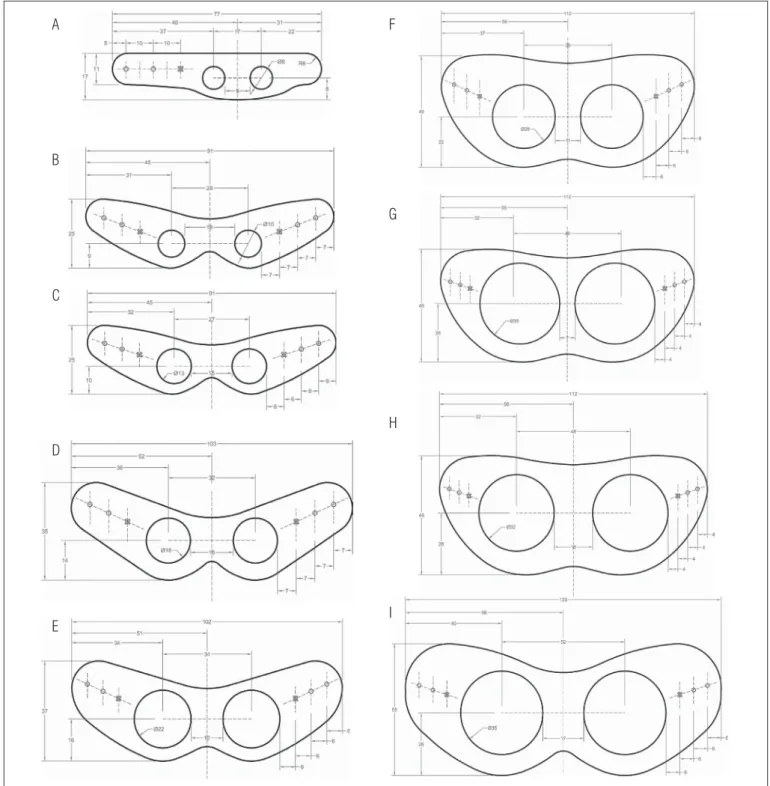
A-scapular 1st week; B-pelvic 1st week and scapular in the 2nd week; C-pelvic 2nd week and scapular in the 3rd week; D-pelvic 3rd week and scapular 4th week; E-pelvic 4th week and scapular 5th and 6th week;
F-pelvic 5th and 6th week and scapular 7th, 8th, 9th and 10th week, G-pelvic 7th and 8th week; H-pelvic 9th and 10th week and scapular 11th, 12th week; I-pelvic 11th and 12th week. The “x” in each vest represents
the fixation point of the pin, and the circles represent the passage point of the plastic retainer to fasten the vest.
A
B
C
D
E
F
G
H
I
Figure 1. Orthosis models attached to the scapular or pelvic vests used for immobilization.
Total protein concentration
A PROTAL kit (Laborlab, cat. n 03800) was used. Its principle is based on the fact that substances containing two or more pep-tide bonds form a violet-colored complex in an alkaline solution when diluted with copper sulfate. Values are expressed in g/dL.
Muscular DNA concentration
The diphenylamine method was used. First, the tis-sue homogenate was submitted to a diphenylamine solu-tion then to a spectrophotometer reading, with 595 nm
wavelength14.
abdominal, 14% of the right intercostal, 33% of the left intercos-tal, 8.5% of the right pectoral, 18% of the left pectoral, 9% of the right paravertebral, and 20% in the left paravertebral muscle (Table 1). When comparing the convex and the concave sides,
in the same table, there was no diference in the CG. In the SG,
however, the values were lower in the convex side involving 10%, 25%, 13%, and 16% of the abdominal, intercostal, pectoral, and paravertebral, respectively.
Figure 2. PVC orthosis in the shape of a vest.
Figure 3. Radiograph of A) scoliotic animals using the PVC vest, B) animals from the scoliotic group (SG) and control group (CG) after removing the PVC vest. Images taken on the 4th, 8th, and 12th week of immobilization and 4 weeks after immobilization (12+4).
A
B
Plasma insulin concentration
he plasma insulin was measured by radioimmunoassay. his technique consists in the transference of an unknown amount of plasma to the tube containing anti-insulin
antibod-ies and insulin marked with a tracer (iodine-125)15.
Statistical analysis
he Kolmogorov-Smirnov test was used, followed by ANOVA and the Tukey test. For all measurements, the level of signiicance was set at 5%.
Results
he radiographic imaging taken every 4 weeks showed the development of scoliosis (Figure 3A). he animals in the CG did not show any deviation, unlike the SG, which developed a “C” curve with an average angle of 40º while using vest. It remained
stable at 28±5º for 4 weeks after immobilization (Figure 3B).
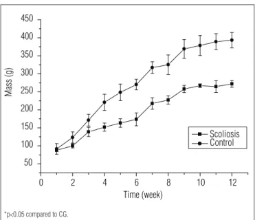
During immobilization, the animal’s body weight was moni-tored for 12 weeks. It was observed that the immobilized animals
had an average weight 29.4±2.7% lower than the animals in the CG
(Figure 4). It is important to consider that the lower weight may be related to the food intake, which was 25% lower than that of the
CG (60±5.0 g/day for the CG vs. 45±3.0 g/day for the SG).
When considering the weight of the vest, a progressive in-crease was observed with the animals’ growth. However, when considering the relationship between the total weight of the
or-thosis and the animals’ body weight, the mean was 4.3±0.2%.
When comparing the PT/DNA relationship between the CG and the SG, which showed alterations in the number of myoibrils, there was a signiicant reduction in the convex side involving 11% of the right abdominal muscle, 16% of the left
he values of glycogen reserves showed that the muscles of the scoliotic animals showed reduction in the right (41%) and left intercostal (67%); right (44%) and left pectoral (59%); abdominal right (45%) and left abdominal (51%), and right and left paravertebral muscles (64%), when compared with the ani-mals in the CG. When comparing the muscles in the concave
side with the convex side, the lower glycogen reservewas40%
lower in the intercostal muscle, 28% in the pectoral, 13% in the abdominal, and 20% in the paravertebral (Table 1).
For a complete chemical and metabolic analysis, the
Homeostatic Model Assessment method was used to estimate the insulin resistance function (HOMA-IR) with
values of 0.81±0.02 in the CG, and 0.66±0.03 in the SG, with
a reduction of 18%.
Discussion
he greatest advantage of this non-invasive scoliosis model was that it allowed the development of scoliosis in animals without the use of any type of surgery or medication as these interventions can afect not only the spine but also the adjacent tissues. Another important aspect is the pres-ervation of all the tissues/structures that can be analyzed after immobilization or even after therapeutic interventions aimed to minimize scoliosis. Additionally, this model can be easily prepared to allow its application on a large number of animals in a short period of time and with low costs, as achieved in the present study.
Our model difered from the ones found in the literature due to its non-invasive approach. An example of an invasive model in rats consisted in a suture of the inferior angle of the scapula to the pelvis ipsilaterally next to the base of the tail. he author
foundthat the ideal time for the induction of a permanent
scoliotic curvature would be 8 weeks. He emphasized that the aim of his study was to evaluate the scoliosis during the period between 1 to 12 weeks after surgery. However, he veriied that
in 6 weeks, the curvature was already permanent16.
In search for an experimental model, we also found those that difered from the invasive (surgical) procedure. However, their appli-cability focused on the alteration of the biochemical and endocrine aspects of the animals through the administration of mutagenic substances during their development stage, osteolathyrism, and
removal of secreting organs such as the pineal gland17.
Another aspect related to the experimental scoliosis lies in the fact that among the animals used in studies, i.e. chickens, goats, rabbits, and rats, the latter had the lowest number of papers published, making it harder to obtain information for comparisons between the induction models in the same
ani-mal model7,18,19.
Table 1. DNA/total protein relationship and muscle glycogen (mg/100mg) after 12 weeks of immobilization of the different muscles of rats in the control group and scoliosis group. The left side (L) is convex, and the right side (R) is concave. Values refer to mean±epm, n=6.
Muscle
DNA/Total Protein Glycogen
Control Group Scoliosis Group Control Group Scoliosis Group
Abdominal R 3.49±0.02 3.10±0.02* 0.58±0.04 0.32±0.05* Abdominal L 3.34±0.04 2.80±0.03*# 0.57±0.01 0.28±0.01*# Intercostal R 3.55±0.02 3.04±0.03* 0.51±0.04 0.30±0.005* Intercostal L 3.40±0.03 2.27±0.02*# 0.55±0.005 0.18±0.009*# Pectoral R 3.28±0.03 3.00±0.03* 0.50±0.06 0.28±0.009* Pectoral L 3.19±0.02 2.60±0.03*# 0.49±0.03 0.20±0.01*# Paravertebral R 3.41±0.03 3.12±0.03* 0.55±0.06 0.20±0.009* Paravertebral L 3.29±0.01 2.62±0.03*# 0.44±0.02 0.16±0.008*#
*p<0.05 compared to control group. #p<0.05 compared to the right side.
Figure 4. Mean±SD of mass (g) of the rats in the control group (CG) and
scoliosis group (SG) between the 1st and 12th week, n=8.
0 2 4 6 8 10 12
50 100 150 200 250 300 350 400 450 Mass (g) Time (week) Scoliosis Control
*p<0.05 compared to CG.
Among the most invasive methodologies, one that stands out involved the removal of the hind limbs and the tail of the
animal so that it could assume the biped position17.However,
the group where the animals were submitted to associated sur-gery pinealectomy had scoliosis, showing a relation with mela-tonin deiciency in the development of idiopathic scoliosis.
In order to verify the efectiveness of the proposed model, the animals were evaluated during the four weeks after immo-bilization, and the scoliotic curve did not return to the level considered normal, even after the vests were removed. To minimize the possible inluence of space in the containment box in relation to the animals’ growth, four rats were placed
in each box20.he reduction in body weight may be due to the
functional limitation exerted by the vest, considering that no alterations were observed in response to the central nervous
system, especially in the hypothalamus with behavior
re-lexes21. he weight of the vest could also be considered
impor-tant; however, the percentage represented by weight of the vest compared to the animal’s weight suggested that this parameter was not relevant.
he evaluation of the DNA/total protein relationship ob-served in the scoliosis group could be represented by the ac-tion of the catabolic systems that follow disuse, suggesting two patterns: reduction in the synthesis process and prevalence of
proteolysis-inducing factors.
It should be considered that the events that could have taken part in the muscle alterations are multi-factorial; therefore, further studies are necessary to provide a better understanding of this interface. he muscle glycogen content is considered an important energy reserve and, when submit-ted to alterations, it can afect performance, i.e. it can im-prove resistance when elevated and, when depleted, it can participate in processes associated with fatigue and
muscu-lar hypertrophy22,23.
In the present study, there was a signiicant reduction
in the glycogen muscle reservesin scoliotic animals. In the
literature, it has been reported that paravertebral muscles had a predominance of type I (red) ibers that were predominantly
oxidative24.his study emphasized that the low glycogen
reserves were involved in the chemical and metabolic balance of the ibers and could be linked to several factors that can
afect the homeostasis of the processes that regulate the
formation of metabolized substrate reserves. Another aspect to consider is that the proposed model creates a process of muscle disuse similar to long periods in bed, limb ixation, microgravity, or the use of orthoses. hese factors trigger a
catabolic state in the skeletal muscles25-27.
With regard to the endocrine proile, it is known that insu-lin is extremely important among the hormones involved in the regulation of carbohydrate metabolism and the modulation of
the skeletal muscles28.
Many authors have been studying the mechanics triggered in the muscle in disuse and have veriied a reduction in insulin signal transduction, which suggests deicit in the insulin recep-tor activation and in the enzymes activated from it, including phosphorylation of IRS-1 (insulin receptor substrate-1) and P13-K activation, decrease in the amount of glucose transporter
protein (GLUT4), and insulin resistance22.Our results showed a
signiicant reduction in glycogen reserves in the paravertebral
muscles of scoliotic animals, simulating the disuse condition created by other immobilization methods.
Finally, due to the situation where homeostasis was dis-turbed, the HOMA-IR was evaluated; it was veriied that in the SG the index was reduced, revealing a disturbance in insulin sensitivity.
It is worth mentioning that the physiological relationships between the phenomena linked to insulin sensitivity and the scoliosis induction process still need further investigation. However, this was a pioneer study as it demonstrated chemi-cal and metabolic alterations in the non-invasive scoliosis induction model. hese alterations were part of the mecha-nisms used concomitantly for the development of scoliosis and relected an anatomical and functional interface that requires further investigation to provide new perspectives on the histophysiological analyses, as well as the applicability and efectiveness of therapeutic methods commonly used for the rehabilitation process of scoliosis.
Conclusion
Considering the experimental aspects of this study, it can be concluded that:
• he PVC vest was efective in the development of scoliotic
curvature during the three-month immobilization period and one-month period free from immobilization, main-taining a stable curvature;
• Some metabolic alterations occurred in the skeletal
mus-cles as observed in the analyses of the glycogen reserves, in the total protein/DNA relationship afected by a negative protein balance, and in the sensitivity to insulin, suggest-ing that the changes in muscle homeostasis followed the scoliosis-induced process.
Acknowledgments
To Conselho Nacional de Desenvolvimento Cientíico e Tec-nológico (CNPq), Brasilia, DF, Brazil, (Institutional Program of Grants for Scientiic Initiation); Research Foundation –
UNIMEP and Fundação de Amparo à Pesquisa do Estado
de São Paulo (FAPESP), São Paulo, SP, Brazil (São Paulo
Research Foundation), Process 2006/60651-1.
References
1. Nogami H, Terashima Y, Tamaki K, Oohira A. Congenital kyphoscoliosis and spinal cord lesion produced in the rat by beta-aminopropionitrile. Teratology. 1977;16(3):351-7.
2. Beguiristain JL, Salis J, Oriaifo A, Canãdell J. Experimental scoliosis by epiphysiodesis in pigs. International Orthopaedics. 1990;3(4):317-21.
3. Tanaka H, Kimura Y, Ujino Y. The experimental study of scoliosis in bipedal rat in lathyrism. Arch Orthop Trauma Surg. 1982;101(1):1-27.
4. Dabney KW, Salzman SK, Wakabayashi T, Sarwark JF, Gao GX, Beckman AL, et al. Experimental scoliosis in the rat. II. Biomechanical analysis of the forces during Harrington distraction. Spine (Phila Pa 1976). 1988;13(5):472-7.
5. Kasuga K. Experimental scoliosis in the rat spine induced by binding the spinous processes. Nihon Seikeigeka Gakkai Zasshi. 1994;68(9):789-807.
6. Lu DS, Luk KD, Lu WW, Cheung KM, Leong JC. Spinal flexibility increase after chymopapain injection is dose dependent: a possible alternative to anterior release in scoliosis. Spine (Phila Pa 1976). 2004;29(2):123-8.
7. Braun JT, Akyuz E, Ogilvie JW. The use of animal models in fusionless scoliosis investigations. Spine (Phila Pa 1976). 2005;30(17 Suppl):S35-45.
8. Rooney GE, Vaishya S, Ameenuddin S, Currier BL, Schiefer TK, Knight A, et al. Rigid fixation of the spinal column improves scaffold alignment and prevents scoliosis in the transected rat spinal cord. Spine (Phila Pa 1976). 2008;33(24):914-24.
9. Stokes IA, McBride CA, Aronsson DD. Intervertebral disc changes in an animal model representing altered mechanics in scoliosis. Stud Health Technol Inform. 2008;140:273-7.
10. Schmid EC, Aubin CE, Moreau AE, Sarwark J, Parent S. A novel fusionless vertebral physeal device inducing spinal growth modulation for the correction of spinal deformities. Eur Spine J. 2008;17(10):1329-35.
11. Schwab F, Patel A, Lafage V, Farcy JP. A porcine model for progressive thoracic scoliosis. Spine (Phila Pa 1976). 2009;34(11):E397-404.
12. Gorman KF, Breden F. Idiopathic-type scoliosis is not exclusive to bipedalism. Med Hypotheses. 2009;72(3):348-52.
13. Lo S, Russell JC, Taylor AW. Determination of glycogen in small tissue samples. J Apll Physiol. 1970;28(2):234-6.
14. Giles KW, Myers A. An improved diphenylamine method for the estimation of deoxyribonucleic acid. Nature. 1965;206:93-9.
15. Scott AM, Atwater I, Rojas E. A method for the simultaneous measurement of insulin release and B cell membrane potential in single mouse islets of Langerhans. Diabetologia 1981;21(5):470-5.
16. Sarwark JF, Dabney KW, Salzman SK, Wakabayashi T, Kitadai HK, Beauchamp JT, et al. Experimental scoliosis in the rat. I. Methodology, anatomic features and neurologic characterization. Spine (Phila Pa 1976). 1988;13(5):466-71.
17. Machida M, Saito M, Dubousset J, Yamada T, Kimura J, Shibasaki K. Pathological mechanism of idiopathic scoliosis: experimental scoliosis in pinealectomized rats. Eur Spine J. 2005;14(9):843-8.
18. Machida M, Murai I, Miyashida I, Dubousset J, Yamada T, Kimura J. Pathogenesis of idiopathic scoliosis. Experimental study in rats. Spine (Phila Pa 1976). 1999;24(19):1985-9.
19. Joe T. [Studies of experimental scoliosis produced by electric stimulation. With special reference to the histochemical properties of the muscle]. Nihon Ika Daigaku Zasshi. 1990;57(5):416-26.
20. Genaro G, Schmidek WR, Franci CR. Social condition affects hormone secretion and exploratory behavior in rats. Braz J Med Biol Res. 2004; 37:833-40.
21. Silva CA, Guirro RRJ, Fonseca V, Arruda EJ, Grassi DO. Assessment of rat behavior with induced scoliosis by polyvinyl chloride vests. Journal Chinese Clinical Medicine. 2008;31:621-6.
22. Hirose M, Kaneki M, Sugita H, Yasuhara S, Martyn JA. Immobilization depresses insulin signaling in skeletal muscle. Am J Physiol Endocrinol Metab. 2000;279(6):1235-41.
23. Coderre L, Vallega GA, Pilch PF, Chipkin SR. Regulation of glycogen concentration and glycogen synthase activity in skeletal muscle of insulin-resistant rats. Arch Biochem Biophys. 2007;464(1)144-50.
24. Delp MD, Duan C. Composition and size of type I, IIA, IID/X and IIB fibers and citrate synthase activity of rat muscle. J Appl Physiol. 1996;80(1):261-70.
25. Stein TP, Schulter MD, Boden G. Development of insulin resistance by astronauts during spaceflight. Aviat Space Environ Med. 1994;65(12):1091-6.
26. Ferrando AA, Lane HW, Stuart CA, Davis-Street J, Wolfe RR. Prolonged bed rest decreases skeletal muscle and whole body protein synthesis. Am J Physiol. 1996;270(4 Pt 1):E627-33.
27. Lima SC, Caierão QM, Durigan JQ, Schwarzenbeck A, Silva CA, Minamoto VB, et al. Short-term immobilization causes morphometric and mechanical alterations in rat muscles. Rev Bras Fisioter. 2007;11(4):297-302.
28. Sesti G. Pathophysiology of insulin resistance. Best Pract Res Clin Endocrinol Metab. 2006;20(4):665-79.