Correction of prominent ears by the cartilaginous
incision technique, deinition of the antihelix with
Mustardé sutures, and ixation of the ear cartilage
at the mastoid
Correção da orelha de abano pela técnica de incisão cartilaginosa, deinição da
antélice com pontos de Mustardé e ixação da cartilagem conchal na mastoide
ABSTRACT
Background: Prominent ear is the most common congenital defect of the ear, with an in
cidence of 5% in Caucasians. Surgical treatment should correct the auriculocephalic and con choscaphal angles as well as protrusion of the lobe when present. This paper aims to re port the experience of our service in the treatment of prominent ears with a combination of several available techniques. Methods: Fortyseven patients operated with a combination
of previously described techniques were evaluated, and cartilaginous incision, Mustardé sutures for antihelix deinition, and conchamastoid ixation were performed. Patients less than 15 years of age were operated under general and local anesthesia, while the remaining patients underwent only local anesthesia. All patients were reassessed on the irst posto pe rative day. Results: The postoperative results were considered satisfactory by both patients
and surgical staff, with no stigma development in the operated ear. Conclusions: The best
treatment of pro minent ears is achieved by a combination of techniques. The approach used on the studied patients has produced naturallooking results with low complication rates, satisfying the surgical staff and, most importantly, the patients.
Keywords: External ear/surgery. Plastic surgery/methods. Ear diseases/surgery.
RESUMO
Introdução: A orelha de abano é o mais comum de todos os defeitos congênitos da orelha,
com incidência de 5% em caucasianos. O tratamento cirúrgico deve corrigir os ângulos au riculocefálico e escafoconchal, bem como a protrusão do lóbulo, quando presente. O objetivo deste trabalho é demonstrar a experiência de nosso serviço no tratamento da orelha de abano com a combinação de diversas técnicas disponíveis. Método: Foram avaliados
47 pacientes, operados com a associação de técnicas já descritas anteriormente, sendo utilizados incisão cartilaginosa, pontos de Mustardé para deinição de antélice e ixação da concha na mastoide. Os pacientes com menos de 15 anos de idade foram operados sob anestesias geral e local, e os demais foram submetidos somente a anestesia local. Todos os pacientes foram reavaliados no primeiro dia de pósoperatório. Resultados: Os resultados pósoperatórios foram considerados satisfatórios pelos pacientes e pela equipe cirúrgica, sem o aparecimento do estigma de orelha operada. Conclusões: O melhor tratamento de orelhas proeminentes é obtido com a associação de diversas técnicas. A abordagem empre gada nos pacientes avaliados tem apresentado resultados naturais e com baixos índices de complicação, satisfazendo a equipe cirúrgica e, principalmente, os pacientes.
Descritores: Orelha externa/cirurgia. Cirurgia plástica/métodos. Otopatias/cirurgia. Study conducted at
Hospital Federal da Lagoa –
Federal Network of
Healthcare at Rio de Janeiro, Rio de Janeiro, RJ, Brazil.
Submitted to SGP (Sistema de
Gestão de Publicações/Manager
Publications System) of RBCP
(Revista Brasileira de Cirurgia Plástica/Brazilian Journal of
Plastic Surgery).
Paper received: June 16, 2011 Paper accepted: October 10, 2011
1. Resident physician in Plastic Surgery at Hospital Federal da Lagoa, Rio de Janeiro, RJ, Brazil.
2. Specialist member of the Brazilian Society of Plastic Surgery (SBCP), former resident physician in Plastic Surgery at Hospital Federal da Lagoa, Rio de
Janeiro, RJ, Brazil.
3. Full member of SBCP and of the Brazilian Association of Surgeons, head of the Plastic Surgery Service of Hospital Federal da Lagoa, Rio de Janeiro,
RJ, Brazil.
Francisco de oliveira
Goulart1
danilo santos vidalde
arruda1
Bruno Menezes Karner2
Pedro loPes GoMes2
or in association, are underdeveloped antihelix, increased conchoscaphal angle, conchal prominence, increased auri culocephalic angle, and lobe protrusion3.
The normal auriculocephalic angle ranges between 25 and 30 degrees; when greater than 40 degrees, it can be con sidered abnormal. Similarly, the normal conchoscaphal angle is ap proximately 90 degrees, and more obtuse angles fre quently require surgical correction4,5. In addition to those with anato
mical alterations in the pavilion itself, patients with signii cantly asymmetrical ears may also beneit from otoplasty6.
This paper aims to demonstrate the experience of our service in the treatment of prominent ears with a combina tion of several available techniques.
METHODS
Fortyseven patients, 2 of whom were unilateral cases, were assessed and operated by a combination of techniques described below.
The surgeries were performed between February 2009 and December 2010 by the Plastic Surgery Service of Hos pital Federal da Lagoa (Rio de Janeiro, RJ, Brazil).
The operated patients were between 7 and 52 years of age (average of 23 years old) and included 18 females and 29 males.
Patients less than 15 years of age (25.5% of the cases) were operated under general and local anesthesia; the remaining patients (74.5% of the cases) underwent local anesthesia only.
Patients operated under general anesthesia were discharged on the day following the procedure, and those who underwent local anesthesia were discharged that same day. All patients were reassessed on the irst postoperative day.
Surgical Technique
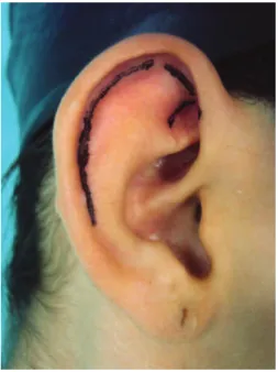
1. Marking of the anterior and posterior surfaces of the ear with a demographic pen. The anterior markings refer to the areas for cartilaginous incision and the posterior markings to the resection of the skin island (Figures 1 and 2).
2. Iniltration of 2% lidocaine solution + epinephrine in 1% saline to give a 1:200,000 epinephrine solu tion, not exceeding 10 mg/kg of local anesthetic.
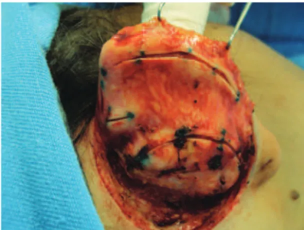
3. Incision and resection of the posterior skin island according to the previous markings, so that the re sulting scar is positioned in the retroauricular crease (Figure 3).
4. Posterior detachment of the ear in the subperichon drial plane until the auricular cartilage is wellex posed and detachment of the mastoid region with re section of the posterior auricular muscle (Figure 4). 5. Marking of the cartilaginous incisions by intro du cing insulin needles according to the previous mar kings on the anterior surface of the ear.
6. Incision of the cartilage at 4 points: at the external edge of the antihelix, a transverse incision between
Figure 1 – Anterior marking.
both, avoiding the irst 2 incisions to be joined and, inally, an incision at the external edge of the concha (Figure 5).
7. Deinition of the antihelix with Mustardé sutures using 4.0 nylon suture between the existing inci sions. The irst suture is placed between the incision at the outer edge of the antihelix and the incision at the inner edge of the upper branch of the antihelix, and 1 or 2 additional sutures are placed between the incision at the outer edge of the antihelix and the incision at the concha to complete the deinition of the antihelix (Figures 6 to 8).
8. Resection of excess conchal cartilage, when neces sary (Figure 9).
9. Rotation of the conchal cartilage and its fixation at the mastoid with 3.0 nylon suture (Figure 10). When required, the upper and lower poles of the ear can be fastened at the mastoid region using 4.0 nylon suture.
Figure 3 – Resected skin island.
Figure 4 – Detachment of the posterior region of the ear and mastoid region.
Figure 5 – Incisions in the auricular cartilage.
Figure 6 – Mustardé suture.
Figure 7 – Mustardé suture.
10. Skin closure with the Greek suture technique using 4.0 nylon suture (Figure 11).
Figure 8 – Mustardé suture.
Figure 9 – Resection of excess conchal cartilage.
Figure 10 – Fixation of the concha at the mastoid.
Figure 11 – Final closure of the skin.
24 hours; thereafter, the patient is assessed on the irst postoperative day.
Postoperative Follow-up
On the irst postoperative day, the surgical dressing was replaced by an elastic bandage for auricular protection; this
was maintained for 45 days and was used only at night for the last 15 days.
Antibiotic therapy was maintained for 7 days, and anal gesics and nonsteroidal antiinlammatory drugs were pres cribed as required.
Outpatient postoperative followup consultations were held 1 day, 1 week, 21 days, 45 days, 3 months, and 6 months after the procedure. The sutures were removed at the con sultation on the 21st postoperative day.
RESULTS
A total of 47 patients underwent correction of prominent ears by a combination of the techniques presented in this study. The techniques used were cartilaginous incision, Mus tardé sutures for antihelix deinition, conchal rotation, and, when required, resection of excess conchal cartilage.
The postoperative results were considered satisfactory by both the patients and the surgical staff, with no development of stigma in the operated ear.
Figures 12 to 15 illustrate the results obtained with the described techniques in patients operated by our service.
DISCUSSION
A surgical procedure for correction of prominent ears was irst described in 1845 by Dieffenbach, who suggested a retroauricular skin resection. Since then, several authors have developed and proposed new surgical techniques, always aiming at more naturallooking and longlasting results7.
A B
Figure 12 – A, immediate preoperative period;
B, immediate postoperative period.
A B
Figure 13 – Patient 1. A, preoperative period, posterior view;
B, postoperative period, posterior view.
A B
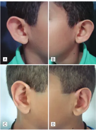
Figure 14 – Patient 2. A, preoperative image, anterior view;
B, postoperative image, anterior view.
A
C
B
D
Figure 15 – Patient 3. A, preoperative image, right proile; B, preoperative image, left proile; C, postoperative image,
right proile; D, postoperative image, left proile.
The cartilaginous incision provides a suitable break of the cartilaginous spring in patients with either thick or thin cartilages, achieving harmonious results without any carti lage break.
For deinition of the antihelix, 2 to 4 Mustardé sutures are used, as needed in each case, avoiding excessive tightening of the sutures so as not to cause aesthetic impairment.
Conchal rotation with ixation at the periosteum of the mastoid region was employed in all patients in this study, with care to avoid clinically relevant closure of the external auditory canal9. In some patients with greater hypertrophy, we also performed resection of excess concha.
Complications of otoplasties are very rare9,10. The most common complications are hematoma and immediate pos toperative infection1. In the late postoperative period, there
may be extrusion of sutures and/or more signiicant compli cations such as hypercorrection or contour irregularities3. We
observed 1 case of suture extrusion and 2 cases of unilateral hematoma in our selected cases; the latter we re promptly resolved by drainage. There was no case of infection.
CONCLUSIONS
Correspondence to: Francisco de Oliveira Goulart