Correlation between electromyographic data and
facial disability index in patients with long-term
facial paralysis: implications for treatment outcomes
Correlação entre eletromiograia e índice de inabilidade facial em pacientes com
paralisia facial de longa duração: implicações para o resultado de tratamentos
ABSTRACT
Background: Several techniques are available for the assessment of facial movement and activity, and facial disability can be evaluated through self-administered questionnaires. Ho-wever, the relationship between these objective and subjective measurements has not been examined to date. The present study examined the relationship between electromyographic data of the levator anguli oris muscle with the facial disability index in patients with long-ter m facial paralysis. We hypothesized that individuals with grealong-ter facial asymmetry have lower facial disability index scores. Methods: Patients were assessed using a clinical scale for the evaluation of facial expression, 2 facial disability index subscales, and the results of surface electromyography (sEMG). Seventeen long-term facial paralysis patients were analyzed and compared to 17 age- and gender-matched healthy controls. Results: Signiicant
differences between right and left hemifaces during smiling and at rest were detected in the experimental group, but not in the controls. Statistical analyses also indicated a weak correlation between sEMG (facial asymmetry) and facial disability index. Conclusions: The use of modern data analysis techniques such as sEMG in combination with self-reported data is of great beneit to clinicians and their patients. The identiication of a combination of measurements from randomized trials that can best determine the most effective treatment for patients with facial paralysis should be the objective of future studies.
Keywords: Facial paralysis. Facial asymmetry. Quality of life. Electromyography.
RESUMO
Introdução: Apesar de o movimento facial e de a atividade muscular poderem ser quanti-icados por meio de diversas técnicas e de a inabilidade facial poder ser quantiicada por meio de questionários de autoavaliação, a relação entre essas medidas objetivas e subjetivas ainda não foi investigada. O objetivo do presente trabalho foi correlacionar dados eletromio-gráicos dos músculos elevadores do ângulo da boca com o índice de inabilidade facial em pacientes com paralisia facial de longa duração. A hipótese do estudo foi de que indivíduos com maior assimetria facial apresentariam escores menores no índice de inabilidade facial.
Método: A avaliação consistiu na aplicação de uma escala clínica para avaliação da mími -Study conducted at the
Speech Therapy Support Unit of the Central Institute of Hospital
das Clínicas da Universidade de
São Paulo and Plastic Surgery Department of Hospital das
Clínicas of the School of Medicine
of Universidade de São Paulo, São Paulo, SP, Brazil.
Submitted to SGP (Sistema de Gestão de Publicações/Manager Publications System) of RBCP (Revista Brasileira de Cirurgia Plástica/Brazilian Journal of Plastic Surgery).
Study conducted with FAPESP support, through scholarship (Process No 2008/02687-5).
Paper received: September 13, 2011 Paper accepted: October 25, 2011
Fernanda Chiarion SaSSi1
Paula nuneS Toledo2
laura daviSon Mangilli3
nivaldo alonSo4
Claudia regina FurquiMde
andrade5
1. Doctor of Sciences from the School of Medicine of Universidade de São Paulo (FMUSP), speech therapist of the Department of Physiotherapy, Speech The rapy and Occupational Therapy of the FMUSP, São Paulo, SP, Brazil.
2. Doctor of Sciences from FMUSP, post-doctoral student at the Department of Physiotherapy, Speech Therapy and Occupational Therapy of FMUSP, São Paulo, SP, Brazil.
3. Master of Sciences from FMUSP, technical assistant of the Support Unit in Speech Therapy at the Central Institute of Hospital das Clínicas da FMUSP,
São Paulo, SP, Brazil.
4. Full professor at the Department of Surgery of FMUSP, associate professor at the Division of Plastic Surgery of the Department of Surgery of FMUSP, São Paulo, SP, Brazil.
ca facial, de duas subescalas do Índice de Inabilidade Facial e da realização do exame de eletromiograia de superfície (EMGs). Foram analisados 17 pacientes com paralisia facial de longa duração e os resultados foram comparados ao grupo controle, composto por 17 in divíduos saudáveis pareados por gênero e idade. Resultados: Os participantes do grupo pesquisa apresentaram diferenças signiicantes entre as hemifaces nas tarefas de repouso e sorriso. O mesmo não foi observado para os participantes do grupo controle. A análise estatística indicou correlação fraca entre os dados da EMGs (assimetria facial) e o Índice de Inabilidade Facial. Conclusões: O uso de técnicas cientíicas modernas de análise de dados,
como a EMGs, combinadas a medidas de autoavaliação oferece grandes possibilidades para clínicos e seus pacientes. A combinação de diferentes medidas em estudos randomizados que veriiquem o tipo de tratamento que oferece melhor resultado aos pacientes com paralisia facial deverá ser abordada em estudo futuro.
Descritores: Paralisia facial. Assimetria facial. Qualidade de vida. Eletromiograia.
INTRODUCTION
Apart from being associated with a wide range of func tional and aesthetic problems, facial nerve paralysis com -monly has negative emotional consequences. Facial nerve paralysis differs from other clinical conditions because a variety of underlying conditions can be the cause of this di -sability, such as cranial base trauma, congenital syndrome, cranial base tumors, and infectious diseases1,2. The incidence
of Bell’s Palsy, which is the most common cause of unilateral facial weakness, is approximately 25 in 100.0001. Although
84% of the cases show full recovery, 16% have chronic facial paralysis or paresis1. The psychological impact of facial disi
-gurement can result in fear of public areas and may adversely affect a patient’s social life2,3.
According to evidence-based clinical practice guidelines, it is important to establish the beneits of assessing facial paralysis and treatment strategies with regard to function, aesthetic appearance, capacity for communication, or a com -bination of these parameters. Facial paralysis patients are usually examined through a subjective assessment of fa ce functionality4, including the use of standardized
photogra-phic documentation and occasional video images2. As a ge
-neral rule, a clinician’s selection of a particular evaluation method is based on his or her training and expertise. In ge neral, the clinical relevance and the effectiveness of the results are not taken into account.
The psychological and social dificulties faced by patients with facial paralysis have already been reported in the litera-ture. Typical symptoms include alterations in emotional wel l-being, decreased self-esteem, anxiety, depression, and alter-native behaviors such as social isolation and addiction2,5,6.
In this sense, self-reported data are extremely important to support the assessment of facial neuromuscular dysfunction. Although the clinical recognition of the problems associated with facial paralysis has been described in the literature, there
are few studies based on self-reporting as a means to evaluate treatment outcome7.
The main obstacle to the improvement of clinical care for facial paralysis patients is the development of an objective and quantitative method for the assessment of facial function. Surface electromyography (sEMG) can provide valuable in formation for clinicians because it provides data on the phy -siological processes that generate muscular strength, produce movement, and mediate the performance of several functions that enable our interaction with the world8.
The beneicial results of neuromuscular training in combi -nation with sEMG have been reported in several studies5,9-12.
The use of sEMG instruments in association with biofeedback techniques enables the provision of continuous information about physiological function such as muscular tension levels12. Neuromuscular training in combination with sEMG
is based on the plasticity of the central nervous system. The brain is able to reorganize itself using visual and/or auditive information provided by sEMG11. The literature indicates that
the therapeutic use of sEMG helps patients develop muscular control and decreases the incidence of synkinesis.
Although facial movement and muscular activity can be quantiied by means of several techniques13-16, and facial di -sability can be quantiied by self-reporting instruments17, the
cor relation between the results of objective and subjective assessment methods has not been described to date. The pur pose of the present study was to determine the correlation bet -ween electromyographic data of the levator anguli oris muscle and the facial disability index in patients with long-ter m facial paralysis. We hypothesized that greater facial asymmetry would be correlated with lower scores on the facial disability index.
METHODS
age, 42 years) were included in the study. All patients had static and/or dynamic facial asymmetry (Table 1). Patients with systemic or neuromuscular disease, cognitive loss, or asymmetry derived from craniofacial deformities were excluded from the research. An age- and gender-matched control group included 17 healthy volunteers.
All participants signed an informed consent form, and the study was approved by the ethics committee of the institution (CAPPesq HCFMUSP No 0201/08).
The inclusion criteria for the research group were as follows: a) a medical diagnosis of longterm unilateral pe -ripheral facial paralysis of more than 2 years from the appearance of symptoms; with or without pre vious surgical treatment (reconstruction or rea -nimation);
b) a score between 4 and 11 on the Facial Paralysis Cli -nical Assessment Scale7.
The inclusion criteria for the control group were as follows: a) absence of a medical history of facial paralysis or
trauma in the head and neck region;
b) a score between 19 and 20 on the Facial Paralysis Clinical Assessment Scale7.
Facial Paralysis Clinical Assessment Scale
Facial symmetry was assessed by two members of the staff. As previously described in the literature, the protocol includes the evaluation of voluntary facial movement on both sides independently and the assessment of involuntary movement, which are graded based on a scale.
The test is composed of three sections: strength of volun-tary movement (raising eyebrows, closing eyelids, upper lip elevation, lateral traction of the upper lip, horizontal trac-tion of the lip, lip closing, lowering the lower lip); strength of involuntary movement (eyelid closure during blinking, and during speaking and smiling); and presence of nega-tive results (eyelid deformity at rest, lip deformity at rest, pre sence of synkinesis/spasms).
Movements are ranked as absent (0), partial (1), or inte-gral (2). The negative indings receive negative scoring as follows: (0) absent, (-1) moderate, or (-2) pronounced. The sum of individual scores provides a inal score that can have a maximum of 20 points.
sEMG Assessment
The elevators of the angle of the mouth (zygomaticus major and minor and levator anguli oris) were the muscular group analyzed in the study. This muscular group was selected because it is directly involved in smiling.
All sEMG assessments were performed using standar-dized surface electrodes (SDS500). The Miotool® 400 (Miotec Equipamentos Biomédicos, Porto Alegre, RS, Brasil) device with four channels was used in association with a computer system and double disposable electrodes (SDS500 Ag/AgCl, 10-mm diameter contact surfaces). This system enables the use of broadband ilter systems, a 20–500 Hz band-pass ilter, and a 60 Hz notch ilter. The gain was set at 100 with low levels of noise (< 5 µV RMS), which is recommended by the International Society of Electrophysiological Kinesio-logy. The system uses active electrodes and a set of compact sensors that include a pre-ampliier, whose location with res pect to the electrodes enables the reduction of artifacts and ampliication of the electromyographic signal, which is trans -mitted to the connection cable (level of noise, <5 µV RMS).
All electromyographic signals were iltered, as previously described. Analysis of means, standard deviation, and mi -nimum and maximum values of muscular activity during each task were evaluated using the software provided. Muscular activity was quantiied in microvolts (µV).
Interelectrode distance was 10 mm. Two sets of bipolar electrodes were applied to the skin on each side of the face, over the levator anguli oris muscle to record electrical activity during voluntary smiling. A third electrode was placed on the right fist to act as a grounding electrode. The electric impedance in the areas of electrode placement was minimized by cleaning the skin with gauze soaked in 70% alcohol.
The baseline activity of the levator anguli oris muscle was recorded irst followed by 3 voluntary smiling tests.
• Step 1 – the participants were instructed to remain in a sitting position, with the head positioned in the Frankfurt horizontal plane. After the placement of electrodes on the skin, each participant was ins -tructed to remain as still and relaxed as possible for one minute. Three independent recordings of 30 seconds each were obtained during resting. • Step 2 – each participant was instructed to smile
for 5 seconds and then keep the muscles relaxed for an additional 5 seconds, with 3 repetitions. During the tests, the participants were instructed to avoid sudden movements of the head. Electromyographic signal recording was started when the participants’ muscle activity was at the baseline level.
Electromyographic Data Analysis
The analysis of the sEMG results was performed using the temporal domain assessment method. The information
Table 1 – Distribution of patients
by type of facial dysfunction.
Category n
Vestibular schwannoma 2
Bell’s Palsy 12
Facial nerve trauma (temporal/cranial base) 3
thus obtained includes when the event occurred and the amplitude (magnitude indicator of muscular activity) of its occurrence. Under resting conditions, the values obtained were the average root mean square (RMS) of the electromyo-graphic activity observed during 30 seconds. The duration of muscular activity during dental pressure tasks (Al and MIC) was obtained by selecting the section of the trace that corresponded to muscle activation (on, peak, and off situation) using the electromyography software cursor and converting into microvolts.
The average action potential during movement was cal culated using the software provided. To compare the results between participants, the sEMG amplitude values were nor malized to the maximum contraction values re -corded (per centage of maximum value for each electrode arrangement).
Electromyographic Data Reliability
Because the literature on the subject indicates subjec-tivity in the interpretation of sEMG readings, a reliability analysis was performed to determine the degree of agree-ment between examiners and thus validate the accuracy of the results. For this purpose, 30 electromyographic sam -ples were randomly selected out of a total of 204. These samples were independently analyzed by two researchers with experience in the field. The correlation coefficient was high for all comparisons [confidence interval of 95% (IC 95%) 0.9788-0.9965], indicating good reliability between the examiners.
Facial Disability Index
The facial disability index is a self-reporting tool for the assessment of physical disability and psychosocial factors related to facial neuromuscular function17. Its objective is to
provide information on the daily experience of patients who live with facial nerve disorder. This questionnaire has two sub-scales: the social well-being sub-scale (IBES), which includes items related to psychological and social aspects, and the physical function subscale (IFF), which assesses daily life activities (for example, tooth brushing, eating or drinking). This questionnaire was administered only to the experimental group.
Data Analysis
Statistical analyses were performed using Wilcoxon’s test and Pearson’s correlation coeficient, and signiicance was set at 0.05. The coeficient of asymmetry between both sides of the face and muscular contraction were calculated in both groups as follows:
• research group – ratio between the non-paralyzed side/paralyzed side;
• control group – right side/left side.
RESULTS sEMG
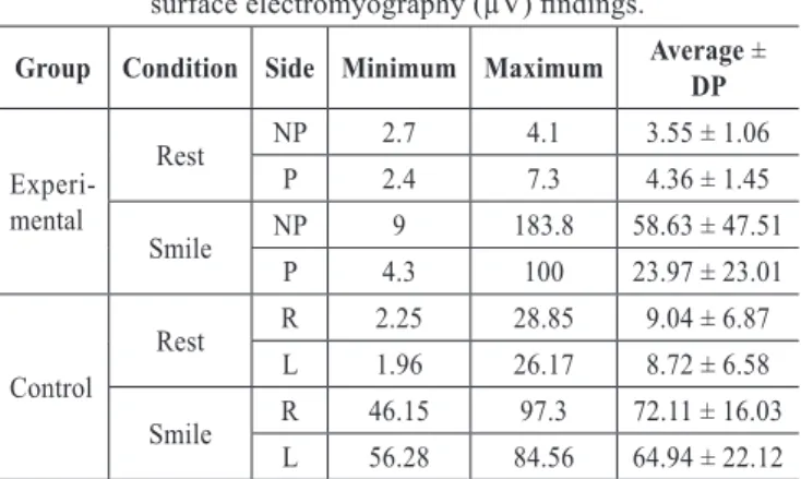
Differences were observed between the research and con -trol groups. Descriptive statistics indicate that the resear ch group showed lower electromyographic signal values during rest and smiling when compared to the control group. A grea -ter variability in the recordings was observed during smiling in both groups (Table 2).
Comparison between the two groups revealed signiicant differences in the electromyographic signal values between the two sides of the face in the experimental group at rest (P = 0.049; Z = 1.96; T = 30) and during smiling (P < 0.001; Z = 3.62; T = 0). The coeficient of asymmetry was high during smiling (average at rest, 0.88 + 0.31; average during smiling, 3.69 + 5.53).
There were no signiicant differences between the sides of the face in the control group at rest (P = 0.57; Z = 0.57; T = 64.5) or during smiling (P = 0.29; Z = 1.07; T = 54). As observed in the experimental group, the control group also showed a high coeficient of asymmetry between both sides of the face during smiling (average at rest, 1.07 + 0.26; average during smiling, 1.3 + 0.67).
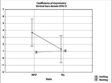
The differences in the facial asymmetry were veriied by comparing the asymmetry coeficient between the groups under both conditions (Figure 1). The statistical analysis demonstrated a signiicant difference between the groups during smiling (P < 0.001; Z = 3.15; T = 10), indicating that the research group had a higher asymmetry index. The groups did not differ when compared during the resting conditions (P = 0.102; Z = 1.63; T = 42).
Facial Disability Index (IFF)
The facial disability scale was only applied to the expe-rimental group. The average of IFF sub-scale values was 70
Table 2 – Descriptive analysis of the surface electromyography (µV) indings.
Group Condition Side Minimum Maximum Average ± DP
Experi-mental
Rest NP 2.7 4.1 3.55 ± 1.06
P 2.4 7.3 4.36 ± 1.45
Smile NP 9 183.8 58.63 ± 47.51 P 4.3 100 23.97 ± 23.01
Control
Rest R 2.25 28.85 9.04 ± 6.87 L 1.96 26.17 8.72 ± 6.58
Smile R 46.15 97.3 72.11 ± 16.03 L 56.28 84.56 64.94 ± 22.12
(minimum 50; maximum 95) and of IBES sub-scale was 68 (minimum 40; maximum 92).
Pearson’s correlation coeficient was used to verify the association between sub-scale values and between sub-scale values and coeficients of asymmetry generated by sEMG. The results indicated a weak correlation between sub-scales and a very weak correlation between sub-scales and the coeficient of asymmetry (Table 3).
DISCUSSION
In the present study, the correlation between the results of electromyography of the levator anguli oris (smile) and the scores obtained from a self-reporting questionnaire (IFF) were analyzed in patients with long-term facial paralysis. The results showed no correlation between the objective examination (sEMG) and self-reporting measures, and our hypothesis was therefore not conirmed. Although there was a great degree of variation in muscular activity among the parti-cipants (sEMG), the results showed signiicant differences between the paralyzed and non-paralyzed sides of the face among patients with facial paralysis, which were not detected in healthy individuals. Reports showing muscular activity differences between the paralyzed and non-paralyzed sides of the face were previously published and indicate that facial nerve integrity is essential for the balance and symmetry of facial expression7,18.
Facial paralysis causes anatomical and physiological changes. Facial asymmetry may be caused not only by weaker muscle contractions on the paralyzed side, but also by hyperactivity of the non-paralyzed side2. Minor alterations
tend to appear approximately 4 months after the lesion and include muscle contracture and muscular hypertrophy associated with synkinesis1. One of the limiting factors is
the viability of the facial muscles because after 12 months without stimulation, the muscles degenerate and can quickly show signs of atrophy. In the present study, a 30% reduction in muscular activity during smiling was observed in the non-pa ralyzed side in the experimental group in comparison to the control group.
The follow-up of long-term facial paralysis patients is a controversial issue. The main reason for surgical and non-sur gical treatments after facial nerve paralysis is to res tore a patient’s ability to smile1,3,5,7,12,19. Nonsurgical treat
-ment techniques include the application of botulinum to xin; physiotherapy; and stimulation/muscular training, such as therapy aimed at improving facial expression through elec-tromyography and biofeedback, and speciic facial exercises. However, even in welldesigned studies, the results vary sig -niicantly likely due to the inherent dificulty in the accurate assessment of recovery2. If a consensus is reached on an
effective procedure for the quantitative analysis of facial function and the comparison of global results in the fol -low-up of facial nerve lesions, the determination of trai ning techniques may become clearer.
The literature indicates that the normal human face exhi-bits an asymmetry of 6% during the production of facial expressions20. This was observed through a combination
of linear measures and invasive electromyography (needle electromyography) studies, and could therefore correspond to anatomical and muscular activity asymmetries caused by muscle imbalance and differences in measurements. These differences were not detected in the present study. The fact that sEMG is not as speciic as invasive electromyography for the measurement of muscle activity likely explains the diffe-rences between the results of our study and those of others.
Although the literature indicates that IIF sub-scales are reliable with respect to the results of self-reporting in patients with facial nerve alterations5 and in enabling the monitoring
of treatment outcomes7, this study did not detect a correlation
Figure 1 – Comparison between groups with regard to the
asymmetry coeficient. IC = conidence interval; NP:P = ratio between non-paralyzed and paralyzed sides;
R:L = ratio between right and left sides.
Table 3 – Pearson’s correlation coeficient for the
facial disability index and facial asymmetry (surface electromyography).
Pearson’s Correlation
IFF vs. smile 0.08
IBES vs. smile -0.09
IFF vs. rest -0.03
IBES vs. rest -0.32
IBES vs. IFF -0.14
between the sub-scales and objective measures. This fact must be further examined.
Effectiveness of treatment is a broad term that encom-passes several questions related to treatment eficacy (What is the purpose of treatment?), treatment eficiency (Is one treatment better than the other?), and the effects of treatment (How does the treatment change behavior?). Objective and self-reporting measures should therefore be seen as comple-mentary. The present study addresses the question of whether the perception of clinicians is the same as that of patients. This fact could potentially improve not only client satisfaction, but also the results of treatment. In this sense, there is a lack of evidence to determine the best techniques for soft tissue and neuromuscular reeducation1,10,11.
CONCLUSIONS
The use of modern data analysis techniques such as sEMG in combination with self-reporting tools is a promising approach for clinicians and their patients. The combination of different methods of analysis in randomized studies and the veriication of the most beneicial therapeutic design for patients with facial paralysis will be the objective of future studies.
REFERENCES
1. Diels HJ. Facial paralysis: is there a role for a therapist? Facial Plast Surg. 2000;16(4):361-4.
2. Hadlock T. Facial paralysis: research and future directions. Facial Plast Surg. 2008;24(2):260-7.
3. Tate JR, Tollefson TT. Advances in facial reanimation. Curr Opin Oto-laryngol Head Neck Surg. 2006;14(4):242-8.
4. Berg T, Jonsson L, Engström M. Agreement between the Sunnybrook, House-Brackmann, and Yanagihara facial nerve grading systems in Bell’s palsy. Otol Neurotol. 2004;25(6):1020-6.
5. VanSwearingen JM, Brach JS. Changes in facial movement and synki-nesis with facial neuromuscular reeducation. Plastic Reconstr Surg. 2003; 111(7):2370-5.
6. Finn JC, Cox SE, Earl ML. Social implications of hyperfunctional facial lines. Dermatol Surg. 2003;29(5):450-5.
7. Salles AG, Toledo PN, Ferreira MC. Botulinum toxin injection in longstanding facial paralysis patients: improvement of facial symmetry ob -served up to 6 months. Aesthetic Plast Surg. 2009;33(4):582-90. 8. De Luca CJ. The use of surface electromyography in biomechanics. J
Appl Biomech. 1997;13(2):135-63.
9. Segal B, Zompa I, Danys I, Black M, Shapiro M, Melmed C, et al. Sym-metry and synkinesis during rehabilitation of unilateral facial paralysis. J Otolaryngol. 1995;24(3):143-8.
10. Brach JS, VanSwearingen JM, Lenert J, Jonhson PC. Facial neuromuscular retraining for oral synkinesis. Plast Reconstr Surg. 1997;99(7):1922-31. 11. Cronin GW, Steenerson RL. The effectiveness of neuromuscular facial
retraining combined with electromyography in facial paralysis rehabili-tation. Otolaryngol Head Neck Surg. 2003;128(4):534-8.
12. Vaiman M, Shlamkovich N, Kessler A, Eviatar E, Segal S. Biofeedback training of nasal muscles using internal and external surface electro-myography of the nose. Am J Otolaryngol. 2005;26(5):302-7. 13. Linstrom CJ. Objective facial motion analysis in patients with facial
nerve dysfunction. Laryngoscope. 2002;112(7 Pt 1):1129-47. 14. Kang TS, Vrabec JT, Giddings N, Terris DJ. Facial nerve grading systems
(1985-2002): beyond the House-Brackmann scale. Otol Neurotol. 2002; 23(5):767-71.
15. Mehta RP, Zhang S, Hadlock TA. Novel 3-D video for quantiication of
facial movement. Otolaryngol Head Neck Surg. 2008;138(4):468-72. 16. Linstrom CJ, Silverman CA, Susman WM. Facial-motion analysis with
a video and computer system: a preliminary report. Am J Otol. 2000; 21(1):123-9.
17. VanSwearingen JM, Brach JS. The facial disability index: reliability and validity of a disability assessment instrument for disorders of the facial neuromuscular system. Phys Ther. 1996;76(12):1288-98. 18. Deleyiannis FW, Askari M, Schmidt KL, Henkelmann TC,
VanSwearin-gen JM, Manders EK. Muscle activity in the partially paralyzed face after placement of a fascial sling: preliminary report. Ann Plast Surg. 2005;55(5):449-55.
19. Coulson SE, Adams RD, O’Dwyer NJ, Croxson GR. Physiotherapy re-habilitation of the smile after long-term facial nerve palsy using video self-modeling and implementation intentions. Otolaryngol Head Neck Surg. 2006;134(1):48-55.
20. Burres SA. Facial biomechanics: the standards of normal. Laryngoscope. 1985;95(6):708-14.
Correspondence to: Claudia Regina Furquim de Andrade