Arq Bras Oftalmol. 2007;70(4):573-6
Retinopexia transconjuntival com drenagem externa do fluido sub-retiniano:
um estudo piloto prospectivo de oito casos consecutivos
Trabalho realizado no Department of Ophthalmology, Catanduva Medicine School, SP and Department of Ophthalmology. School of Medicine of Ribeirão Preto, University of São Paulo USP Ribeirão Preto (SP) -Brazil.
1CERV - Retina and Vitreous Specialized Centerof HORP - Eye Hospital of Rio Preto, São José do Rio Preto (SP) Brazil.Retina and Vitreous Section, Department of Oph-thalmology, School of Medicine of Ribeirão Preto, Uni-versity of São Paulo - USP - Ribeirão Preto (SP) - Brazil. 2Retina and Vitreous Section, Department of Ophthal-mology, School of Medicine of Ribeirão Preto, University of São Paulo - USP - Ribeirão Preto (SP) - Brazil. 3Departments of Ophthalmology and Health Evaluation
Sciences, Penn State College of Medicine Hershey -Pennsylvania - USA.
Correspondence to: Rubens Camargo Siqueira. Av. José Munia, 4500 - São José do Rio Preto (SP) Zip Code 15090-500
E-mail: [email protected] Recebido para publicação em 05.11.2006 Última versão recebida em 26.02.2007 Aprovação em 22.04.2007
The authors have no proprietary interests in any of the products cited in this paper.
Rubens Camargo Siqueira1
Rodrigo Jorge2
Ingrid Ursula Scott3
INTRODUCTION
Although superior rhegmatogenous retinal detachments can be repai-red surgically using nondrainage techniques, removal of subretinal fluid remains an important surgical step in retinal detachments with inferior breaks. Drainage of subretinal fluid has been reported to be the most hazardous step in retinal detachment surgery(1). The most common
compli-cations of subretinal fluid drainage include subretinal hemorrhage, retinal incarceration, and retinal perforation(2-3). Several external drainage
techni-ques have been described(4-6), such as using a suture needle and argon
laser for scleral and choroidal perforation(2). In 1985, McLeod described a
technique for posterior transcleral drainage of subretinal fluid monitored by indirect ophthalmoscopy to drain posteriorly sequestrated subretinal fluid following vitrectomy(3). Drainage monitoring was also described by
Char-Transconjunctival retinopexy with active external
drainage of subretinal fluid: a prospective pilot
study of eight consecutive cases
Keywords: Retina; Retinal detachment/surgey; Drainage/methods; Body fluids
Purpose: To describe an alternative surgical technique for the management of retinal detachment with no or minimal proliferative vitreoretinopathy (grade B) using transconjunctival retinopexy with active external drainage of subretinal fluid. Methods: In a prospective, interventional study, eight consecutive patients with retinal detachment with no or minimal pro-liferative vitreoretinopathy (grade B) underwent transconjunctival reti-nopexy with active external drainage of subretinal fluid. Transconjunctival external drainage of subretinal fluid was achieved by using a 29 gauge needle placed in the subretinal space under indirect ophthalmoscopic monitoring. Active suction was performed (500 mmHg vacuum) using a vitrectomy line coupled to the needle. After retinal reattachment, cryo-therapy was applied to the scleral region corresponding to the area of the retinal break(s). Results: In all cases there was retinal attachment at the end of surgery. Retinal redetachment occurred in four pseudophakic patients who then underwent pars plana vitrectomy. The four phakic patients maintained retinal attachment during follow-up (13-20 months). Conclusion: Transconjunctival retinopexy with active external drainage of subretinal fluid represents a useful, faster, and cheaper alternative to scleral bucklingfor retinal detachments with no or minimal proliferative retinopathy in phakic patients and, unlike scleral buckling, is not associated with induced myopia.
Arq Bras Oftalmol. 2007;70(4):573-6
574Transconjunctival retinopexy with active external drainage of subretinal fluid: a prospective pilot study of eight consecutive cases
les, who used a 25-gauge needle and an automated system for external drainage, associated with conventional scleral bu-ckling(4). In the current report, we describe a new technique of
transconjunctival retinopexy with active external drainage of subretinal fluid.
METHODS
The study was approved by the Institutional Review Bo-ard of the University of São Paulo and written informed con-sent was obtained from all study participants. Eight conse-cutive patients with retinal detachment with minimal or no proliferative retinopathy (grade B) underwent transconjunc-tival retinopexy with active external drainage of subretinal fluid. Inclusion criteria for the study included:
1) Primary rhegmatogenous retinal detachment;
2) All retinal breaks located within 1 clock hour of the fundus; and
3) No or minimal proliferative vitreoretinopathy (grade B). All patients were followed for a minimum of 10 months (10-20 months). Preoperative data collected included age, gender, race, involved eye, previous ocular surgery or trauma, duration of symptoms, presenting best-corrected visual acuity, status of the macula (attached or detached), circumferential extent of retinal detachment (number of clock hours), location of retinal breaks (in the superior 4 clock hours [10 to 2 o’clock, inclusive]; temporally or nasally [2 to 4 o’clock or 8 to 10 o’clock, nonin-clusive]; or in the inferior 4 clock hours [4 to 8 o’clock, inclusi-ve]), lens status (phakic, pseudophakic, aphakic).
Surgical technique
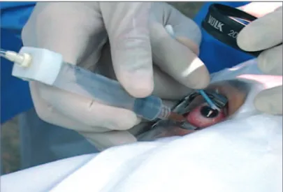
All patients underwent surgery by the same surgeon (RCS). Peribulbar anesthesia with 5 ml 2% lidocaine and 5 ml 0.5% bupivacainewas administered.The eye and surgical field were prepped with povidone-iodine and draped with a sterile drape. One drop 2% aqueous povidine was instilled into the fornix before surgery. Using indirect ophthalmoscopy, the drainage site was determined and marked on the sclera over the con-junctiva using bipolar cautery. A 29-gauge needle was cou-pled to the aspiration line (Figure 1) from the Accurus vitrec-tomy machine (Alcon, Fort Worth, Texas, USA). The distal 3 mm of the needle was bent and inserted through the con-junctiva and sclera at the point marked previously next to the biggest retinal elevation but non near the tear and the needle tip was visualized by indirect ophthalmoscopy as “flashing steel” in the subretinal space (Figure 2). Five hundred mil-limeters of mercury aspiration was employed to remove the subretinal fluid slowly under indirect ophthalmoscopic mo-nitoring. During active aspiration, the needle tip was redi-rected in the subretinal space according to the level of sub-retinal fluid in order to avoid sub-retinal perforation. As the sub-retinal detachment became shallower, the aspiration was reduced by using the foot pedal control. The needle tip was withdrawn when the retina was in close proximity to the tip. Intravitreal
injection of balanced salt solution (BSS, Alcon, Fort Worth, Texas, USA) was necessary in very extensive retinal detach-ments in order to avoid hypotony. After retinal reattachment, cryotherapy was applied to the scleral region that correspon-ded to the retinal break(s).
Postoperative examinations
Postoperative examinations were conducted on days 1, 7, and 14 and then months 1, 3, 6, and 12 after surgery. Each postoperative examination included measurement of best-cor-rected visual acuity, slit-lamp examination, applanation tono-metry, and dilated fundus examination.
RESULTS
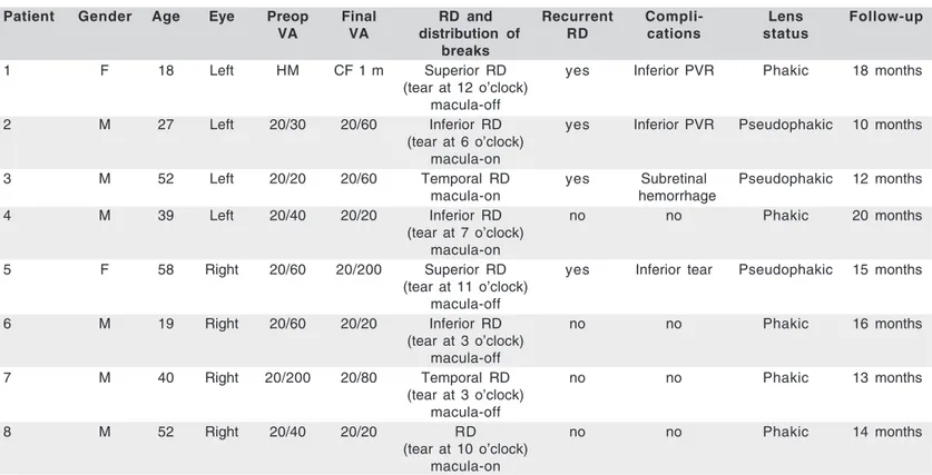
Clinical characteristics of the study population and study results are summarized in table 1. The mean follow-up duration
Figure 1 - Transconjuctival retinopexy. An external photograph show-ing monitorshow-ing of subretinal fluid drainage, after needle insertion
through the sclera.
Arq Bras Oftalmol. 2007;70(4):573-6 Transconjunctival retinopexy with active external drainage of subretinal fluid: a prospective pilot study of eight consecutive cases 575
was 15 months, a period considered sufficient for analysis of results, since proliferative vitreoretinopathy typically deve-lops between 6 and 8 weeks after surgery. The average age of the patients was 60 years (range, 35-86 years).
In all cases, there was complete retinal attachment at the end of surgery. One patient developed a subretinal hemor-rhage during drainage that resolved spontaneously within 4 weeks. Retinal redetachment occurred in four of eight patients (all four patients who developed recurrent retinal detachment were pseudophakic with inferior proliferative vitreoretino-pathy) at a mean of 17 days (range, 7-40 days) after initial retinal detachment repair; these patients then underwent suc-cessful retinal detachment repair using a standard three-port
pars plana vitrectomy technique. The four phakic patients
maintained retinal attachment during follow-up (13-20 mon-ths). None of the patients developed elevated intraocular pressure during the follow-up period.
DISCUSSION
Various techniques of subretinal fluid drainage have been described using passive and active drainage with or without indirect ophthalmoscopic monitoring(3-4,7-11). Passive needle
drainage of subretinal fluid without monitoring is the most extensively used technique. Our technique consists of a trans-conjunctival approach with active external drainage of subre-tinal fluid under indirect ophthalmoscopic monitoring.
Scleral buckling requires partial or 360 degrees of
conjunc-tival and Tenon’s capsule reflection and rectus muscle isola-tion. Our technique does not require conjunctival, Tenon’s or rectus muscle manipulation.
Ophthalmoscopic monitoring of subretinal fluid drainage was reported(3) and this technique consisted of external
drai-nage of posteriorly-sequestrated subretinal fluid following vitrectomy and fluid/gas or fluid/silicone oil exchange, there-by avoiding the need to create a drainage retinotomy site. These authors emphasized the reduced risk of retinal incarce-ration and prolapse with ophthalmoscopic monitoring. Ho-wever, monitoring of subretinal fluid drainage requires clear media for optimal visualization of the needle tip in the sub-retinal space. For this reason, scleral buckling may be more appropriate for patients with moderate or advanced cataract.
Subretinal hemorrhage is a potential complication of sub-retinal fluid drainage(5). One patient in the current series
deve-loped this complication. Subretinal hemorrhage is less likely to occur during active monitored drainage because there is a sharp choroidal penetration with a very thin 29-gauge needle and there is no choroidal congestion induced by prior cryo-therapy. In addition, if there is bleeding into the subretinal space, the blood tends to be aspirated with the subretinal fluid while choroidal vascular thrombosis occurs(3).
Hypotony is another potential complication of subretinal fluid drainage. To avoid this complication, some surgeons perform drainage with concomitant injection of vitreous subs-titutes(3) or tighten the scleral buckle prior to drainage(6). In the
current series, intravitreal injection of balanced salt solution Table 1. Clinical characteristics of study population and study results
Patient Gender Age Eye Preop Final RD and Recurrent Compli- Lens Follow-up
VA VA distribution of RD cations status
breaks
1 F 18 Left HM CF 1 m Superior RD yes Inferior PVR Phakic 18 months
(tear at 12 o’clock) macula-off
2 M 27 Left 20/30 20/60 Inferior RD yes Inferior PVR Pseudophakic 10 months
(tear at 6 o’clock) macula-on
3 M 52 Left 20/20 20/60 Temporal RD yes Subretinal Pseudophakic 12 months
macula-on hemorrhage
4 M 39 Left 20/40 20/20 Inferior RD no no Phakic 20 months
(tear at 7 o’clock) macula-on
5 F 58 Right 20/60 20/200 Superior RD yes Inferior tear Pseudophakic 15 months
(tear at 11 o’clock) macula-off
6 M 19 Right 20/60 20/20 Inferior RD no no Phakic 16 months
(tear at 3 o’clock) macula-off
7 M 40 Right 20/200 20/80 Temporal RD no no Phakic 13 months
(tear at 3 o’clock) macula-off
8 M 52 Right 20/40 20/20 RD no no Phakic 14 months
(tear at 10 o’clock) macula-on
Arq Bras Oftalmol. 2007;70(4):573-6
576Transconjunctival retinopexy with active external drainage of subretinal fluid: a prospective pilot study of eight consecutive cases
was sometimes performed to restore intraocular pressure and avoid corneal folds, which may prevent good visualization of the posterior segment.
In the absence of severe vitreous traction on the retina, chorioretinal adhesion promoted by cryotherapy scars may be sufficient to maintain the retina attached without scleral in-dentation, as with pneumatic retinopexy(7). The absence of
scleral indentation avoids buckle-induced myopia.
All patients who developed recurrent retinal detachment were pseudophakic. The distinct category of pseudophakic re-tinal detachment compared with phakic rere-tinal detachment has a higher prevalence of missed breaks (5%-20%), resulting from the smaller size and anterior location of breaks in these eyes as well as from the incomplete peripheral fundus view due to anterior or posterior capsule fibrosis, cortical remnants, small pupil, vitreous opacities, and optical aberrations at the intra-ocular lens rim(8,12-14). All phakic patients in the current series
maintained retinal attachment during follow-up (13-20 months).
CONCLUSION
Transconjunctival retinopexy with active external drainage of subretinal fluid represents a useful alternative technique for retinal detachment repair in phakic patients with little pro-liferative vitreoretinopathy; this technique is quicker and cheaper compared with scleral buckling, and is not associated with induced myopia.Further studies are needed to determine the safety and efficacy of this new technique to repair phakic retinal detachments with no or minimal proliferative vitreoreti-nopathy (grade B).
RESUMO
Objetivo: Descrever uma técnica cirúrgica alternativa para o tratamento de descolamento da retina sem ou com mínima vitreorretinopatia proliferativa (grau B) usando uma retino-pexia transconjuntival com drenagem externa do fluido sub-retiniano. Métodos: Prospectivo estudo intervencional, com oito pacientes consecutivos com descolamento da retina com nenhum ou mínima vitreorretinopatia proliferativa (grau B) que foram submetidos a retinopexia transconjuntival com dre-nagem externa ativa do fluido sub-retiniano. A dredre-nagem exter-na transconjuntival do fluido sub-retiniano foi realizada com agulha calibre 29 colocada no espaço sub-retiniano e
moni-torada pela oftalmoscopia binocular indireta. A sucção ativa foi realizada (vácuo de 500 mmHg) usando a linha de extração do vitreófago conectado a agulha. Após a colagem da retina, crioterapia foi aplicada na região escleral correspondente a área da(s) ruptura(s). Resultados: Em todos os casos a retina aplicou no final da cirurgia. O redescolamento da retina ocor-reu em 4 pacientes pseudofácicos, que foram submetidos a seguir a vitrectomia pars plana. Os 4 pacientes fácicos
man-tiveram a retina aplicada durante o seguimento (13 a 20 meses). Conclusão: A retinopexia transconjuntival com drenagem ex-terna ativa do fluido sub-retiniano representa alex-ternativa útil, rápida e barata a cirurgia do descolamento da retina com im-plante escleral em pacientes com nenhuma ou mínima vitreor-retinopatia proliferativa, fácicos e diferente do implante escle-ral não está associada a indução da miopia.
Descritores: Retina; Descolamento retiniano/cirurgia; Drena-gem/métodos; Líquidos corporais
REFERENCES
1. Ferguson EC. Drainage of subretinal fluid in scleral bucklings. Int Ophthal-mol Clin. 1962;2:181-205.
2. Bovino JA, Marcus DF, Nelsen PT. Argon laser choroidotomy for drainage of subretinal fluid. Arch Ophthalmol. 1985;103(3):443-4.
3. McLeod D. Monitored posterior transcleral drainage of subretinal fluid. Br J Ophthalmol. 1985;69(6):433-4.
4. Charles ST. Controlled drainage of subretinal and choroidal fluid. Retina. 1985;5(4):233-4.
5. Wilkinson CP, Bradford RH Jr. Complications of draining subretinal fluid. Retina. 1984;4(1):1-4.
6. Jaffe GJ, Brownlow R, Hines J. Modified external needle drainage procedure for rhegmatogenous retinal detachment. Retina. 2003;23(1):80-5.
7. Tornambe PE, Hilton GF. Pneumatic retinopexy and buckling. Arch Ophthalmol. 1990.108(3):318-9. Comment on: Arch Ophthalmol. 1989.107(19):1469-71. 8. Brazitikos PD, Androudi S, Christen WG, Stangos NT. Primary pars plana
vitrectomy versus scleral buckle surgery for the treatment of pseudophakic retinal detachment: a randomized clinical trial. Retina. 2005;25(8):957-64. 9. Azad RV, Talwar D, Pai A. Modified needle drainage of subretinal fluid for
conventional scleral buckling procedures. Ophthalmic Surg Lasers. 1997;28 (2):165-7.
10. Peyman GA, Charles H, Tawakol ME, Federman J, Ando F. External drainage of subretinal fluid with a contact Nd: YAG laser. Int Ophthalmol. 1987;11(2):77-8. 11. Sonoda Y, Yamakiri K, Sonoda S, Uchino E, Doi N, Sakamoto T. Endos-copy-guided subretinal fluid drainage in vitrectomy for retinal detachment. Ophthalmologica. 2006;220(2):83-6.
12. Yoshida A, Ogasawara H, Jalkh AE, Sanders RJ, McMeel JW, Schepens CL. Retinal detachment after cataract surgery. Surgical results. Ophthalmology. 1992;99(3):460-5.
13. Rosen PH, Wong HC, McLeod D. Indentation microsurgery: internal sear-ching for retinal breaks. Eye. 1989;3(Pt 3):277-81.