Cirurgia de descolamento de retina com injeção de óleo de silicone no sistema
de vitrectomia transconjuntival sem sutura de 23-gauge
Rubens Camargo Siqueira1 Aline Degasperi Cote Gil2 Rodrigo Jorge3
in transconjunctival sutureless 23-gauge vitrectomy
ABSTRACT
Trabalho realizado na Faculdade de Medicina de Catanduva e Faculdade de Medicina da Universidade de São Paulo USP - Ribeirão Preto (SP) - Brasil. 1Doutor em Oftalmologia pela Universidade de São
Pau-lo USP - Ribeirão Preto (SP) - Brasil; Responsável pePau-lo Departamento de Oftalmologia da Faculdade de Medi-cina de Catanduva - Catanduva (SP) - Brasil; Médico colaborador em pesquisa do Departamento de Retina da Universidade de São Paulo - USP - Ribeirão Preto (SP) - Brasil; Responsável pelo Centro Especializado Retina e Vítreo do Hospital do Olho de São José do Rio Preto - HORP - São José do Rio Preto (SP) - Brasil. 2Fellow do Centro Especializado Retina e Vítreo do
HORP - São José do Rio Preto (SP) - Brasil. 3Responsável pelo Departamento de Retina e Vítreo da
USP - Ribeirão Preto (SP) - Brasil; Doutor em Oftalmo-logia pela Universidade de São Paulo - USP - Ribeirão Preto (SP) - Brasil.
Corresponding author: Rubens Camargo Siqueira. Av. José Munia, 4500 - São José do Rio Preto (SP) Zip Code 15090-500
E-mail: [email protected] Recebido para publicação em 24.09.2006 Última versão recebida em 13.06.2007 Aprovação em 22.08.2007
Nota Editorial: Depois de concluída a análise do arti-go sob sigilo editorial e com a anuência do Dr. Cláudio Renato Garcia sobre a divulgação de seu nome como revisor, agradecemos sua participação neste processo.
Purpose: To report a surgical technique for retinal detachment surgery using transconjunctival sutureless 23-gauge vitrectomy with silicone oil injection. Methods: Thirty-one patients with retinal detachment underwent vitreoretinal surgery using a transconjunctival sutureless 23-gauge vi-trectomy system. At the end of the procedure silicone oil was injected into all eyes through a microcannula. After removing the microcannula, a bipolar cautery was used in the conjunctiva over the wound to prevent silicone oil reflux. Results: In all patients retinal reattachment and injection of silicone oil through transconjunctival sutureless 23-gauge vitrectomy system was possible. Retinal re-detachment occurred in 8 patients (25.80%) who were submitted to a new vitreoretinal surgery by the technique described above, however, more extensive retinotomy was necessary in all cases, specially inferiorly. Three patients (9.67%) pre-sented silicone oil leakage in subconjuntival compartment. Another surgical procedure was necessary to remove the subconjuntival silicone oil. Conclusions: The retinal detachment surgery with silicone oil injection in transconjunctival sutureless 23-gauge vitrectomy is a safe and efficient technique to repair retinal detachment and it has the advantage of being minimally invasive.
Keywords: Retinal detachment/surgery; Vitrectomy; Ophthalmologic surgical procedures/ methods; Silicone oils/therapeutic use; Surgical procedures, minimally invasive
INTRODUCTION
One of the most innovative vitreoretinal surgical techniques introdu-ced in recent years is transconjunctival sutureless vitrectomy(1-2). In this procedure, three polyamide microcannulae are inserted transconjunc-tivally through the sclera in pars plana. The vitreoretinal instruments and infusion line are then introduced through these cannulas into the vitreous cavity. Because a thin 25-gauge instrumentarium is used, the small in-cisions left in the sclera after removal of the cannulas are self-sealing without suturing.
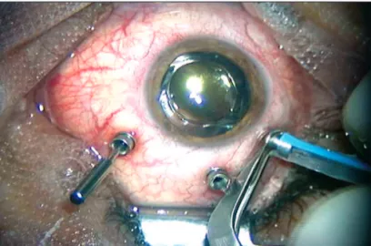
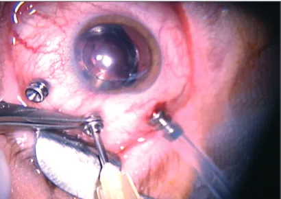
Figure 2 - Silicone oil injected through the second infusion line connected to the microcanula; another microcannula is used for infusion line Figure 1 - Silicone oil injected through the infusion line
Eckardt introduced transconjunctival sutureless 23-gauge vitrectomy. He used this technique in patients with macular pucker, macular hole, proliferative or non-proliferative diabe-tic retinopathy, rhegmatogenous retinal detachment and cen-tral retinal vein occlusion. In some eyes the author used gas tamponade at the end of surgery but not silicone oil(5).
A method for retinal detachment surgery with silicone oil injection in transconjunctival sutureless 23-gauge vitrecto-my is presented.
METHODS
Thirty-one patients with rhegmatogenous retinal detach-ment in different stages due to proliferatative vitreoreti-nopathy (PVR) were submitted to three-port pars plana trectomy using transconjunctival sutureless 23-gauge vi-trectomy system (DORC, Zuidland, Holland). Surgeries were performed under peribulbar anesthesia by the same surgeon (RCS).
The procedure starts by pushing the conjunctiva 1 mm to 2 mm laterally (i.e., parallel to the corneal limbus) in the in-ferotemporal, superotemporal, and superonasal quadrants using a special pressure plate to hold it firmly to the sclera. A 23-gauge stiletto blade (45º angle; BD Medical-Ophthalmic Systems, Franklin Lakes, NJ) is then inserted at a 30º to 40º angle through the conjunctiva, sclera, and pars plana (3.5 mm from the corneoscleral limbus). To obtain scleral tunnels parallel to the corneoscleral limbus, the scleral incisions are made parallel to the corneoscleral limbus. The incision with the 23-gauge stiletto blade is 0.72 mm wide. Constant pres-sure is applied to the prespres-sure plate while the incision is made and during withdrawal of the stiletto blade to prevent slippage of the conjunctiva against the sclera(5).
The microcannula is then inserted through the conjunc-tival incision and into the scleral tunnel using a specially designed blunt inserter.
The pneumatic vitreous cutter was attached to the vi-trectomy unit (ACCURS vivi-trectomy machine -Alcon, Fort Worth, Texas, USA). A cutting rate of up to 1200 per minute and suction of up to 500 mmHg were used.
Limited core vitrectomy and removal of the vitreous clo-se to the sclerotomy uclo-sed for the inclo-sertion of the vitreous cutter were initially performed. In phakic eyes, vitrectomy at the vitreous base from the 6- to 12-o’clock position was performed with the vitreous cutter held with the right hand, and that from the 12- to 6-o’clock position was performed with the left hand.
Initially, the central vitreous was removed (core vitrec-tomy), with latter peeling of dense epiretinal membranes. Vitreous shaving was performed with scleral indentation by an assistant using a muscle hook. The remaining traction points like star folds with intraretinal fibrosis were identi-fied; and a puncture retinotomy was performed using
endo-diathermy, instead of removing membranes with the use of forceps. After relaxing the traction areas, subretinal fluid was aspirated with a long extensible silicone-tipped cannula, ta-king advantage of the retinotomy created. The entire retina was checked with indirect ophthalmoscopy and, if not at-tached, additional retinotomies were made to release the trac-tion followed by aspiratrac-tion of subretinal fluid until retina got flattened against the retinal pigment epithelium (RPE).
Figure 4 - Bipolar cautery in the conjunctiva over the wound, prevent the
little leakage of the silicone oil Figure 5 - Silicone oil leakage in subconjuntival compartment
RESULTS
Thirty-one eyes of 31 patients with retinal detachment with proliferative vitreoretinopathy (PVR) at different stages were submitted to vitreoretinal surgery using high pressure air infu-sion and puncture retinotomies to relax the points of traction caused by PVR. In the total group 17 patients were males (54.84%) and 14 females (45.16%); the age range was 15-85 years (mean age of 53.70). The outcomes are shown on table 1. It was possible to reattach the retina and inject silicone oil in all patients enrolled in the study. Retinal re-detachment occurred in 8 patients (25.80%). These patients were submitted to a new vitreoretinal surgery with the same technique initially descri-bed; however, more extensive retinotomies were necessary in all cases, mainly inferiorly.
The use of cannulas was free of complications in all cases; there were no cannula-induced lesions of the posterior capsule in phakic or pseudophakic eyes.
In the first patient the infusion line disconnected of the microtrocar during oil infusion and it required reconnection. Therefore the infusion line fixation using forceps was neces-sary to prevent line escape.
Postoperative wound leaks occurred in three eyes with silicone oil draining into the subconjuntival space (Figure 5). Another surgical procedure to remove the subconjuntival silicone was needed.
Sutures were not used in any case to close the conjunctival or scleral incisions. At follow-up examination, the intraocular pressure (measured with Haag-Streit Goldmann applanation tonometry) varied from 8 to 62 mmHg (mean= 18.32 mmHg).
Approximately three months after surgery all patients we-re submitted to silicone oil we-removal using the 23-gauge sys-tem. Suction was performed through the second line infusion (Figure 2) with active aspiration.
DISCUSSION
This study was designed to retrospectively revise safety data, including visual acuity, incidence of re-detachment, si-licone oil leakage as well as the need to use suture due to silicone oil draining to the subconjuntival space. The efficacy of the system was evaluated by comparing preoperative and postoperative VA results and evaluating its efficiency.
Sutureless self-sealing sclerotomies for pars plana vitrec-tomy were first described by Chen(6), in 1996, and since then other researchers reported their experience with the technique and its modifications(7-12). Instead of the usual right-angled incision through the sclera, a tunnel-like tangential incision is made at a 30º to 45º angle through the sclera. Suture closure is not required because the wound borders are pressed together by the intraocular pressure. The tunnels can be made in the posterior-anterior direction (i.e., in the direction to the cor-neoscleral limbus)(6-7) in the anterior-posterior direction(8), or
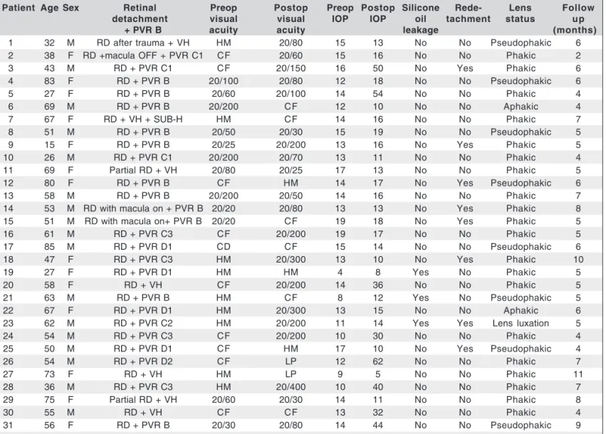
Table 1. Results of retinal detachment surgery with silicone oil injection in transconjunctival sutureless 23-gauge vitrectomy. Visual acuity is best-corrected Snellen acuity
Patient Age Sex Retinal Preop Postop Preop Postop Silicone Rede- Lens Follow
detachment visual visual IOP IOP oil tachment status up
+ PVR B acuity acuity leakage (months)
1 32 M RD after trauma + VH HM 20/80 15 13 No No Pseudophakic 6
2 38 F RD +macula OFF + PVR C1 CF 20/60 15 16 No No Phakic 2
3 43 M RD + PVR C1 CF 20/150 16 50 No Yes Phakic 6
4 83 F RD + PVR B 20/100 20/80 12 18 No No Pseudophakic 6
5 27 F RD + PVR B 20/60 20/100 14 54 No No Phakic 4
6 69 M RD + PVR B 20/200 CF 12 10 No No Aphakic 4
7 67 F RD + VH + SUB-H HM CF 14 16 No No Phakic 7
8 51 M RD + PVR B 20/50 20/30 15 19 No No Pseudophakic 5
9 15 F RD + PVR B 20/25 20/200 13 16 No Yes Phakic 5
10 26 M RD + PVR C1 20/200 20/70 13 11 No No Phakic 4
11 69 F Partial RD + VH 20/80 20/25 17 13 No No Phakic 5
12 80 F RD + PVR B CF HM 14 17 No Yes Pseudophakic 6
13 58 M RD + PVR B 20/200 20/50 14 16 No No Phakic 7
14 53 M RD with macula on + PVR B 20/20 20/80 13 13 No Yes Phakic 8
15 51 M RD with macula on+ PVR B 20/20 CF 19 18 No Yes Phakic 5
16 61 M RD + PVR C3 CF 20/200 19 17 No No Phakic 5
17 85 M RD + PVR D1 CD CF 15 14 No No Pseudophakic 6
18 47 F RD + PVR C3 HM 20/300 13 10 No Yes Phakic 10
19 27 F RD + PVR D1 HM HM 4 8 Yes No Phakic 5
20 58 F RD + VH CF 20/200 14 36 No No Phakic 5
21 63 M RD + PVR B HM CF 8 12 Yes No Pseudophakic 5
22 67 F RD + PVR D1 HM 20/300 13 15 No No Aphakic 6
23 62 M RD + PVR C2 HM 20/200 11 14 Yes Yes Lens luxation 5
24 54 M RD + PVR C3 CF 20/200 10 30 No No Phakic 4
25 50 M RD + PVR D1 CF HM 17 10 No Yes Pseudophakic 4
26 54 M RD + PVR D2 CF LP 12 62 No No Phakic 7
27 73 F RD + VH HM LP 9 5 No No Phakic 11
28 36 M RD + PVR C3 HM 20/400 10 40 No No Phakic 7
29 75 F Partial RD + VH 20/60 20/30 14 11 No No Phakic 8
30 55 M RD + VH CF CF 13 32 No No Phakic 4
31 56 F RD + PVR B 20/30 20/80 14 44 No No Pseudophakic 9
LP= light perception; CF= finger counting; HM= hand movements; RD= retinal detachment; VH= vitreous hemorrhage
parallel to the corneoscleral limbus(7,11). We performed parallel incisions in all cases. Unlike conventional sclerotomies, which are always accompanied by temporary postoperative astig-matism secondary to suture closure(5), tunnel incisions rarely results in astigmatism and lead only to a slight postoperative inflammatory reaction.
Although the conjunctiva is always opened in most su-tureless self-sealing sclerotomy techniques, transconjuncti-val vitrectomy requires merely pin points for
microcannu-las(3,5). Sclerotomies in 25-gauge vitrectomy require no
su-turing because they are only 0.5 mm in diameter as compared to 1.15-mm width of the sclerotomies in conventional 20-gauge vitrectomy. If 23-20-gauge instruments are used in com-bination with microcannulas, the sclerotomies are too large to allow perpendicular scleral incisions without suture clo-sure(1-3,5).
The cannulas can be placed in tunnel incisions (0.72 mm wide) running tangentially to the scleral surface(5). In
preli-minary investigations on eyes from eye banks, we first used a beveled trocar(1,4). However, the trocar did not cut well enou-gh to make a tunnel incision and we used a stiletto blade. The cannulas were then inserted into the tunnel with the aid of a special blunt inserter and remained firmly in place in all eyes, even during the whole procedure requiring extensive vitrectomy on the retinal periphery under circular scleral indentation. In a few eyes, a slight accumulation of infusion liquid drained from under the conjunctiva when the cannulas were withdrawn at the end of the surgery(1-2,4).
We reported retinal detachment surgery with silicone oil injection in transconjunctival sutureless 23-gauge vitrec-tomy. In all patients it was possible to reattach the retina and inject silicone oil through the transconjunctival sutureless 23-gauge vitrectomy system.
ble-eding, need for vitreous volume enhancement, or re-operations to suture the wounds. Intraoperative wound leaks through the cannula can result in bleeding, vitreous prolapse with secondary vitreoretinal traction, miosis due to hypotony, or even retinal prolapse(4). We used bipolar cautery in the conjunctiva over the wound to prevent any leakage of silicone oil. However, three cases presented silicone oil leakage in subconjuntival space without other complications (choroidal detachments or blee-ding) and a new procedure was necessary for remove the sub-conjuntival silicone and to suture the wounds.
In conclusion, the retinal detachment surgery with sili-cone oil injection in transconjunctival sutureless 23-gauge vitrectomy is a safe and efficient technique to repair retinal detachment and offers a special advantage of being minimal-ly invasive.
RESUMO
Objetivos: Relatar técnica cirúrgica para descolamento de retina utilizando sistema de 23-gauge com injeção de óleo de silicone. Métodos: Trinta e um pacientes com descolamento da retina foram submetidos a cirurgia vitreorretiniana usando o sistema 23-gauge de vitrectomia transconjuntival sem su-tura. Ao final do procedimento o óleo de silicone foi injetado em todos os olhos através de uma microcânula. Após a retira-da retira-da microcânula, foi utilizado cautério bipolar na incisão conjuntival para prevenir o vazamento do óleo de silicone.
Resultados: Em todos os pacientes foi possível reaplicar a retina e injetar o óleo de silicone através do sistema 23-gauge de vitrectomia transconjuntival. Redescolamento da retina ocorreu em 8 pacientes (25,80%) os quais foram submetidos a uma nova cirurgia vitreorretiniana com a mesma técnica, entretanto, uma retinotomia mais extensa foi necessária em todos estes casos especialmente na parte inferior que foi mais comprometido. Em 3 casos (9,67%) houve extravasamento do óleo de silicone para o compartimento subconjuntival. Novo procedimento foi necessário para remover o silicone sub-conjuntival. Conclusões: A cirurgia do descolamento da
reti-na com injeção de óleo de silicone utilizando o sistema 23-gauge de vitrectomia transconjuntival é uma técnica segura e eficiente para o reparo do descolamento da retina e oferece a vantagem de ser um sistema minimamente invasivo.
Descritores: Descolamento retiniano/cirurgia; Vitrectomia; Procedimentos cirúrgicos oftalmológicos/métodos; Óleo de silicone/uso terapêutico; Procedimentos cirúrgicos minima-mente invasivos
REFERENCES
1. Fujii GY, De Juan E Jr, Humayun MS, Pieramici DJ, Chang TS, Awh C, et al. A new 25-gauge instrument system for transconjunctival sutureless vitrec-tomy surgery. Ophthalmology. 2002;109(10):1807-12; discussion 1813. Erratum in: Ophthalmology. 2003;110(1):9. Comment in: Ophthalmology. 2003;110(1):9.
2. Fujii GY, de Juan E Jr, Humayun MS, Chang TS, Pieramici DJ, Barnes A, Kent D. Initial experience using the transconjunctival sutureless vitrectomy system for vitreoretinal surgery. Ophthalmology. 2002;109(10):1814-20. Comment in: Ophthalmology. 2003;110(12):2427-8; author reply 2428. Oph-thalmology. 2003;110(12):2427; author reply 2427. OphOph-thalmology. 2003;110(12):2428-9; author reply 2429.
3. Packo K. Early experience reveals benefits of 25-Ga. Technology. Rev Ophthalmol [serial on the Internet]. 2004; [cited 2006 Dec 12] 11: [about 8 p.] Available from: http://www.revophth.com/index.asp?page=1_563.htm 4. Charles S, Calzada J, Wood B. Vitreous microsurgery. 4th ed. Philadelphia.
Lippincott Williams & Wilkins; 2006. p.85-94
5. Eckardt C. Transconjunctival sutureless 23-gauge vitrectomy.Retina. 2005; 25(2):208-11.
6. Chen JC. Sutureless pars plana vitrectomy through self-sealing sclerotomies. Arch Ophthalmol. 1996;114(10):1273-5.
7. Kwok AK, Tham CC, Lam DS, Li M, Chen JC. Modified sutureless sclero-tomies in pars plana vitrectomy. Am J Ophthalmol. 1999;127(6):731-3. 8. Assi AC, Scott RA, Charteris DG. Reversed self-sealing pars plana
sclero-tomies. Retina. 2000;20(6):689-92.
9. Jackson T. Modified sutureless sclerotomies in pars plana vitrectomy. Am J Ophthalmol. 2000;129(1):116-7.
10. Rahman R, Rosen PH, Riddell C, Towler H. Self-sealing sclerotomies for sutureless pars plana vitrectomy. Ophthalmic Surg Lasers. 2000;31(6):462-6. 11. Theelen T, Verbeek AM, Tilanus MA, van den Biesen PR. A novel technique for self-sealing, wedge-shaped pars plana sclerotomies and its features in ultrasound biomicroscopy and clinical outcome. Am J Ophthalmol. 2003; 136(6):1085-92.
12. Eckert T, Eckardt C. Verhalten des Hornhautastigmatismus nach Pars-plana-Vitrektomie mit oder ohne gleichzeitiger Kataraktoperation. Ophthalmologe. 1996;93(1):38-44.