3 6 8 Arq Bras Oftalmol. 2011;74( 5): 368-70
R
ELATO DEC
ASO|
CA S E RE P O R TLong term follow-up of acute multifocal hemorrhagic retinal vasculitis
(Blumenkranz syndrome): case report
Síndrome de vasculite retiniana hemorrágica multifocal aguda (síndrome de Blumenkranz)
com seguimento a longo prazo: relato de caso
MIGUEL HAGE AMARO1, AARON BROCK ROLLER2, CESAR TAVARES MOTTA3, MARIO MARTINSDOS SANTOS MOTTA4
Submitted for publication: November 18, 2010 Accepted for publication: February 18, 2011
Study carried out at Instituto de Olhos e Laser de Belém (PA), Brazil.
1Physician, Instituto de Olhos e Laser de Belém (PA), Brazil. 2Physician, Retina Service, University of Iowa, USA.
3Physician, Universidade do Rio de Janeiro - UNIRIO - Rio de Janeiro (RJ), Brazil. 4Professor, Universidade do Rio de Janeiro - UNIRIO - Rio de Janeiro (RJ), Brazil.
Funding: No specific financial support was available for this study.
Disclosure of potential conflicts of interest: M.H.Amaro, None; A.B.Roller, None; C.T.Motta, None; M.M.S.Motta, None.
Correspondence address: Miguel Hage Amaro. Rua Quintino Bocaiuva, 516 - Belém (PA) - 66053-240 - Brazil - E-mail: [email protected]
ABSTRACT
Purpose: To report a 16-year long-term follow-up of a patient with acute multifocal hemorrhagic retinal vasculitis (Blumenkranz syndrome).A 21-year old male was seen in 1994 with acute multifocal hemorrhagic retinal vasculitis (Blumenkranz syndrome), first in the left eye, and later in the right eye. He was treated with retinal photocoagu-lation in areas of retinal ischemia and oral steroids, followed by sequential annual fundus examination and photography for 16 years. Vision improved to 20/25 in both eyes after retinal ischemic areas photocoagulation and oral steroids, and his vision has been maintained for 16 years.Photocoagulation of retinal ischemia and oral steroids are effective for the treatment of acute multifocal hemorrhagic retinal vasculitis (Blumenkranz syndrome).
Keywords: Purpura, Schoenlein-Henoch; Retinal vasculitis; Vasculitis; Light coagula-tion; Retinal hemorrhage; Case report
RESUMO
Relato de caso com acompanhamento por 16 anos de um paciente com a vasculite hemorrágica multifocal aguda (síndrome de Blumenkranz).Um paciente de 21 anos de idade foi diagnosticado em 1994 com a vasculite hemorrágica multifocal aguda (síndrome de Blumenkranz), primeiro no olho esquerdo e depois no olho direito. Foi tratado com fotocoagulação retiniana nas áreas retinianas isquêmicas e corticosteroide oral e segui-do por exames complementares da retina por 16 anos.A visão melhorou para 20/25 em ambos os olhos após a fotocoagulação retiniana nas áreas isquêmicas da retina e corticosteroide oral permanecendo assim até o momento por 16 anos.A fotocoagulação retiniana nas áreas isquêmicas e o uso de corticosteróide oral são tratamentos efetivos para a vasculite hemorrágica multifocal aguda (síndrome de Blumenkranz).
Descritores: Púrpura de Schoenlein-Henoch; Vasculite retiniana; Vasculite; Fotocoagula-ção; Hemorragia retiniana; Relato de caso
INTRODUCTION
Acute multifocal hemorrhagic retinal vasculitis (Blumenkranz syn-drome) is an uncommon disease that was first described in seven patients over a 5-year period to 1988(1).
These patients have features including subtle hemorrhagic mul-tifocal retinal vasculitis, frequently in the posterior segment and predo-minantly in men. The syndrome is bilateral, but with a degree of asymmetric involvement that causes abrupt visual loss in the affec-ted eye without systemic symptoms or a proven systemic etiology. Other signs of this disease are retinal hemorrhage that appears to result from vasculitic occlusion of the retinal veins, papillitis, non-confluent posterior retinal infiltrates, vitreous cellular inflamma-tion and late complicainflamma-tions such as secondary vitreous hemorrhage, neovascularization, epiretinal membrane, rubeosis iridis, and neo-vascular glaucoma.
These symptoms are similar to those in a variety of other disor-ders such as Behçet’s disease(2,3), ocular syphilis(4), ocular
toxoplasmo-sis(5), Eales’ disease(6), sarcoidosis(7,8) and viral diseases(9-12), but
Blumen-kranz syndrome occurs in otherwise healthy patients.
Final visual acuity in patients with Blumenkranz syndrome ran-ges from 20/20 to light perception. Seven of 14 eyes in the first description of the disease had visual acuity of 20/40 or better.
The aim of this case report is to show the most long-term follow-up of a patient with Blumenkranz syndrome, who was initially reported in 1994(13).
CASE REPORT
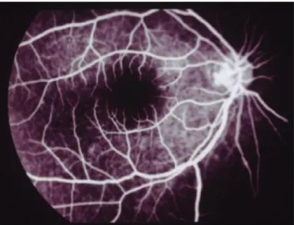
A 21-year-old man was seen in 1994 and referred for further evaluation after abrupt visual loss in the left eye. Examination at that time demonstrated visual acuity of 20/400 in the left eye and 20/20 in the right. Slit-lamp examination demonstrated clear cornea with 1+ cell and flare in the anterior chamber of the right eye. No keratic precipitates were seen, and intraocular pressure was 16 mmHg in the left eye and 14 mmHg in the right. The left eye showed intra-retinal hemorrhages in the posterior pole. Fluorescein angiography demonstrated a combination of blocked fluorescence and retinal capillary non-perfusion in areas of hemorrhage (Figure 1). Fluores-cein angiography of the right eye was normal (Figure 2).
Laboratory evaluation included a normal leukocyte count and hematocrit, Wintrobe’s sedimentation rate of 41, a non-reactive rapid plasma reagin test and fluorescent antibody titer, and normal SMA 6, chest X-ray, liver function tests, lumbar puncture, and elec-trocardiography. The patient had negative IgG and IgM titers for toxoplasmosis. Fluorescent antinuclear antibody and VDRL test were negative for syphilis. Total T and B cell counts and helper/suppressor cell ratio were within normal limits. Serum antibodies to herpes simplex virus, cytomegalovirus and Epstein-Barr virus were not found. Results of serum immunoelectrophoresis were normal, and skin biopsy after histamine administration was negative.
AMARO MH, E T A L.
3 6 9
Arq Bras Oftalmol. 2011;74( 5): 368-70 We decided to treat areas of retinal ischemia with
photocoagula-tion and to maintain therapy with oral steroids. After 1 month, the final visual acuity in the left eye was 20/25. After 1 month of treat-ment, a new subtle visual loss occurred in the right eye and visual acuity was 20/400. Slit-lamp examination revealed the same cli-nical findings as in the right eye: clear cornea, 1+ cell and flare in the anterior chamber, no keratic precipitates, and intraocular pres-sure of 15 mmHg in the right eye and 15 mmHg in the left. Intraretinal hemorrhages were seen in the posterior pole. Fluorescein angiogra-phy demonstrated a combination of blocked fluorescence and retinal capillary non-perfusion in areas of hemorrhages (Figure 3).
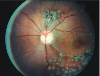
We decide to treat the patient with oral prednisone (40 mg/day) and areas of retinal ischemia with photocoagulation, resulting in final visual acuity of 20/25 after 1 month. Figures 4, 5 and 6 show the scars of retinal photocoagulation.
DISCUSSION
We present an unusual case of the most long-term follow-up of 16 years of a 21-year-old man with acute multifocal hemorrhagic
retinal vasculitis, after treatment with photocoagulation of retinal ischemia and oral steroids.
In our patient, oral prednisone and retinal photocoagulation were effective, but acyclovir was ineffective. This represents the best model to treat the initial form of this disease. Final visual acuity improved in both eyes to 20/25, and our results agree with those obtained in 50% of the cases in the first description of this disease(1).
The clinical features of this disease resemble the ocular findings of several other diseases, including Behcet’s disease, Eales’ disease, sarcoidosis, syphilis, toxoplasmosis, and herpes simplex and other viral diseases. However, laboratory results for this patient were ne-gative at initial presentation and remained so at follow-up, indica-ting that he was otherwise healthy and had no other ocular or systemic involvement.
If our patient had Behçet’s disease, his clinical and systemic fin-dings failed to satisfy the diagnostic criteria. The cause of Behçet’s disease is unknown. Numerous causes have been proposed, inclu-ding infection, toxins, and genetic factors. No virus has been isolated to date. An autoimmune and genetic cause has gained acceptance. Figure 1. Fluorescein angiography demonstrated a combination of blocked fluorescence
and retinal capillary non-perfusion in areas of hemorrhage due multiples hemorrhagic vaso-occlusions.
Figure 2. Fluorescein angiography of the right eye was normal.
Figure 3. Right eye 30 days after, the fluorescein angiography demonstrated a combi-nation of blocked fluorescence and retinal capillary non-perfusion in areas of hemorrha-ges due multiple hemorrhagic retinal vaso-occlusions.
LO N G T E R M F O L L O W-U P O F A C U T E M U L T I F O C A L H E M O R R H A G I C R E T I N A L V A S C U L I T I S (BL U M E N K R A N Z S Y N D R O M E): C A S E R E P O R T
3 7 0 Arq Bras Oftalmol. 2011;74( 5): 368-70
Figure 5. Multiples laser scars 16 years later in the left eye.
Figure 6. Multiples laser scars 16 years later in the right eye.
There is no specific diagnostic test for Behçet’s disease, and its diagnosis is based on features that are present during history taking and examination. Five different sets of criteria for the diagnosis of
Behçet’s disease have been used, and this lack of agreement has hindered the interpretation of different studies. A new internatio-nal study group(14) has agreed upon diagnostic criteria for Behçet’s
disease. These criteria require the presence of oral ulceration plus any two of the following: genital ulceration, typical eye and skin lesions, a positive skin test for pathergy. None of these was present in our patient. Moreover, if this was a viral disease, the failure of acyclovir was contradictory.
Blumenkranz syndrome appears to be a specific disease with unknown etiology that responds well to photocoagulation of retinal ischemia and oral steroids.
The present case with long-term follow-up of Blumenkranz syndrome has helped us to establish until now the best model for initial treatment and supports the conclusions of those who first described this illness.
ACKNOWLEDGEMENTS
Lawrence Yannuzzi, MD saw the complete pictures of this case and Mark Blumenkranz, MD saw the initial pictures sent by letter in 1994 and the final pictures sent by e-mail in the 2010.
REFERENCES
1. Blumenkranz MS, Kaplan HJ, Clarkson JG, Culbertson WW, Williams GA, Kleiner RC, Meissner RH. Acute multifocal hemorrhagic retinal vasculitis. Ophthalmology. 1988; 95(12):1663-72.
2. Colvard DM, Robertson DM, O’Duffy JD. The ocular manifestations of Behçet’s disease. Arch Ophthalmol. 1977; 95(10):1813-7.
3. James DG, Spiteri MA. Behçet’s disease. Ophthalmology. 1982;89(11):1279-84. 4. Lobes LA Jr, Folk JC. Syphilitic phlebitis simulating branch vein occlusion. Ann
Ophthal-mol. 1981;13(7):825-7.
5. Gaynon MW, Boldrey EE, Strahlman ER, Fine SL. Retinal neovascularization and ocular toxoplasmosis. Am J Ophthalmol. 1984;98(5):585-9.
6. Sptiznas M, Meyer-Schwickerath G, Stephan B.The clinical picture of Eales’ disease. Albrecht Von Graefes Arch Clin Exp Ophthalmol.1975;194(2):73-85.
7. Chumbley LC, Kearns TP. Retinopathy of sarcoidosis. Am J Ophthalmol. 1972;73(1):123-31. 8. Sanders MD, Shilling JS. Retinal, choroidal, and optic disc involvement in sarcoidosis.
Trans Ophthalmol Soc UK. 1976;96(1):140-4.
9. Willerson D Jr, Aaberg TM, Reeser FH. Necrotizing vaso-occlusive retinitis. Am J Ophthal-mol. 1977;84(2):209-19.
10. Grutzmacher RD, Henderson D, McDonald PJ, Coster DJ. Herpes simplex chorioretinitis in a healthy adult. Am J Ophthalmol. 1983;96(6):788-96.
11. Aaberg TM, Cesarz TJ, Rytel MW. Correlation of virology and clinical course of cytomega-lovirus retinitis. Am J Ophthalmol. 1972;74(3):407-15.
12. Wong KW, D’Amico DJ, Hedges TR 3rd, Soong HK, Schooley RT, Kenyon KR. Ocular invol-vement associated with chronic Epstein-Barr virus disease. Arch Ophthalmol. 1987;105(6): 788-92.
13. Amaro MH, Barja JL. Síndrome de vasculite retiniana hemorrágica multifocal aguda; relato de um caso. Rev Bras Oftalmol. 1994;53(3):65-8.