405
DOI: 10.1590/0004-282X20160041
ARTICLE
3-D simulation of posterior fossa reduction
in Chiari I
Simulação em 3D da redução da fossa posterior no Chiari do tipo I
Yvens Barbosa Fernandes1,3, Pedro Fábio Mendonça Perestrelo2, Pedro Yoshito Noritomi2, Roger Neves
Mathias3, Jorge Vicente Lopes da Silva2, Andrei Fernandes Joaquim1,3
Chiari I (CI) is one of the most common congenital craniocervical disorders and its diagnosed when there is tonsillar herniation into the foramen magnum of up to 3–5 mm1,2. The impaction of the tonsils at the level of the
foramen magnum can lead to interruption of the cerebro-spinal fluid flow and clinical symptoms, with upper cer-vical spine compression2,3,4. The incidence of CI in a
gen-eral population is estimate in about 0.1–0.5%, even though most of them are asymptomatic5,6.
One of the potential reasons for CI is decreased poste-rior fossa, secondary to shortness of the clivus and platy-basia, commonly found in patients with this anomaly3,4,7.
However, the role of loss of posterior fossa volume (PFV)
secondary to clivus shortness and platybasia were not clearly measured yet. By this reason, in the current man-uscript, we described and proposed a 3D computational model to simulate changes in the PFV using different cli-vus and basal angles lengths.
METHODS
A CT scan of a normal (without tonsillar herniation) control male adult subject was used as a model. The im-ages were saved in a Digital Imaging and Communications in Medicine (DICOM) format and were treated using the
1Hospital Municipal Mário Gatti, Departamento de Neurocirurgia, Campinas SP, Brasil;
2Centro de Tecnologia de Informação Renato Archer, Three Dimensional Technologies Division, Campinas SP, Brasil; 3Universidade Estadual de Campinas, Departamento de Neurologia, Campinas SP, Brasil.
Correspondence: Andrei F. Joaquim; Departamento de Neurologia, Univesidade Estadual de Campinas; Cidade Universitária Zeferino Vaz; 13090-610 Campinas SP, Brasi; E-mail: andjoaquim@yahoo.com
Conflict of interest: There is no conlict of interest to declare.
Received 27 October 2015; Received in inal form 08 February 2016; Accepted 24 February 2016.
ABSTRACT
We proposed a 3D model to evaluate the role of platybasia and clivus length in the development of Chiari I (CI). Using a computer aided design software, two DICOM iles of a normal CT scan and MR were used to simulate different clivus lengths (CL) and also different basal angles (BA). The inal posterior fossa volume (PFV) was obtained for each variation and the percentage of the volumetric change was acquired with the same method. The initial normal values of CL and BA were 35.65 mm and 112.66º respectively, with a total PFV of 209 ml. Ranging the CL from 34.65 to 29.65 – 24.65 – 19.65, there was a PFV decrease of 0.47% – 1.12% – 1.69%, respectively. Ranging the BA from 122.66º to 127.66º – 142.66º, the PFV decreased 0.69% – 3.23%, respectively. Our model highlights the importance of the basal angle and clivus length to the development of CI.
Keywords: Chiari I; clivus length; platybasia; tonsillar herniation; 3D computational model.
RESUMO
No presente estudo, propusemos a criação de um modelo computacional em 3D com elaboração de software onde dois arquivos em formato DICOM com uma TC e RNM de crânio foram usados para simular diferentes mensurações na extensão do clivus (EC) e no ângulo basal (AB). O volume inal da fossa posterior (VFP) foi obtido em cada variação, bem como a percentagem de volume alterada. O tamanho inicial da EC era de 35,65 mm e o do AB era de 112.66º, com um VFP de 209 ml. Variando a EC de 34,65 para 29,65 – 24.65 e 19.65, houve uma diminuição do VFP de 0.47%, 1.12% e 1.69%, respectivamente. Variando o AB de 122,66º para 127,66º e 142,66º, o VFP diminui para 0.69% e 3.23%, respectivamente. Nosso modelo enfatiza a importância da patogênese do aumento do AB e do encurtamento do clivus no desenvolvimento do Chiari I.
406 Arq Neuropsiquiatr 2016;74(5):405-408
software InVesalius®, to remove imperfections and unnec-essary tissues for volume evaluation. After that, the files were exported to the software Rhinoceros®, a computer aided design (CAD) program used to formulate the com-putational model. We could reproduce, in a virtual envi-ronment, the changes necessary for estimating the PFV. Two basic measurements were used as variables:
1) Basal angle – formed by a line extending from the na-sion to the posterior tubercullum sellae and then another line from the posterior tubercullum sellae to the basion and 2) Clivus length – calculated from the distance in millime-ters (mm) from the posterior tubercullum sellae to the basion.
Finally, successive measurements of the PFV were esti-mated based on changing these two variables.
RESULTS
The initial basal angle measured was 112.66º and the clivus length was 34.65 mm, with an initial PFV of 209 ml. In Table, we reported the PFV calculated in three differ-ent situations:
• decreasing the clivus length (5 mm progressively), • increasing the basal angle (5o progressively) and,
• a combination of decreasing clivus lengths and con-comitant increasing the basal angles.
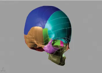
Figures 1, 2, 3 and 4 illustrate our computational model for calculating posterior fossa volume.
DISCUSSION
he real cause of CI remains elusive despite more than 100 years since its initial description by Hans Chiari8,9. Recently, we
proposed a new hypothesis to explain the origin of CI based on the evolution of the skull base of the Homo clade in the Pleistocene Epoch (beginning about 2,6 million years ago)7.
It is well known by anthropological data that the basal angle has been constantly decreasing during hominins evolution to give extra volume to the remarkable brain size increase (at least threefold in the last 4 million years)7. In the most
primi-tive hominins, such as in the Homo erectus, the basal angle was estimated to be much higher than, for instance, in Homo sapiens, suggesting its importance to the modern shape of the human skull base7,10,11.
In patients with CI a higher incidence of platybasia and shortness of clivus has been described by some authors2,12,13.
Karagöz et al.13 performed a craniometrical study in 22
pa-tients with CI, comparing them with 21 normal subjects as a control group. hey reported that, in CI group, the depth of the posterior fossa and the length of the clivus were shorter than in normal subjects (p < 0.001 and p = 0.007, respectively). he ratios of the depth of the posterior fossa to the height of the su-pratentorial region were also signiicantly smaller in the Chiari group compared with controls (p < 0.001) and patients with CI had a tendency for platybasia. hey concluded that their ind-ings strongly supported the existence of underdevelopment of the posterior fossa and the occipital bone in patients with CI.
Table. Posterior fossa volume according to changing in the clivus length and the basal angle. A total of 19 combinations were obtained.
Parameters Volume (cm3) Volume (%)
Original Craniometrical values 122.66o e 34.65 mm 209.650 100
Clivus Shortness
34.65 mm to 19.65 mm 206.118 98.315
34.65 mm to 24.65 mm 207.308 98.883
34.65 mm to 29.65 mm 208.658 99.527
Increasing basal angle
122.66o to 127.66o 208.204 99.310
122.66o to 132.66o 207.074 98.771
122.66o to 137.66o 205.168 97.862
122.66o to 142.66o 202.884 96.773
Shortness of clivus and Increasing the basal angle
34.65 mm to 19.65 mm 122.66o to 127.66o 205.056 97.809
34.65 mm to 19.65 mm 122.66o to 132.66o 203.206 96.926
34.65 mm to 19.65 mm 122.66o to 137.66o 202.210 96.451
34.65 mm to 19.65 mm 122.66o to 142.66o 201.164 95.952
34.65 mm to 24.65 mm 122.66o to 127.66o 205,164 97.860
34.65 mm to 24.65 mm 122.66o to 132.66o 204.298 97.447
34.65 mm to 24.65 mm 122.66o to 137.66o 203.190 96.919
34.65 mm to 24.65 mm 122.66o to 142.66o 202.232 96.462
34.65 mm to 29.65 mm 122.66o to 127.66o 206.364 98.433
34.65 mm to 29.65 mm 122.66o to 132.66o 204.608 97.595
34.65 mm to 29.65 mm 122.66o to 137.66o 202.958 96.808
407
YB Fernandes et al. 3D simulation in Chiari I
Similar results were reported by Heiss et al., who compared the craniometrical indings of 48 CI patients with 18 healthy volunteers as a control group12. Patients with C I had a
small-er clivus length (38.6 ± 3.4 mm vsmall-ersus 43.2 ± 3.5 mm in the control group; p < 0.001) and also a smaller basiocciput (19.7 ± 3.3 versus 26.3 ± 4.4 mm in the control group; p < 0.0001). Considering this evidences, we proposed a 3D computational model to measure the loss of volume secondary to a progres-sive increase in the basal angle and also shortening the clivus to estimate the changes in the inal PFV.
Our obtained data suggested that the PFV is afected by changing both variables leading to tonsillar herniation and clinical symptoms. Curiously, in our computational model, we demonstrated that the herniated estimated tonsillar vol-ume was quite similar to the amount of decreased PFV.
Limitations
Our study is limited by a single model of a normal sub-ject and also for not considering all complex geometry of
Figure 1. The virtual model created based on the normal subject model and his DICOM images of the CT scan .
Figure 3. Computational model with the posterior fossa volume (red) and the supratentorial content (green).
Figure 2. The virtual model created for measurement of the posterior fossa volume based on the normal subject model and his DICOM images of a CT scan.
Figure 4. Computational rendering of the posterior fossa.
the posterior fossa, since just two parameters were changed. However, our preliminary computational model was success-fully designed for initial volumetric evaluation of the poste-rior fossa, potentially opening perspectives for further re-inements and future studies, such as a genetic link between ancient skulls and the incidence of tonsillar herniation sec-ondary to clivus shortness and platybasia.
In conclusion we proposed a 3D computational model that was able to predict the inal PFV after changing the bas-al angle and clivus length. Our model seems to highlight the importance of the basal angle and clivus length to the devel-opment of the CI. Tonsillar herniation may be explained by slight changes on both variables. Further studies addressing the role of clivus and basal angle might be warranted to bet-ter understand the inluence of anthropological evolution and anatomical changes involved in the etiology of CI.
408 Arq Neuropsiquiatr 2016;74(5):405-408
References
1. Guo F, Wang M, Long J, Wang H, Sun H, Yang B et al. Surgical management of Chiari malformation: analysis of 128 cases. Pediatr Neurosurg. 2007;43(5):375-81. doi:10.1097/00013414-200306000-00005
2. Joaquim AF, FernandesYB, MathiasRN, Batista UC, Ghizoni E, Tedeschi H et al. Incidence of Basilar invagination in patients with Tonsillar herniation: a case control craniometrical study. Arq Neuropsiquiatr. 2014;72(9):706-11. doi:10.1590/0004-282X20140113
3. Joaquim AF, Ghizoni E, Giacomini LA, Tedeschi H, Patel AA. Basilar invagination: Surgical results. J Craniovertebr Junction Spine. 2014;5(2):78-84. doi:10.4103/0974-8237.139202
4. Goel A. Basilar invagination, Chiari malformation, syringomyelia: a review. Neurol India. 2009;57(3):235-46. doi:10.4103/0028-3886.53260
5. Speer MC, Enterline DS, Mehltretter L, Hammock P, Joseph J, Dickerson M et al. Chiari type I malformation with or without syringomyelia: prevalence and genetics. J Genet Couns. 2003;12(4):297-311. doi:10.1023/A:1023948921381
6. Meadows J, Kraut M, Guarnieri M, Haroun RI, Carson BS. Asymptomatic Chiari Type I malformations identiied on magnetic resonance imaging. J Neurosurg. 2000;92(6):920-6. doi:10.3171/jns.2000.92.6.0920
7. Fernandes YB, Ramina R, Campos-Herrera CR, Borges G. Evolutionary hypothesis for Chiari type I malformation. Med Hypotheses. 2013;81(4):715-9. doi:10.1016/j.mehy.2013.07.035
8. Chiari H. Über Veränderungen des Kleinhirns infolge von Hydrocephalie des Grosshirns. Dtsch med Wschr. 1891;17:1172-75.
9. Chiari H. Über Veränderungen des Kleinhirns, des Pons und der Medulla Oblongata in Folge von congenitaler Hydrocephalie des Grosshirns. Dtsch Akd Wissensch. 1895;63:71-125.
10. Lieberman DE, Ross CF, Ravosa MJ. The primate cranial base: ontogeny, function and integration. Am J Phys Anthropol Suppl 2000;31:117-69.
11. Lieberman DE. Sphenoid shortening and the evolution of modern human cranial shape. Nature. 1998; 393:158-62. doi:10.1038/30227
12. Heiss JD, Suffredini G, Bakhtian KD, Sarntinoranont M, Oldield EH. Normalization of hindbrain morphology after decompression of Chiari malformation Type I. J Neurosurg. 2012;117(5):942-6. doi:10.3171/2012.8.JNS111476