https://doi.org/10.1590/0004-282X20170071
ARTICLE
Cranio-vertebral transition assessment by
magnetic resonance imaging in a sample of a
northeast Brazilian population
Avaliação da transição crânio-vertebral por ressonância magnética em uma amostra da
população do nordeste brasileiro
Heitor Cabral Frade1, Caio César Nuto Leite França1, José Jailson Costa do Nascimento1,
Maurus Marques de Almeida Holanda2, Eulâmpio José da Silva Neto3, Severino Aires Araújo Neto2
he cranio-vertebral transition (CVT) is composed by the base of the occipital bone and the irst two cervical vertebrae, atlas and axis (C1 and C2, respectively). his region is suscep-tible to conformational abnormalities, such as platybasia – lattening of skull base – and basilar invagination – protru-sion of the tooth of the C2 to the posterior fossa. hey are mutually associated, and both may also relate to cerebellar tonsil herniation. Clinical repercussions are varied and derive
from compression of nervous structures or obstruction of cerebrospinal luid circulation, with consequent hydrocepha-lus and syringomyelia1.
Imaging exams are indispensable in the assessment of CVT, and the gold-standard is magnetic resonance imaging (MRI), especially because it can demonstrate both musculoskeletal and nervous elements involved. Craniometric imaging param-eters were irst established last century, notably from German
1Universidade Federal da Paraíba, João Pessoa PB, Brasil;
2Universidade Federal da Paraíba, Departamento de Medicina Interna, João Pessoa PB, Brasil;
3Universidade Federal da Paraíba, Departamento de Morfologia, João Pessoa PB, Brasil;
Correspondence: Severino Aires Araújo Neto; Departamento de Medicina Interna da UFPB; Cidade Universitária, s/n; 58051-900 João Pessoa PB, Brasil; E-mail: [email protected]
Conlict of interest: There is no conlict of interest to declare.
Received 26 July 2016; Received in inal form 28 February 2017; Accepted 14 March 2017.
ABSTRACT
Platybasia and basilar invagination are important alterations of the cranial-vertebral transition. Neuroimaging-based platybasia parameters include the Welcker basal angle, distance between the apex of the odontoid and Chamberlain’s line, and the clivus-canal angle. This study aimed to measure and correlate these parameters in a sample from northeast Brazil. Methods: Cross-sectional analysis of craniometric parameters from individuals submitted to magnetic resonance at an outpatient imaging center between 2011 and 2012. Results: Of 181 analyzed cases, the Welcker basal angle averaged 128.96º (SD 6.51), median distance between apex of the odontoid and Chamberlain’s line was 2.27 mm (IQR -1.23–4.47) and the median clivus-canal angle was 150.5º (IQR 143.2–157.3). The Welcker basal angle was inversely correlated to the clivus-canal angle, and correlated to the distance between the apex of the odontoid and Chamberlain’s line. Conclusion: There was a tendency to platibasia, basilar invagination and narrowing of the cranio-vertebral transition.
Keywords: platybasia; magnetic resonance imaging; cephalometry
RESUMO
Platibasia e invaginação basilar são importantes alterações da transição craniovertebral. Existem parâmetros importantes obtidos pela neuroimagem, como o ângulo basal de Welcker, distância do ápice do odontoide à linha de Chamberlain e o ângulo clivo-canal. Este estudo procurou medir e correlacioná-los em uma amostra do Nordeste Brasileiro. Métodos: Estudo transversal com medidas de indivíduos submetidos a ressonância magnética craniana em um centro de diagnóstico por imagem entre 2011 e 2012. Resultados: Dos 181 casos analisados, o ângulo basal de Welcker teve média 128.96º (DP 6.51), a distância do ápice do odontoide à linha de Chamberlain obteve mediana 2.27 mm (IIQ -1.23–4.47) e o ângulo clivo-canal mediano foi 150.5º (IIQ 143.2–157.3). O ângulo basal de Welcker foi inversamente correlacionado com o ângulo clivo-canal e diretamente correlacionado com a distância do ápice do odontoide à linha de Chamberlain.
Conclusão: Houve uma tendência a platibasia, invaginação basilar e estreitamento da transição craniovertebral, que poderiam ser
inluenciados pela natureza multirracial e por fatores antropológicos da população estudada.
radiograph-based studies, but some are still used for evaluat-ing CVT by computerized tomography (CT) or MR1.
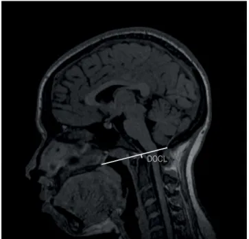
Chamberlain’s line extends from the posterior portion of the hard palate up to the posterior margin of the foramen magnum (ostium). he distance between the apex of the odontoid and the Chamberlain’s line (DOCL) is used to mea-sure the projection of the axis into the posterior fossa to diag-nose basilar invagination.
he Welcker basal angle (WBA) and the clivus-canal angle (CCA) are also largely used for evaluation of platybasia1.he
WBA measures angulation of the skull base and is obtained by two lines: one traced from the nasion up to the tubercle of sella turcica and another line from the latter to the anterior margin of the foramen magnum (basion). Values above a normal limit of 140º have been considered diagnostic of platybasia1,2. he CCA is
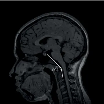
measured between the line that follows parallel to the clivus and another that borders the posterior surface of the odontoid pro-cess2. Values lower than 150º are considered abnormal1. A
nar-rower angle increases the risk nervous compression.
For several decades, researchers have reported a high prev-alence of platybasia and basilar invagination in the north-eastern Brazil population when compared with other regions within and beyond Brazilian borders3,4,5. Studies conducted
in northeast Brazil by Silva et al.3,4, and Alves et al.6
, have also found an association with brachycephaly3,4,6,7 – a condition in
which the lateral diameter of the skull is disproportionately large. In a corpse-based study, also conducted in northeast-ern Brazil, there was a surprisingly high rate of brachycephaly (77.6%), higher than that reported in other populations asso-ciated with brachycephaly8
. his condition is also popularly referred to as “cabeça-chata” (lat head), a term often used in a
pejorative manner to deine the northeastern population9 . Brachycephaly and its association with platybasia may not be pathological, but rather a phenotypic tendency of a population, apparently related to ethnic-anthropological fac-tors in northeast Brazil10. hus, diagnosing platybasia and
basilar invagination with craniometric parameters derived from overseas samples may not be appropriate. However, there have not previously been imaging studies assessing CVT parameters in the northeastern population.
he authors aimed to assess craniometry in a sample from the northeastern Brazil population via MR imaging. Speciic objectives were: 1) outline a proile of the sample for the DOCL, WBA and CCA measures; 2) verify if there were correlations between these three parameters.
METHODS
Data was obtained from head MRIexaminations, conducted at an imaging service at outpatient level in “Sertão” (drylands) of the state of Paraíba (NE). he MR imaging was carried out by spontaneous demand on that service between January 2011 and December 2012. All patients aged 18 years or above were
included. Exclusion criteria were: manifested clinical suspicion of CVT disease by the referring physician, surgery to the base of the skull or important previous cranio-vertebral trauma, techni-cal diiculties in the identiication of the parameter structures, resulting from positioning errors, misangulation and imaging artifacts, such as those generated by metallic material or patient movement. If a patient had more than one examination during the period of the study, only the irst one was included.
All examinations were carried out in an open device, of 0.35 T (Magnetom C!, Siemens Medical Solutions, Erlangen, Germany, 2011). he parameters were outlined in the mid sagittal plane of T1 3D sequence (magnetization-prepared rapid gradient-echo or MP-RAGE) acquired at the sagital plane, with thickness varying from 0.9 to 1.1 mm. Images were analyzed in digital DICOM format, by means of an open version of the visualization software Osirix®
in its free version 3.9.2 (Mac-Apple®
platform).
he DOCL, CCA and WBA were measured according to the techniques described by Smoker1 and Amaral et al.2
(Figures 1, 2 and 3).
he data was analyzed by means of the statistical soft-ware R3.2.0. he Shapiro-Wilk test was used to assess nor-mality. he comparison between genders was made using the unpaired Wilcoxon’s test. Spearman’s correlation was used to verify the relation between the variables, with the p-value corrected by Holm’s method. he value of p < 0.05 was con-sidered as statistically signiicant.
he study was substantiated on secondary data (tests), guaranteeing privacy and conidentiality of the patients, ensuring the non-use of the information to the detri-ment of the people, according to Resolution 466/12 of the National Council of Health. he project was approved by
DOCL
the Ethics in Research Committee of the institution (CAAE: 30255914.1.0000.5188), with substantiated exemption of the consent term. he use of the images was authorized by the direction of the service. here was no interference in the pro-tocol of the examinations or any clinical or surgical interven-tion in funcinterven-tion solely for this research.
RESULTS
A total of 181 head MRIs were studied. Eighty participants (44.2%) were men and 101 (55.8%) were women. he median age was 46 years (IQR = 32–63). he craniometric variables are described in Table.
here was no signiicant diference in craniometric parameters for gender and age.
he WBA showed a parametric distribution, with an aver-age of 128.96º (conidence interval of 95%, 128.01–129.91).
here was no signiicant correlation between the CCA and DOCL (p = -0.215; p = 0.26), but there was a positive
correla-tion between the WBA and the DOCL (p = 0.366; p < 0.0001),
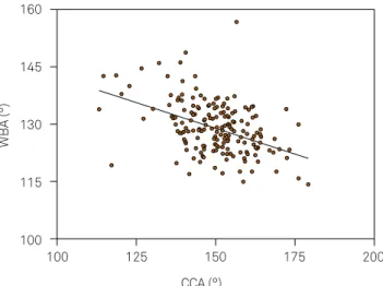
as well as an inverse correlation between the WBA and the CCA (p = -0.415; p < 0.0001) (Figures 4, 5 and 6).
DISCUSSION
he use of lines, distances and angles that relate to ana-tomical landmarks of a CVT helps in the diagnosis of altera-tions such as platybasia and basilar invagination2,11. Despite
most of these measures having been designed when radio-graphs were the only imaging method available1,12,13,14, they
are currently adopted for CT and MR imaging analysis. here has been considerable variation in craniomet-ric parameters across studies. he WBA has varied broadly, with one reported average of 115.41° (±8.45°) for Netto et al.7.
CCA
Figure 2. Clivus-canal angle (CCA).
WBA
Figure 3. Welcker basal angle (WBA).
DOCL (mm)
CCA (°)
200
175
150
125
100
-12 -4 4 12 20
Figure 4. Relationship between clivus-canal angle (CCA) and distance between odontoid apex and Chamberlain’s line (DOCL).
Table. Craniometric parameters.
Craniometry
Percentiles Mean
(SD)
2.50 25 50 75 97.50
WBA (°) 117.1 124.3 128.5 133.4 142.65 128.96
(6.51)
DOCL (mm) -7.55 1.23 2.27 4.47 15.5 2.25
(5.06)
CCA (°) 121.7 143.2 150.5 157.3 172.5 150.14
Other authors have reported relatively large amplitudes: 103.5° to 131.5° for Boogard15; 115° a 140° for List16; 115° to
150° for Walsh et al.17; 120° to 147° for McGreger14; 121.5° to
148.5° for Brailsford13; 115º to 140º Royo-Salvador18; and 97º
to 133.2º for Batista et al.19
here is still no consensus over the upper limit for DOCL values, which range from 2.0 to 6.6 mm1,20,21. his diference is
not insigniicant when compared to the small dimensions of CVT and hence could lead to considerable changes in speci-icity in the diagnosis of basilar invagination, as well as in the estimated prevalence11,22.
Taking into consideration the DOCL upper limit of 2 mm20, more than half of the participants in this study
would be diagnosed with basilar invagination, which would be unlikely for individuals who come to imaging services by spontaneous demand.
According to Smoker, a CCA value lower than 150º is con-sidered abnormal1. Botelho and Ferreira11 analyzed the CCA via
MRI, obtaining an average of 148º ± 9.8º (with a range from 129º to 179º). Batista et al.19, analyzing with the use of CT, obtained
an average of 153.6º ± 7.6º (varying from 132.3º to 173.9º). Our study showed a CCA with an average of 150.14º ± 11.37º – very close to the values cited by Botelho and Ferreira11 and
Batista et al.19, which is apparently higher than that suggested
by Smoker1. Despite Smoker1 having demonstrated
signii-cant correlation between the CCA and DOCL, none was found in this study. hus, it can be speculated that the values of the CCA obtained by MRI or CT of asymptomatic patients seem to be higher than the ones published using radiographs, from which normality standards were deined1,19; or,
alterna-tively, it is possible that each population naturally presents with diferent values.
Northeastern Brazilian population tendency toward brachycephaly as well as other CVT peculiarities have been addressed in previous clinical4,5,23, and anthropological
studies9
. A strong relationship between brachycephalia and
alterations of the CVT in patients submitted to surgeries in this region was also found by other researchers 4,5.
Nonetheless, some of the variance between samples may be also attributed to diferent imaging methods used in each study, as well as to a lack of validation and standardization of the old craniometric techniques as they are adapted to new imaging resources, such as CT and MRI.
In this study, an inverse correlation between the WBA and CCA suggests that lattening of the skull base may accentuate thebulbo-pontine transition in its passage in the CVT. A simi-lar correlation was referred to by Joaquim et al.22, who
demon-strated that the presence of platybasia and a decreased clivus seem to favor a protrusion of the cervical spine to the inside of the skull base (basilar invagination)24. Previous studies have
already hypothesized that the presence of platybasia may be associated with compression of posterior fossa structures by mechanical opposition to the apex of the odontoid process1,4,5.
CVT imaging analysis is still poorly-described in the north-eastern population, especially those from the countryside. Despite all participants included in this studywere chosen from a single reference imaging center in the countryside of the state of Paraíba, their place of birth was not veriied.hus, it is not possible to know how much of the sample was indeed born in that region and how accurately this sample represents Paraíba or Brazilian Northeast populations as a whole. herefore, this study could provide further evidence to help deine northeast-ern craniometric standards and its peculiarities.
In conclusion, the inverse relation of the WBA and the CCA, demonstrated here, suggests that platybasia, despite not being directly involved in nervous compressions, may be a factor associated with this, as it can contribute to the nar-rowing of the passage of the neuroaxis in the CVT. he cor-relation found between the WBA and DOCL demonstrates the relationship between platibasya and basilar invagination, already referred to in literature, and reinforces the clinical importance of platybasia.
DOCL (mm)
WBA (°)
160
145
130
115
100
-12 -4 4 12 20
Figure 5. Relationship between Welcker basal angle (WBA) and distance between odontoid apex and Chamberlain’s line (DOCL).
CCA (°)
WBA (°)
160
145
130
115
100
100 125 150 175 200
Descriptive results of all three parameters also suggest that their normal ranges may be diferent from those previ-ously shown in international studies, which demands further
research involving larger sampling of normal individuals, not only to conirm a diference, but also to propose new appro-priate limits for people from Northeast region.
References
1. Smoker WR. Craniovertebral junction: normal anatomy, craniometry, and congenital anomalies. Radiographics. 1994;14(2):255-77. https://doi.org/10.1148/radiographics.14.2.8190952
2. Amaral DT, Amaral LL, Filho GH, Puertas E. Craniovertebral junction: normal anatomy and craniometry. Coluna/Columna. 2004;3(2):100-3.
3. Silva AB, Moraes AA, Bessa IC, Sesana WE. [Cerebello-pontine angle syndrome associated with cranio-vertebral malformation: report of two cases]. Arq Neuropsiquiatr. 1972 ;30(1):84-7. Portuguese. https://doi.org/10.1590/S0004-282X1972000100009
4. Silva JA, Brito JC, Nóbrega PV, Costa MD, Souza AB. Achados cirúrgicos em 260 casos de impressão basilar. Arq Neuropsiquiatr. 1994;52(3):363-9.
5. Silva JA, Holanda MM. Basilar impression, Chiari malformation and syringomyelia: a retrospective study of 53 surgically treated patients. Arq Neuropsiquiatr. 2003;61(2B):368-75. https://doi.org/10.1590/S0004-282X2003000300009
6. Alves HA, Santos MI, Melo FC, Ribeiro W. Comparative study of the cephalic index of the population from the regions of north and south of Brazil. Int J Morphol. 2011;29(4):1370-4. https://doi.org/10.4067/S0717-95022011000400050
7. Netto DS, Nascimento SR, Ruiz CR. Metric analysis of basal sphenoid angle in adult human skulls. Einstein (Sao Paulo). 2014;12(3):314-7. https://doi.org/10.1590/S1679-45082014AO2933
8. Nascimento JJ, Macêdo AE, Ribeiro EC, Souza NA, Silva Neto E. Estimativa do sexo e mensuração de índices cranianos do acervo pertencente ao departamento de morfologia da UFPB. In: Anais do 26º Congresso Brasileiro de Anatomia, 2º Encontro de Ligas Estudantis de Morfologia; 29 set-2 out 2014; Curitia, PR. São Paulo: Sociedade Brasileira de Anatomia; 2014. p. 781.
9. Queiroz AC. Politicamente correto e direitos humanos. Brasília: SEDH; 2004.
10. Carvalho OA. Análise das anormalidades de desenvolvimento na população pré-história do Sítio Furna do Estrago, Pernambuco. Rio de Janeiro: Escola Nacional de Saúde Pública 1995.
11. Botelho RV, Ferreira ED. Angular craniometry in craniocervical junction malformation. Neurosurg Rev. 2013;36(4):603-10. https://doi.org/10.1007/s10143-013-0471-0
12. Chamberlain WE. Basilar impression (platybasia): a bizarre developmental anomaly of the occipital bone and upper cervical spine with striking and misleading neurologic manifestations. Yale J Biol Med. 1939;11(5):487-96.
13. Brailsford JF. The radiology of bones and joints. Am J Med Sci. 1936;192(2):281.
14. McGregor M. The signiicance of certain measurements of the skull in the diagnosis of basilar impression. Br J Radiol. 1948;21(244):171-81. https://doi.org/10.1259/0007-1285-21-244-171
15. Boogard JA. Basilar impression: its causes and consequences. Nederl Tysdschr v Geneesk Tweede, Afedling. 1865;1:81-108.
16. List CF. Neurologic syndromes accompanying developmental anomalies of occipital bone, atlas and axis. Arch Neurol Psychiatry. 1941;45(4):577-616. https://doi.org/10.1001/archneurpsyc.1941.02280160009001
17. Walsh MN, Camp JD, Craig WM. Basilar invagination of the skull (so-called platybasia): report of a case with operation. Mayo Clin Proc. 1941:449-52.
18. Royo-Salvador MB. [Platybasia, basilar groove, odontoid process and kinking of the brainstem: a common etiology with idiopathic syringomyelia, scoliosis and Chiari malformations]. Rev Neurol. 1996;24(134):1241-50. Spanish.
19. Batista UC, Joaquim AF, Fernandes YB, Mathias RN, Ghizoni E, Tedeschi H. Computed tomography evaluation of the normal craniocervical junction craniometry in 100 asymptomatic patients. Neurosurg Focus. 2015;38(4):E5. https://doi.org/10.3171/2015.1.FOCUS14642
20. Goel A. Basilar invagination, Chiari malformation, syringomyelia: a review. Neurol India. 2009;57(3):235-46. https://doi.org/10.4103/0028-3886.53260
21. Smith JS, Shaffrey CI, Abel MF, Menezes AH. Basilar invagination. Neurosurgery. 2010;66(3 Suppl):39-47. https://doi.org/10.1227/01.NEU.0000365770.10690.6F
22. Joaquim AF, Fernandes YB, Mathias RN, Batista UC, Ghizoni E, Tedeschi H et al. Incidence of basilar invagination in patients with tonsillar herniation? A case control craniometrical study. Arq Neuropsiquiatr. 2014;72(9):706-11. https://doi.org/10.1590/0004-282X20140113
23. Silva JAG. Impressão Basilar. In: Silva JAG. Malformações occipitocervicais: impressão basilar, malformação de Chiari, siringomielia, platibasia. Recife: Ed Universitária; 2003. p. 169-300.