https://doi.org/10.1590/0004-282X20170060
ARTICLE
Cerebrospinal fluid drainage options
for posthemorrhagic hydrocephalus in
premature neonates
Opções de drenagem liquórica em neonatos prematuros com hidrocefalia pós hemorrágica
José Roberto Tude Melo1, Rosane Klein Passos2, Marcelo Liberato Coelho Mendes de Carvalho1
1Hospital Pediátrico Martagão Gesteira, Unidade de Neurocirurgia Pediátrica, Salvador BA, Brasil; 2Hospital Pediátrico Martagão Gesteira, Unidade de Radiologia, Salvador BA, Brasil.
Correspondence: José Roberto Tude Melo; Rua Jose Duarte, 114 / 2° andar; 40050-050 Salvador BA, Brasil; Email: [email protected]
Conflict of interest: There is no conlict of interest to declare.
Received 20 January 2016; Received in inal form 05 January 2017; Accepted 14 March 2017.
ABSTRACT
Objective: The literature describes various cerebrospinal luid (CSF) drainage techniques to alleviate posthemorrhagic hydrocephalus in preterm newborns; however, consensus has not been reached. The scope of this study was describing a case series of premature neonates with posthemorrhagic hydrocephalus and assessing the outcomes of different approaches used for CSF diversion. Methods: A consecutive review of the medical records of neonates with posthemorrhagic hydrocephalus treated with CSF drainage was conducted. Results: Forty premature neonates were included. Serial lumbar puncture, ventriculosubgaleal shunt, and ventriculoperitoneal shunt were the treatments of choice in 25%, 37.5% and 37.5% of the cases, respectively. Conclusion: Cerebrospinal luid diversion should be tailored to each case with preference given to temporary CSF drainage in neonates with lower age and lower birth-weight, while the permanent ventriculoperitoneal shunt should be considered in healthier, higher birth-weight neonates born closer to term.
Keywords: cerebral hemorrhage; hydrocephalus; cerebrospinal luid.
RESUMO
Objetivo: A literatura descreve várias opções de drenagem liquórica (DL) para alivio da hidrocefalia pós-hemorrágica (HPH) em neonatos prematuros; contudo, não existe um consenso sobre a melhor abordagem. O escopo deste estudo foi descrever uma série de casos de neonatos prematuros, portadores de HPH, veriicando os resultados de diferentes técnicas utilizadas para DL. Métodos: Revisão consecutiva dos prontuários de neonatos com diagnostico de HPH submetidos a DL. Resultados: Quarenta recém-nascidos prematuros foram incluídos. A punção lombar seriada (PL), a derivação ventriculosubgaleal (VSG) e a derivação ventrículo peritoneal (VP) foram o tratamento escolhido em 25%, 37,5% e 37,5% dos casos, respectivamente. Conclusão: As opções de DL devem ser avaliadas caso a caso, sendo dada preferência às drenagens temporária em prematuros com idade e peso mais baixos ao nascer, enquanto o shunt deinitivo (derivação VP) pode ser considerado naqueles prematuros mais saudáveis, com idade e peso superiores.
Palavras-chave: hemorragia cerebral; hidrocefalia; líquido cefalorraquidiano.
Intraventricular hemorrhage (IVH) has been a major cause of mortality among premature neonates for more than 40 years1,2 and is associated with neonatal encephalopathy, subsequent subtle apnea, and death1,2,3,4. Low birth-weight pre-mature neonates are more vulnerable to IVH and, depending on the IVH grade, to posthemorrhagic hydrocephalus (PHH). Posthemorrhagic hydrocephalus can evolve to progressive PHH, and in more severe cases, to periventricular hemor-rhagic infarct, hemorhemor-rhagic cerebral injury, and periventric-ular leukomalacia1,4,5. Between 15% to 20% of neonates born with a weight less than 1,500 g are estimated to develop IVH. Further, 75% of those with Papile grade III or IV hemorrhages develop progressive PHH and need a permanent shunt 4,6.
METHODS
Study design and inclusion criteria
his single-center study was approved by the Brazilian Research Ethics Committee (registration number 38819114.7.0000.5557). his retrospective review and obser -vational study included all premature neonates admitted with a diagnosis of PHH to the neonatal intensive care unit at a Reference Public Pediatric Hospital in Salvador da Bahia, Brazil between December 2009 and December 2014. he diagnosis of PHH identiied by a transcranial ultrasonography, and treated with a CSF drainage procedure, and a minimum follow-up of three months for the assessment of treatment outcomes.
Definitions of prematurity, IVH and PHH
Premature neonates were deined as those born before 37 weeks of gestation and as low birth weight when weight-at-birth was less than 1.500g4. In all patients, tran-scranial ultrasonography was performed by a senior radiolo-gist with more than ten years of experience using a classi-cal transfontanellar approach with a 1.9–6-MHz curvilinear transducer (Toshiba Aplio™ 100 with color Doppler).
he Papile system was used for grading IVH by transcra -nial ultrasonography. Briely, grade I was deined as hemor -rhage restricted to the ventricular subependymal matrix occupying a maximum of 10% of the ventricles. Grade II and III were deined as hemorrhage comprising 10–50% and more than 50% of the ventricular system, respectively. If the hemorrhage extended to the periventricular, i.e., parenchy-mal, regions, it was considered as a grade IV IVH14.
A diagnosis of PHH secondary to IVH was made when the anterior horns width of the lateral ventricles was ≥ 6 mm as mea -sured in the anterior coronal plane at the level of the septum pel-lucidum (median line), with the midpoint in the lateral wall of the lateral ventricle (at the level of caudate nucleus and the foramen of Monro) (Figure)4,15,16.Comorbidities considered as severe were global hypotonia associated to bradycardia, cardiorespiratory arrest, respiratory failure, cutaneous cyanosis, sepsis and other infections associated with clinical and laboratory worsening.
Surgical intervention
Conditions that were included that required a neurosur -gical evaluation were bulging fontanelles, an increase in the cranial circumference, bradycardia, and other signs of intra-cranial hypertension, associated with transintra-cranial ultraso-nography showing ventriculomegaly (anterior horns width showing progressive increase) and IVH grades III and IV.
he CSF drainage is commonly indicated in cases of progres -sive increases in cranial circumference (≥ 2 standard deviations above the patient’s age group during the irst week), bulging fonta -nelles, or changes in respiratory patterns, associated with progres-sive widening of the ventricular system detected by transcranial ultrasonography13,17,18. In cases of ventriculomegaly from ex vacuo hydrocephalus (i.e. without bulging fontanelles, increase in the
cranial circumference, bradycardia, or other signs of intracranial hypertension), a nonsurgical treatment was proposed.
he study did not assess the preferred CSF drainage method among transcutaneous transfontanellar puncture, external ventricular drainage, or ventriculostomy, as our pediatric neu-rosurgical team prefers not use them, in cases of neonates with PHH. As described in previous studies8,10,13,17,19,20,21,22,23, the follow-ing techniques were employed for CSF drainage to resolve PHH: Serial LP was performed at the L3-L4 or L4-L5 level, with the ana-tomical landmark as an imaginary line traced from the iliac crest to the lumbar column. A valveless ventriculosubgaleal shunt with subcutaneous reservoirs was placed with an incision near the external angle of the anterior fontanelle, followed by the detach-ment of subgaleal space and the introduction of a 3-cm catheter into the anterior ventricular horn; the catheter was then con-nected to the reservoir and the scalp. he VP shunt was placed as follows: an incision was made near the lambdoid suture, and a mini-laparotomy was performed for tunneling of the distal cath-eter of the VP shunt. A burr-hole was made for the osseous and dural exposition, the dura was opened, and a 5-cm catheter was introduced into the posterior ventricular horn. he proximal and distal catheters were connected and ixed in the periosteum, and the distal catheter was introduced into the peritoneal cavity under direct vision. Finally, the musculoaponeurotic, subcutane-ous, and cutaneous layers were closed. In this study, the serial LPs and ventriculosubgaleal shunts were considered to be temporary CSF drainage options, while the VP shunt was considered as the only permanent CSF drainage approach.
CORONAL ANTERIOR
VLE VD
Follow-up and complications
Patients were evaluated for the persistence or enlarge-ment of hydrocephalus, CSF leak, shunt infection, failure, or mechanical dysfunctions for at least three months after CSF diversion. Hydrocephalus was assessed by the enlarge -ment of cranial circumference or bulging fontanelles during follow-up and transcranial ultrasonography showing anterior horns width ≥ 6 mm measured in the anterior coronal plane.
Statistical analysis
Epi InfoTM version 7, a public domain statistical software for epidemiology developed by the US Centers for Disease Control and Prevention, was used for database analysis. Some results were presented as descriptive statistics. Measures of central tendency (mean, mode, and median) were calculated and pre-sented where relevant. he chi-squared test was used to com -pare ratios with a conidence interval of 95%. he diferences were considered statistically signiicant if the p-value was < 0.05.
RESULTS
Forty-nine preterm neonates with PHH were treated by CSF drainage during the study period; however, nine patients were excluded because they were lost to follow-up. hus, 40 preterm neonates were included in the inal analysis. he median gestational age and birth weight were 28 weeks (range; 24–35 weeks) and 1,105 g (range; 600–2,800 g), respec-tively. Patient characteristics are shown in Table 1.
Twenty-ive patients (62.5%) were treated with tempo -rary CSF drainage approaches: 10 patients (25%) with serial LP, and 15 cases (37.5%) with a ventriculosubgaleal shunt as the irst option to alleviate PHH. Ventriculoperitoneal shunts were used in 15 neonates (37.5%) to treat PHH as the irst option, generally with low-pressure valves.
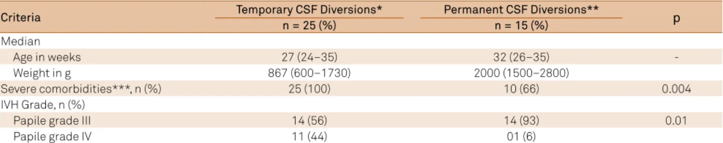
he median age and weight of patients treated with tempo -rary drainage methods were 27 weeks (range; 24–35 weeks) and 867 g (range; 600–1730g), respectively, while the median age and weight of patients treated with VP shunts were 32 weeks (range; 26–35 weeks) and 2000 g (range; 1500–2800g), respectively (Table 2). All of the patients treated with temporary CSF drainage meth -ods were considered clinically more severely afected, had previ -ous infections, or sufered from severe comorbidities. In contrast, signiicantly fewer patients treated with the VP shunt had severe comorbidities (66%; p = 0.004) (Table 2), and none in the latter group had comorbidities related to sepsis or other infections.
Regarding the initial method chosen for PHH treatment, 50% (5/10) of those undergoing serial LP and 46% (7/15) of those undergoing ventriculosubgaleal shunt required VP shunts dur-ing the course of PHH evolution (12/25, 48%) (Table 3). Durdur-ing the follow-up period of all enrolled patients, 68% (27/40) were considered as permanent ventricular drainage dependents and were using VP shunts. he incidence of progressive PHH was higher in case of IVH grade III (79% vs 42%; p = 0.02).
Among the 27 patients considered as permanent ventricular drainage dependents, 44% (12/27) had infectious complications or mechanical dysfunction (obstruction or overdrainage). Ten of these patients had received a VP shunt as the irst option for PHH treatment (Table 3). his group that received a VP shunt as the irst option for PHH treatment (n = 15) had a higher incidence of complications than those treated for progressive PHH with a VP shunt during the follow-up period (n = 12) (66% vs 16%, p = 0.01). here was a total of ive deaths (5/40; 13%). he average and median time of follow-up was 11.8 months (DP +/- 16.2 months) and three months (range; 3–60 months).
DISCUSSION
Intraventricular hemorrhage is the leading cause of hydrocephalus in premature neonates, especially those
Table 1. General characteristics of preterm neonates treated for CSF diversion due to PHH (Salvador, Brazil, 2009–2014).
General characteristics n = 40
Male sex, n (%) 22 (55)
Median gestational age in weeks 28 (24–35) Median weight at birth in g 1,105 (600–2,800) IVH grade, n (%)
Papile grade III 28 (70)
Papile grade IV 12 (30)
Complications after birth*, n (%) 35 (88)
CSF: cerebrospinal luid; IVH: Intraventricular hemorrhage; PHH: posthemorrhagic hydrocephalus. *hypotonia, bradycardia, cardiac arrest, respiratory failure, skin cyanosis, sepsis and other infections.
Table 2. Characteristics of preterm neonates according to the initial PHH treatment approaches (Salvador, Brazil, 2009–2014).
Criteria Temporary CSF Diversions*n = 25 (%) Permanent CSF Diversions**n = 15 (%) p
Median
Age in weeks 27 (24–35) 32 (26–35)
-Weight in g 867 (600–1730) 2000 (1500–2800)
Severe comorbidities***, n (%) 25 (100) 10 (66) 0.004
IVH Grade, n (%)
Papile grade III 14 (56) 14 (93) 0.01
Papile grade IV 11 (44) 01 (6)
with low birth-weight, and generally occurs within the irst four days of life11.Low gestational age and low birth-weight increase the risk for developing IVH4,6,11,17, a inding consis -tent with the median age (28 weeks) and weight (1,105g) of the patients enrolled in this study. here is a minor male predominance reported in previous studies17.
Most of the patients included in the current study were diagnosed with Papile grade III IVH. his inding is in agree -ment with previously reported risk of PHH in this patient population6,13,23. hese patients are commonly referred to pediatric neurosurgery for either temporary or permanent CSF diversion to relieve PHH-induced intracranial hyper -tension6,13,17,22,23, while grade I and II patients are usually not referred for neurosurgical evaluation due to the lower inci-dence of PHH and intracranial hypertension. he high num -ber of patients with clinical complications such as hypoto-nia, bradycardia, cardiac arrest, respiratory failure, skin cyanosis, sepsis, and other infections, highlight the suscep-tibility of these patients as well as the challenges in their management4,17, both of which are clinically considered in deciding the appropriate treatment options for CSF diver -sion in PHH cases23.
he method chosen for CSF drainage in PHH cases does not follow a speciic protocol, and varies with the neurosur -geon’s experience, the age and weight of the premature neo-nate, the presence of associated comorbidities, the char-acteristics of the ventricular system assessed by imaging studies, and the clinical presentation. In this study, none of the patients received an initial treatment with transcutane-ous transfontanellar puncture, external ventricular drain-age, or ventriculostomy. Transcutaneous transfontanellar puncture is not used as a irst choice at our hospital due to the associated risks, which include CSF leak, porencephalic cysts, and multiloculated hydrocephalus4. External ventric-ular drainage is not used for temporary relief of hydroceph-alus as a irst option in premature neonates due to the risks associated with infection and accidental removal of the external ventricular drainage, which have been observed in other reference centers as well4,6,10,13,17,19,22,23. While still con-sidered as a controversial method in neonates with IVH, several groups have recommended the irrigation of ventri-cles for the removal of blood clots, the coagulation of cho-roid plexus, and the opening of the third ventricle loor by
ventricular endoscopy, with the aim of reducing progressive PHH rates or, at a minimum, of delaying the use of perma-nent shunts in select cases20,21. At our hospital, we do not have experience in the use of ventriculostomy in premature neonates; rather, this technique is reserved for use in chil-dren over two years of age who are diagnosed with obstruc-tive and noncommunicating hydrocephalus, or congenital cerebral cysts (arachnoid cysts).
In this study, we found a tendency to use temporary drainage methods in infants with lower gestational age and birth weight, while the VP shunt was reserved for older, heavier and healthier children, a inding that corroborates previously published data regarding CSF diversion options23. he serial LP was chosen in low birth-weight premature neo -nates with more severe and clinically unstable comorbidi-ties, as it is the fastest method that does not require general anesthesia or transportation to the operating room; the pro-cedure can be performed in the neonatal intensive care unit. In cases of more stable clinical conditions, the ventriculosub-galeal shunt as an alternative temporary drainage approach is considered, especially in infants with grade III IVH. As seen in Table 2, in addition to age and weight, additional factors including associated morbidities and the IVH grade, aid the neurosurgeon in deciding between a temporary and a perma-nent CSF drainage.
Ventriculoperitoneal shunt was chosen as the irst option for relief in 37.5% of patients, similar to that found in pre-vious studies (34%)17. In several reference centers, the VP shunt is often chosen as a irst-line treatment modality for IVH-associated PHH (range; 53–72%)13,23.his divergence in published studies on the subject shows the lack of a stan-dardized protocol across diferent institutions. Our indings corroborate earlier studies suggesting that the VP shunt should be chosen as the initial therapy in select cases, spe-ciically in older preterm neonates who are heavier, healthier, and are without infections4,17,23.
As previous studies6, among the temporary ventricular drainage options, we found that serial LP in up to 50% of the cases resolved PHH. he ventriculosubgaleal shunt achieved resolution in 54% of the cases, comparable to the serial LP. he advantages of the ventriculosubgaleal shunt over the serial LP include the likelihood and the ease of serial punctures in the reservoir if intracranial hypertension develops10,13,23.
Table 3. CSF drainage approaches and complications in preterm neonates with PHH (Salvador, Brazil, 2009–2014).
Variable
First option for therapeutic relief of hydrocephalus n = 40
LP (n = 10)
VSG shunt (n = 15)
VP shunt (n = 15)
Retreatment due to persistence or enlargement of hydrocephalus*, n (%) 5 (50) 7 (46)
-CSF leak, n (%) - 4 (26)
-Shunt malfunction or infection, n (%) - - 10 (66)
CSF: cerebrospinal luid; PHH: posthemorrhagic hydrocephalus; VSG: ventriculosubgaleal. *Enlargement of cranial circumference or bulging fontanelles
In this study, 48% of the patients were initially treated with temporary methods for PHH resolution and were sub-sequently treated by the VP shunt based on monitoring for the clinical signs of intracranial hypertension, such as bulg-ing fontanelles and increased head circumference. he pre -dictive factors that can aid in the replacement of a temporary ventricular drainage method with a permanent one are not yet established4,13,17. In general, depending on the study sam-ple, the percentage of patients depending on a permanent shunt in the follow-up period ranges from 30% to 75% 4,6,15, in agreement with our indings that show 68% of our patients needing the permanent shunt.
Ventriculoperitoneal shunt complications in these patients are often higher than those observed in the gen-eral pediatric population. In certain series, more than 50% of VP shunt recipients need further VP shunt exchanges and revisions6,23. Our data corroborate these earlier indings; the patients treated with a VP shunt for initial PHH management had higher complication rates than those treated with a VP shunt as a second option during the follow-up period. In a series of children with congenital hydrocephalus described previously by our team24, we observed a much lower inci-dence of VP shunt-related complications than those observed in the current series, which conirms the need for extreme care in the selection of PHH patients for a VP shunt as a irst CSF drainage option.
he overall mortality rates of IVH in preterm neonates may vary from 30% to 58%, and are higher in those with Papile grade IV IVH4,6,25.he mortality rate in this study refers to only those patients undergoing neurosurgical
evaluation. Moreover, nine patients were excluded from the inal analysis due to the lack of the minimum stipulated follow-up period of three months. he mortality rate in our cohort (13%) is similar to the previously-published studies that included only the IVH patients with PHH undergoing neurosurgical treatment23.
Only the CSF drainage methods used for PHH treatment at the hospital where the study was conducted were ana-lyzed, and do not necessarily relect the reality of other ref -erence centers. Finally, we would also like to indicate that only those patients assessed by a pediatric neurosurgeon were included in this study, while those determined by the neonatologist not to qualify for neurosurgical evaluation were excluded.
In conclusion, temporary CSF diversion methods should be the irst option for PHH treatment in premature neonates, especially in those small for gestational age and those with low birth-weight. A serial LP should be considered in more severe cases as general anesthesia, transportation, or exces-sive handling of the patients are not required. he ventricu -losubgaleal shunt is another option, especially in those with more stable clinical conditions and controlled comorbidities. Only in the older, heavier, and healthier newborns, especially in those with Papile IVH grade III, should the VP shunt be considered as the irst option for PHH treatment. As relected in our indings, which showed complications in patients who were treated with the VP shunt as the irst option, ventricular endoscopy should be considered and evaluated as an alter-native option in PHH treatment to delay the implantation of a permanent shunt.
References
1. Bassan H. Intracranial hemorrhage in the preterm infant: understanding it, preventing it. Clin Perinatol. 2009;36(4):737-62. https://doi.org/10.1016/j.clp.2009.07.014
2. Harcke HT, Naeye RL, Storch A, Blanc WA. Perinatal cerebral intraventricular hemorrhage. J Pediatr. 1972;80(1):37-42. https://doi.org/10.1016/S0022-3476(72)80450-5
3. Lozano R, Naghavi M, Foreman K, Lim S, Shibuya K, Aboyans V et al. Global and regional mortality from 235 causes of death for 20 age groups in 1990 and 2010: a systematic analysis for the Global Burden of Disease Study 2010. Lancet. 2012;380(9859):2095-128. https://doi.org/10.1016/S0140-6736(12)61728-0
4. Robinson S. Neonatal posthemorrhagic hydrocephalus from prematurity: pathophysiology and current treatment concepts. J Neurosurg Pediatr. 2012;9(3):242-58. https://doi.org/10.3171/2011.12.PEDS11136 5. Fadzli F, Ramli NM, Rahmat K, Ganesan D. Neonatal
post-hemorrhagic hydrocephalus resulting in foraminal
septae-radiological technique and surgical implications. Childs Nerv Syst. 2013;29(1):159-62. https://doi.org/10.1007/s00381-012-1923-5 6. Zanten SA, Haan TR, Ursum J, Sonderen L. Neurodevelopmental
outcome of post-hemorrhagic ventricular dilatation at 12 and 24 months corrected age with high-threshold therapy. Eur J Paediatr Neurol. 2011;15(6):487-92. https://doi.org/10.1016/j.ejpn.2011.04.011 7. Bassan H, Eshel R, Golan I, Kohelet D, Ben Sira L, Mandel D et al.
Timing of external ventricular drainage and neurodevelopmental
outcome in preterm infants with posthemorrhagic hydrocephalus. Eur J Paediatr Neurol. 2012;16(6):662-70. https://doi.org/10.1016/j.ejpn.2012.04.002
8. Karas CS, Baig MN, Elton SW. Ventriculosubgaleal shunts at Columbus Children’s Hospital: neurosurgical implant placement in the neonatal intensive care unit. J Neurosurg. 2007;107(3 Suppl):220-3. https://doi.org/10.3171/PED-07/09/220 9. Limbrick DD Jr, Mathur A, Johnston JM, Munro R,
Sagar J, Inder T et al. Neurosurgical treatment of progressive posthemorrhagic ventricular dilation in preterm infants: a 10-year single-institution study. J Neurosurg Pediatr. 2010;6(3):224-30. https://doi.org/10.3171/2010.5.PEDS1010
10. Melo JR, Di Rocco F, Bourgeois M, Puget S,
Blauwblomme T, Sainte-Rose C et al. Surgical options for treatment of traumatic subdural hematomas in children younger than 2 years of age. J Neurosurg Pediatr. 2014;13(4):456-61. https://doi.org/10.3171/2014.1.PEDS13393
11. Okazaki M, Fukuhara T, Namba Y. Delayed germinal matrix hemorrhage induced by ventriculoperitoneal shunt insertion for congenital hydrocephalus. J Neurosurg Pediatr. 2013;12(1):67-70. https://doi.org/10.3171/2013.4.PEDS12599
13. Wellons JC 3rd, Shannon CN, Kulkarni AV, Simon TD, Riva-Cambrin J, Whitehead WE et al. A multicenter retrospective comparison of conversion from temporary to permanent cerebrospinal luid diversion in very low birth weight infants with posthemorrhagic hydrocephalus. J Neurosurg Pediatr. 2009;4(1):50-5.
https://doi.org/10.3171/2009.2.PEDS08400
14. Papile LA, Burstein J, Burstein R, Kofler H. Incidence and evolution of subependymal and intraventricular hemorrhage: a study of infants with birth weights less than 1,500 gm. J Pediatr. 1978;92(4):529-34. https://doi.org/10.1016/S0022-3476(78)80282-0
15. Brouwer A, Groenendaal F, Haastert IL, Rademaker K, Hanlo P, Vries L. Neurodevelopmental outcome of preterm infants with severe intraventricular hemorrhage and therapy for post-hemorrhagic ventricular dilatation. J Pediatr. 2008;152(5):648-54.
https://doi.org/10.1016/j.jpeds.2007.10.005
16. Brouwer MJ, Vries LS, Groenendaal F, Koopman C, Pistorius LR, Mulder EJ et al. New reference values for the neonatal cerebral ventricles. Radiology. 2012;262(1):224-33. https://doi.org/10.1148/radiol.11110334 17. Riva-Cambrin J, Shannon CN, Holubkov R, Whitehead WE,
Kulkarni AV, Drake J et al. Center effect and other factors inluencing temporization and shunting of cerebrospinal luid in preterm infants with intraventricular hemorrhage. J Neurosurg Pediatr. 2012;9(5):473-81. https://doi.org/10.3171/2012.1.PEDS11292 18. Melo JR, Pacheco P, Melo EN, Vasconcellos A, Passos RK. Clinical
and ultrasonographic criteria for using ventriculoperitoneal shunts in newborns with myelomeningocele. Arq Neuropsiquiatr. 2015;73(9):759-63. https://doi.org/10.1590/0004-282X20150110 19. Alan N, Manjila S, Minich N, Bass N, Cohen AR,
Walsh M et al. Reduced ventricular shunt rate in very preterm
infants with severe intraventricular hemorrhage: an institutional experience. J Neurosurg Pediatr. 2012;10(5):357-64.
https://doi.org/10.3171/2012.7.PEDS11504
20. Chamiraju P, Bhatia S, Sandberg DI, Ragheb J. Endoscopic third ventriculostomy and choroid plexus cauterization in
posthemorrhagic hydrocephalus of prematurity. J Neurosurg Pediatr. 2014;13(4):433-9. https://doi.org/10.3171/2013.12.PEDS13219 21. Smyth MD, Tubbs RS, Wellons JC 3rd, Oakes WJ, Blount JP,
Grabb PA. Endoscopic third ventriculostomy for hydrocephalus secondary to central nervous system infection or intraventricular hemorrhage in children. Pediatr Neurosurg. 2003;39(5):258-63. https://doi.org/10.1159/000072871
22. Tubbs RS, Smyth MD, Wellons JC 3rd, Blount JP,
Grabb PA, Oakes WJ. Alternative uses for the subgaleal shunt in pediatric neurosurgery. Pediatr Neurosurg. 2003;39(1):22-4. https://doi.org/10.1159/000070875
23. Willis B, Javalkar V, Vannemreddy P, Caldito G, Matsuyama J, Guthikonda B et al. Ventricular reservoirs and ventriculoperitoneal shunts for premature infants with posthemorrhagic hydrocephalus: an institutional experience. J Neurosurg Pediatr. 2009;3(2):94-100. https://doi.org/10.3171/2008.11.PEDS0827
24. Melo JR, Melo EN, Vasconcellos AG, Pacheco P. Congenital hydrocephalus in the northeast of Brazil: epidemiological aspects, prenatal diagnosis, and treatment. Childs Nerv Syst. 2013;29(10):1899-903. https://doi.org/10.1007/s00381-013-2111-y 25. Clark CE, Clyman RI, Roth RS, Sniderman SH, Lane B,