Case Report
Keywords
Heart ventricle/injuries; ventricle, right/injuries; wounds and injuries; wounds, stab.
Subacute ventricular perforation is a rare complication of pacemaker or implantable-cardioverter defibrillator implantation. However, it can be life threatening. The development of small-diameter active fixation leads may be associated with increased risk for delayed right ventricular perforation. Additionally, the management of this complication has been poorly described. We report an unusual case of subacute right ventricular perforation caused by a passive fixation lead.
Right Ventricular Perforation by a Passive-fixation Pacemaker Lead
Diego Chemello, Anandaraja Subramanian, Benedict M Glover, Douglas Ing
Toronto General Hospital, University Health Network
Mailing address: Douglas Ing •
200, Elizabeth Street, 3rd floor, room 503A - Toronto General Hospital,
Toronto, ON. M5G 2C4 E-mail: [email protected]
Manuscript received September 14, 2009; revised manuscript received November 18, 2009; accepted March 12, 2010.
and 0.5/0.5 in the atrial and ventricular leads, respectively. Impedances were in the normal range (atrial lead 429 ohms and ventricular lead 727 ohms). Thirty days post-implant she presented to the emergency department due to sudden onset chest pain in the lower right quadrant. There was no evidence of acute coronary syndrome. Pacemaker interrogation revealed ventricular undersensing and loss of capture with high pacing output (7.5 volts at 1.5 ms), as well as intermittent diaphragmatic stimulation. The chest X-ray obtained in the postero-anterior view revealed the RV lead outside the heart silhouette (Figure 1C). Additionally, the lateral view showed the lead in an unusual posterior aspect (Figure 1D). A chest computed tomography with tridimensional reconstruction confirmed RV lead perforation through the RV apex, with 7 cm of lead positioned outside of the heart. There was no pericardial effusion (Figure 2A and 2B). Patient underwent lead removal and repositioning in a slightly different place in the RV apex. A pericardial drain was also inserted to monitor bleeding. The lead sensed R waves at 21.1 millivolts with an impedance of 707 ohms and pacing threshold of 0.5 volts at 0.6 milliamperes. The post-operative course was uneventful. The pericardial drain was removed after minimal drainage and patient was discharged home.
Discussion
Lead perforation is a relatively rare (0.3-1%) complication of pacemakers and ICD. It usually occurs within the first 24 hours after implantation, more commonly with active fixation leads and in the atrial aspect5. Late perforation is believed to be very rare. The clinical course is extremely variable with some patients presenting completely asymptomatic, while others can develop cardiac tamponade and hemodynamic instability6,7. The clinical predictors associated with lead perforation are use of temporary pacemaker, helical screw leads and steroids3.Potential predictors for late perforation, particularly associated with passive fixation leads, include smaller lead diameters, septal or apical positioning, as well as high degree of slack on the ventricular lead. In the present case the chest X-Ray after the initial implant (Figure 1A) showed a lead positioned in the RV apex and apparently with ideal slack. However, the lateral view shows a distal angle in the RV lead, which could increase the tension in the lead, causing perforation (Figure 1B).
In most patients, the leads can safely be removed under fluoroscopic guidance and close monitoring. Although controversial, the insertion of a prophylactic pericardial drain is based on professional judgment. The emergent risk of pericardial effusion and tamponade in these situations, as well the presence of surgical backup can favors the decision of prophylactic drain insertion. An interesting aspect
Introduction
Myocardial perforation is a rare complication of pacemaker or implantable-cardioverter defibrillator (ICD) implantation and usually happens at the time of lead insertion1,2. Previous use of temporary pacemaker lead seems to increase the risk of myocardial perforation3. The development of small diameter leads and active fixation mechanisms have resulted in increased stiffness at the tips of leads, potentially increasing the rate of this uncommon event4. We describe an unusual case of subacute myocardial perforation caused by a passive fixation lead. In a hemodynamically stable patient, signs and symptoms suggestive of perforation are presented, as well as the therapeutic management.
Case report
A 77-year-old woman underwent a dual-chamber pacemaker implantation for episodes of sinus pauses and syncope (Zephyr XL, St Jude Medical Inc, St Paul, MN, USA). A 7-French passive fixation lead (St Jude IsoFlex S 1646-T) was inserted via the left subclavian vein approach and positioned in the right ventricular (RV) apex without any immediate complications (Figure 1A and 1B). The atrial and ventricular sensing were measured at 1.0 and 10.4 millivolts, respectively. The pacing thresholds (volts/milliseconds) were 1.0/0.5
Case Report
Chemello et al RV perforation by a passive-fixation lead
Arq Bras Cardiol 2011;96(5):e95-e97
Figure 1 -Chest x–ray in postero-anterior (A) and lateral view (B) immediately after pacemaker implant, showing right atrial and right ventricular leads in standard
positions. The lateral view (B) shows a sharp angle in the distal aspect of the right ventricular lead. Chest x–ray 30 days after pacemaker implant in postero-anterior view (C) showing the right ventricular lead outside the heart silhouette. The lateral view (D) shows the right ventricular lead in an atypical posterior aspect.
observed is that progressive technology has resulted in the development of small diameter leads with modified designs and increased stiffness at the tip3,8, which are related to the increased number of perforation in patients who receive pacemaker or ICD9,10.
The present case describes a patient with recent passive-fixation lead perforation and no hemodynamic instability. It is important for the general cardiologist to pay attention for minor signs that can suggest lead perforation, as major signs such as pericardial effusion are not necessarily present. Chest or upper abdominal pain and a pacemaker interrogation that shows lower impedance, as well as undersensing or failure to capture the involved chamber are suspicious findings.
Finally, the decision to implant leads based only on their size is not justified, as most recent complications described
in the literature occurred in the newer models of pacemaker or ICD leads.
Potential Conflict of Interest
No potential conflict of interest relevant to this article was reported.
Sources of Funding
This study was partially funded by CAPES.
Study Association
This article is part of the thesis of doctoral submitted by Diego Chemello, from Universidade Federal do Rio Grande do Sul.
Case Report
Chemello et al
RV perforation by a passive-fixation lead
Arq Bras Cardiol 2011;96(5):e95-e97
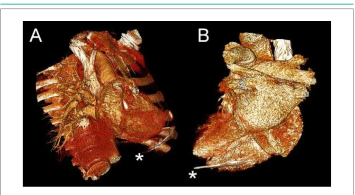
Figure 2 -Chest computed tomography (volume-rendering technique) demonstrating reconstruction of the right ventricular lead perforation (A and B). The tip of the
pacemaker lead is marked with asterisk (*).
References
1. Chauhan A, Grace AA, Newell SA, Stone DL, Shapiro LM, Schofield PM, et al. Early complications after dual chamber versus single chamber pacemaker implantation. Pacing Clin Electrophysiol. 1994; 17 (11 Pt 2): 2012-5.
2. Ellenbogen KA, Wood MA, Shepard RK. Delayed complications following pacemaker implantation. Pacing Clin Electrophysiol. 2002; 25 (8): 1155-8.
3. Mahapatra S, Bybee KA, Bunch TJ, Espinosa RE, Sinak LJ, McGoon MD, et al. Incidence and predictors of cardiac perforation after permanent pacemaker placement. Heart Rhythm. 2005; 2 (9): 907-11.
4. Laborderie J, Barandon L, Ploux S, Deplagne A, Mokrani B, Reuter S, et al. Management of subacute and delayed right ventricular perforation with a pacing or an implantable cardioverter-defibrillator lead. Am J Cardiol. 2008; 102 (10): 1352-5.
5. Hirschl DA, Jain VR, Spindola-Franco H, Gross JN, Haramati LB. Prevalence and characterization of asymptomatic pacemaker and ICD lead perforation on CT. Pacing Clin Electrophysiol. 2007; 30 (1): 28-32.
6. Ramirez MF, Ching CK, Ho KL, Teo WS. “The attack of the 52 cm lead”: an unusual case of late cardiac perforation by a passive-fixation permanent pacemaker lead. Int J Cardiol. 2007; 115 (1): e5-7.
7. Selcuk H, Selcuk MT, Maden O, Ozeke O, Celenk MK, Turkvatan A, et al. Uncomplicated heart and lung perforation by a displaced ventricular pacemaker lead: a case report. Pacing Clin Electrophysiol. 2006; 29 (4): 429-30.
8. Danik SB, Mansour M, Singh J, Reddy VY, Ellinor PT, Milan D, et al. Increased incidence of subacute lead perforation noted with one implantable cardioverter-defibrillator. Heart Rhythm. 2007; 4 (4): 439-42.
9. Yavari A, Khawaja ZO, Krishnamoorthy S, McWilliams ET. Perforation of right ventricular free wall by pacemaker lead detected by multidetector computed tomography. Europace. 2009; 11 (2): 252-54.
10. Tziakas D, Alexoudis A, Konstantinou F, Chalikias G, Stakos D, Bougioukas G. A rare case of late right ventricular perforation by a passive-fixation permanent pacemaker lead. Europace. 2009; 11 (7): 968-9.