Arquivos Brasileiros de Cardiologia - Volume 85, Nº 4, October 2005
Case Report
Case Report
Case Report
Case Report
Case Report
Case Report
Biventricular Endomyocardiofibrosis Associated
with Renal Amyloidosis
M a i l i n g A d d r e s s : J o s é R a m o s F i l h o • R u a B e n j a m i n A r r u d a , 1 2 6 / 1 - 1 2 9 1 4 - 5 6 0 - B r a g a n ç a P a u l i s t a , S P - B r a z i l E-mail: [email protected] Received on 09/02/04 • Accepted on 03/30/05
José Ramos Filho, Carlos Alberto Fontes de Souza, Enzo Magrini, Marcelo de Macedo, Roberto Bauab Isolato
Universidade São Francisco - Bragança Paulista, SP - Brazil
Endomyocardiofibrosis is a restrictive cardiomyopathy characterized by fibrotic involvement of the endocardium and adjacent myocardium, and by diastolic dysfunction caused by changes in distensibility making ventricular filling inadequate while preserving the systolic function. Clinically, it appears as heart failure, but etiological symptomatic discernment, suspicion and a clinical examination would be necessary in order to make a correct etiological diagnosis. The case of a patient with biventricular endomyocardial fibrosis associated with renal amyloidosis is presented.
Endomyocardial fibrosis, characterized by the fibrotic involvement of the endocardium and adjacent myocardium is an unknown etiological illness and is the main form of restrictive cardiomyopathy found in our midst1. As in the other forms of restrictive cardiomyopathy,
the systolic function is usually preserved to the detriment of the diastolic, caused by a reduction in distensibility, thereby impairing the adequate filling of one or both ventricles2. The possibility of a diagnosis of restrictive
syndrome should always be raised whenever a patient presents symptoms of limited physical effort with tiredness or fatigue and dyspneia, jugular congestion, hepatomegaly, edema in lower members or anasarca. The Doppler echocardiogram is essential and a cardiac biopsy can be healpfull, the anatomophatological study of necropsy hearts is able to make the diagnosis.
C
ASE
REPORT
A forty-nine year old black woman, native of Ilhéus-Bahia (Brazil), sought medical help at Emergency with a history of fatigue, anasarca, dyspnea with slight exertion and night paroxistics. These symptoms had appeared about a year previously and there had been a remarked worsening in the last six months. At first, the patient had a discrete edema in her lower members in the evening.
Arquivos Brasileiros de Cardiologia - Volume 85, Nº 4, October 2005
BIVENTRICULAR ENDOMYOCARDIOFIBROSIS ASSOCIATED WITH RENAL AMYLOIDOSIS
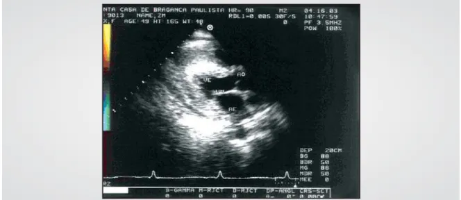
Fig. 1 - Longitudinal paraesternal transthoracic echocardiogram showing acoustic texture with thick granulations and apical obliteration due to a deposit of fibrotic tissue. AO- aorta; AE- left atrium; VE- left ventricle; VM- mitral valve
restrictive cardiac insufficiency was reached, with the possibility of establishing three etiologies: cardiac amyloidosis, endomyocardial fibrosis or endomyocardial filbroelastosis. A myocardial scintigram with MIBI-Tc99 was performed evidencing normal perfusion and biventricular hypertrophy.
In order to make the definitive diagnosis, the patient underwent a myocardial biopsy and the histological examination proved endomyocardial fibrosis.
When using a polarized light technique, the Congo red stain was negative for amyloidosis, and Orcein for elastic fiber. With Masson’s Trichroma stain, the fibrotic conjunctive tissue showed positiveness (figures 3 and 4). Because of renal function loss a biopsy was carried out and associated amyloidosis was diagnosed; 24-hour proteinuria (4.9g) was investigated, as was the absence of erithrocitarian dismorphism. Hepatitis B, C and HIV, serical complements C3 and C4 were investigated and all negative. The patient has heart failure, functioning class III according to NYHA, and is waiting for cardiac surgical procedure.
D
ISCUSSION
Endomyocardial fibrosis is characterized by the deposit of fibrotic tissue covering the endocardium initially at the apex, and then, proceeding towards the atrioventricular valvar plane, leading to an increase in end diastolic pressure of the affected ventricle. It consequently leads to enlargement of the chamber and an increase in either systemic or pulmonary venal pressure. The final result is a hardening of the endocavity causing consequently ventricular dysfunction because of restriction of its filling2.
In the case of the preferential affecting of the right ventricle the following was observed: an obstruction of the pointed end (associated or not with a thrombus), dilation in the outflow tract and paradoxical movement
of the interventricular septum; The right chambers may be enlarged and the systemic veins dilated3; Tricuspid
regurgitation in varying degrees and an increase in the end diastolic pressure of the right ventricle usually without the association of pulmonary hypertension.
If the left ventricle is predominantly affected the alterations are repeated, in relation to the left ventricular cavities3. The increase in pressures is transmitted to the
left atrium and, consequently, to the pulmonary venal bed, thereby causing pulmonary hypertension. This increase in pulmonary pressure could, in turn, be transmitted over the tricuspid valve causing regurgitation, even without affecting the right cavities. Mitral or tricuspid valve regurgitation may or may not be expressed through cardiac murmurs, especially when the end diastolic pressure of the ventricular cavity is very similar to the average pressure of the chamber 2,3.
In relation to the electrocardiogram, the affecting of the right ventricle shows a decrease in the extent of the QRS complexes, due to the almost-constant pericardial effusion. Other signs found are the changes in the ST segment and T wave and right chamber overload criteria with the classic qR pattern in V1. In the affected left ventricle, it is already possible to find changes similar to those of the right ventricle, including atrial fibrillation and overloading of the left chambers. The EKG associated with cardiac amyloidosis disease may present low QRS voltage and evidence of sinusal nodule disease and/or a blockage of the atrioventricular tract with abnormal Q waves suggesting chronic myocardial infarct1.
When the thorax was X-rayed, an enlargement of the heart area in different degrees was found, associated with signs of pulmonary venocapillar congestion4.
Arquivos Brasileiros de Cardiologia - Volume 85, Nº 4, October 2005
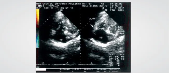
Fig. 2 - A modified apical echocardiogram of the right cavities during ventricular diastole and ventricular systole showing apical obliteration of the right ventricle by fibrotic tissue. DERR - pericardial effusion; AD - right atrium; VD - right ventricle; VT- tricuspid valve
BIVENTRICULAR ENDOMYOCARDIOFIBROSIS ASSOCIATED WITH RENAL AMYLOIDOSIS
Fig. 3 - Panoramic view of the biopsy demonstrating the cardiac muscle with extensive areas of fibrosis intermingling with myocardial fibers (arrows). HE stain increased 40 times
Arquivos Brasileiros de Cardiologia - Volume 85, Nº 4, October 2005
R
EFERENCES
1. Brauwald E, Zipes DP, Libby P. Heart Disease: a Textbook of Cardiovascular Medicine. In: Restrictive and Infiltrative Cardiomyopathies. Philadelphia: W.B. Saunders, 1997: 1433-4. 2. Guimarães AC, Esteves JP, Filho AS, Macedo V. Clinical aspects of endomyocardiofibrosis in Bahia, Brazil. Am Heart J. 1971, 81: 7-19. 3. Davies JN, Ball JD. The pathology of endomyocardiofibrosis in
Uganda. Br Heart J. 1955; 17: 337-59.
4. Fernandes F, Mady C, Vianna CB et al. Aspectos radiológicos da endomiocardiofibrose. Arq Bras Cardiol. 1997; 68: 269-72. 5. Lira VM En domiocardiofibrose. Patologia. Arq Bras Cardiol 1996;
67: 273-8.
6. Moraes CR, Rodrigues JV, Gomes CA et al. Dez anos de cirurgia da
endomiocardiofibrose: o que aprendemos. Rev Bras Cir Cardiovasc 1987; 2: 42-52.
7. Moraes CR, Rodrigues JV, Gomes CA et al. A cirurgia da endomiocardiofibrose revisitada. Rev Bras Cir Cardiovasc 1998; 13: 100-4.
8. Balakrishnan KB, Jaiswal PK, Tharakan JM,Venkitachalam CG, Ghosh. Clinical course of patients in Kerala. In: Vatalian MS, Sommers K, Kartha CC, eds. Endomyocardial Fibrosis. Delhi: Oxford University Press; 1993: 20-8.
9. Oliveira SA, Barreto ACP, Mady C et al. Surgical treatment of endomyo-cardial fibrosis: a new approach. J Am Coll Cardiol 1990; 16: 1246-51. 10. Moraes CR, Buffolo E, Lima R et al. Surgical treatment of endomyocardial fibrosis. J Thorac Cardiovasc Surg 1983; 85: 738-45.
BIVENTRICULAR ENDOMYOCARDIOFIBROSIS ASSOCIATED WITH RENAL AMYLOIDOSIS
tissue showed obliteration. Fibrosis is more ecogenic than the myocardial walls and this makes different from other forms of restrictive cardiomyopathies1,2.
The Doppler shows an aspect which is quite characteristic of restrictive myocardiopathologies, especially of endomyocardial fibrosis. The flow through the mitral or tricuspid valves, depending on which ventricle is damaged, is presented as practically forming only one velocity peak in the protodiastole (E wave) with an acute reduction in deceleration time, because the A wave is absent or greatly reduced. This aspect indicates a pattern of rapid flow immediately after the valvar opening, then followed by an abrupt disruption due to the existence of cavity obliteration and, therefore, a smaller cavity volume, or a restriction to the entry of blood due to the reduction in distensibility.
Biopsy endomyocardial helps in the differential diagnosis with other restricted cardiophaties that, with the clinical and complemental examination make the diagnosis.
The mitral flow is of great prognostic value. So, patients who present a mitral flow with restrictive characteristics (decelerating time less than 150 ms) have only a 49% chance of survival for one year against 92% in those whose flow indicates only a relaxation abnormality. The reduction in relaxation can be seen by means of the tricuspid and/or mitral valves1,2.
During the cardiac catheterization, the angiogram shows areas of obliteration in the ventricular apexes. The size of the cavities can be reduced and the myocardial wall thicker than normal. The pressure curves may be similar to those found in constrictive pericarditis, with depression and plateau wave form recorded during the diastole in the right ventricle 1,2. It can also show evident
mitral and tricuspid regurgitations.
The study of the ventriculography, using radionuclear substances, shows a normal-sized or reduced ventricular cavity, as well as myocardial wall relaxation abnormalities which can also be identified1.
The complimentary test to finalize the diagnosis is, inexorably, the endomyocardial biopsy, which makes it possible to differentiate between the various modalities of restrictive myocardiopathy3,5.
In the studied case, the myocardial and renal biopsies both confirmed the diagnosis of endomyocardial fibrosis and renal amyloidosis, respectively.
Patients suffering from restrictive myocardiopathies, in particular those with endomyocardial fibrosis could first be labeled as carriers of congestive cardiac insufficiency without establishing a more definitive etiological diagnosis. Clinical treatment is frustrating and unsatisfactory1,6.
Surgical treatment is the principal therapeutic option7.
Diuretics are necessary to reduce both pre and after loading. However, it is unsafe to increase the diuresis to the point of reducing the pressures of cardiac filling2.
Therapy with corticosteroids did not prove beneficial.
The prognosis varies from two to ten years depending on the gravity of the clinical condition, after the first signs8. The alternations which negatively influence the
prognosis are: the intensity of the apical fibrosis, signs of diastolic restriction, damaging of the systolic ventricular functioning, involvement of the subvalvar system, presence of pulmonary hypertension and intercavity thrombi3,5. It is, however, the duty of both the physician
and the cardiologist to undertake a careful examination, complemented by subsidiar y exams such as an electrocardiogram, thorax X-ray, a Doppler echocardiogram, angiogram, myocardial ventriculography and biopsy, in order to make a correct diagnosis so as to be able to indicate the necessary treatment as quickly as possible. Surgical treatment has been prematurely indicated immediately after diagnosis7,9. This has led to
endocardial resection, but however, with a high morbid-mortality rate, even with experienced surgeons10.
In some patients the tricuspid and mitral valves are substituted with prothetic6,7 valves and a heart transplant