Risk Factors for Cerebrovascular Disease and Cognitive Function in
the Elderly
Nicole de Liz Maineri, Flávio Merino de Freitas Xavier, Maria Cristina Cachapuz Berleze, Emílio Hideyuki Moriguchi
Hospital São Lucas da PUCRS, Instituto de Geriatria e Gerontologia, Hospital Mãe de Deus - Porto Alegre, RS - BrazilSummary
Objective: To determine whether stroke risk factors and cognitive dysfunction are concomitant in individuals over the age of 60.
Methods: The cognitive abilities of elderly individuals with different degrees of risk as per the Framingham stroke risk profile were compared. The Framingham stroke risk profile was used to calculate the risk of an ischemic cerebral event. Neuropsychological tests included the Buschke Selective Reminding Test, verbal fluency (animals), clock drawing, Rey Auditory Verbal Learning, digit span and vocabulary. A random and typical population sample was used in the study, selected from the 200 elderly residents in the area surrounding the primary health care facility (Morada das Flores Clinic, Porto Alegre). Forty-six elderly individuals were included in the study.
Results: The elderly individuals with a risk profile, had lower memory test performance levels (NF, p=0.02) and planning capacity (clock drawing test, p=0.03). Diabetes proved to be related to delayed recall performance in the Rey auditory verbal learning test (p=0.04).
Conclusion: The presence of stroke risk factors in the elderly was associated with worse cognitive performance for memory and executive functions. (Arq Bras Cardiol 2007;89(3):142-146)
Key words: Framingham’s stroke risk profile, cognitive functions, neuropsychological tests.
Mailing address: Nicole de Liz Maineri •
Rua Jaraguá, 518/202– Porto Alegre, RS - Brazil E-mail: [email protected]
Manuscript received April 11, 2005; revised manuscript received December 22, 2006; accepted March 23, 2007.
Introduction
It is agreed that cerebrovascular disease (CVD) represents a significant risk factor for vascular dementia. Recent evidence has indicated that CVD is also related to a higher risk for Alzheimer dementia (AD). Findings such as greater cortical senile plaque density in non-demented patients with severe coronary artery disease1, a significant positive association between the
atherosclerotic index and the AD diagnosis2, a positive association
between elevated homocysteine levels and the prevalence of AD3, a negative association between the use of statins and AD
prevalence4, and less cognitive decline in AD patients using
atorvastatina5 have led to the belief that there is an association
between cardiovascular risk factors and the AD pathogenesis. Isolated studies indicate that CVD could also be a risk factor for minimal cognitive loss (MCI), and more specifically, it could be associated with the mild cognitive impairment subtype called “multiple domains”6. Given the associations between CVD and
the various dementia syndromes (Alzheimer, vascular) or “pre-dementia” (MCI), two recent studies attempted to determine whether CVD risk factors such as hypertension and diabetes mellitus – in an isolated manner - are associated with cognitive impairment7-8. According to these studies, elderly individuals
with stroke risk factors, but without CVD or dementia at the beginning of the longitudinal follow-up, were inclined to suffer greater cognitive loss over time. It is speculated that individuals with stroke risk factors, who have not suffered a marked vascular event, present microscopic cerebrovascular lesions that can be identified in neuroimage studies, even though the patient does not present any clinical repercussions other than mild cognitive loss9,10.
These two initial studies conducted in 2004, indicated some potentially important implications in relation to prevention and therefore should be validated in various populations, particularly the elderly, since the mean participant age in one of the studies was of only 60 years.
The objective of the present study is to verify whether the association indicated in these two studies between stroke risk factors and cognitive loss is reproducible.
Methods
stroke over the next 10 years. The Cerebrovascular Risk Table determines a score for cerebrovascular events over the next 10 years, based on the vascular risk factor data of the Framingham Study that comprises the following determinant risk factors: age, gender, total and HDL cholesterol levels, systolic and diastolic blood pressure, presence or not of diabetes mellitus and whether or not the individual is a smoker.
Using the score, we determined the “presence” of risk for an ischemic event during the next 10 years as: a score greater than or equal to 15 on the Framingham table for females and a score greater than or equal to 10 for males.
Results
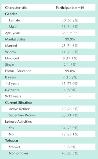
The socio-demographic characteristics of the sample are shown in table 1. The percentile of women was 65.2% and the mean age was 68.6 years (minimum of 60 years and maximum of 81 years). The level of education for most of the participants (63%) was low (1-5 years of formal education). Ex-smokers were classified as non-smokers.
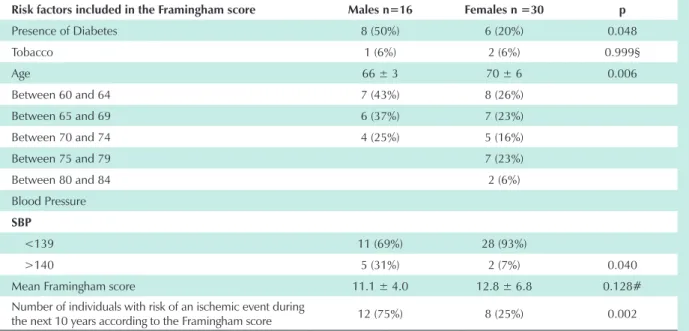
A comparison of the risk profiles for cerebrovascular events for males and females is shown in table 2. This table demonstrates the profile for males and females in relation to Statistics - The computer program, Statistical Package for
the Social Sciences (SPSS) version 11.5 was used to process and analyze the data. The Student’s t-test was used for the statistical analysis of the parametric variables. The Mann-Whitney U test (two independent samples) and chi square test (analysis of the association between different variables) were used for the non-parametric variables. In all cases, statistical significance was established as the probability of a type I error (p) less than 5%.
Ethical aspects - All study participants or their designated representatives were required to sign a free and informed consent form. The project was submitted to the Research Ethics Committee of the Pontifical Catholic University of Rio Grande do Sul and was approved in November 2004.
Procedures - The interviews were conducted at the clinic and lasted, on average, for one hour. Study participants who were unable to travel to the clinic, were assessed in their homes. In addition to the tests listed below, questionnaires on the participants’ medical histories, socio-demographic variables, alcohol and cigarette use were used.
The tests were used for both the cognitive investigation and the presence of cerebrovascular risk.
Mini-Mental Status Exam11 (MMSE): This exam is a short
mental status examination which evaluates orientation, memory, attention and calculation, language and constructive praxis (maximum score 30).
Free and Cued Selective Reminding Test12 (Buschke):
This test evaluates memory storage, retention and recall (maximum score 96).
Clock Drawing Test13: This test assesses visual constructive
abilities, planning and visual neglect. The individual is asked to draw a clock with the numbers and afterwards to draw the hands to depict a time of a quarter to three (maximum score 4).
Verbal Fluency for Category (animals)14 : This test assesses long
term memory recall and retrieval, which requires organizational, self-regulation and operational memory abilities.
Vocabulary (WAIS-III subtest)13: This test evaluates language
development and word knowledge. It is considered to be the best individual measurement of overall intelligence.
Digit Span (WAIS-III subtest)15: This test assesses attention
and immediate memory, and is divided in two stages. During the first stage the individual is asked to repeat increasingly larger series of numbers in the same order spoken by the examiner (direct order). During the second stage the individual is asked to repeat the series in the opposite order from the presentation (reverse order).
Rey’s Auditory Verbal Learning Test16: This test checks
the ability to learn and retain a series of words, the extent of the verbal memory, susceptibility to distractions and recall memory. A list of fifteen words (list A) is read slowly, five times. The individual should repeat the words in any order, after each reading of the list. In this study, only list Aand delayed recall of list A were used.
The Framingham Stroke Risk Profile (Cerebrovascular Risk Table)17,18 is a validated score that combines the greatest
cardiovascular risk factors, giving them different weights, in order to produce a risk score of the probability to have a
Table 1 – Socio-demographic data of the sample
Characteristic Participants n=46 Gender
Female 30 (65.2%)
Male 16 (34.8%)
Age, years 68.6 ± 5.9
Marital Status 99.9%
Married 25 (54.3%)
Widow 11 (23.9%)
Divorced 8 (17.4%)
Single 2 (4.3%)
Formal Education 99.8%
0 years 7 (15.2%)
1-5 years 35 (76.0%)
6-8 years 4 (8.6%)
9-11 years
-Current Situation
Active Retiree 13 (28.3%)
Sedentary Retiree 33 (71.7%)
Leisure Activities
Yes 34 (73.9%)
No 12 (26.1%)
Tobacco
Smoker 3 (6.5%)
the Framingham score risk components, the mean score based on gender and the percentile of men and women with high risk as per the score. Even though the mean risk for cerebrovascular events was the same for both males and females over the next ten years, a breakdown of the Framingham score by components, revealed different gender risk profiles for each item. Even though the mean age of the males was lower (therefore a lower age factor risk) there was a significantly greater number of males with altered blood pressure and diabetes when compared to the females. A greater percentile of males, when compared to the females, had a high risk for stroke (p=0.002): in the sample, 75% of the males presented a risk of stroke in comparison to 25% of the females.
Through the multivariate analysis, we investigated if any of the Framingham score components (smoking, diabetes, blood pressure and age) had a more significant impact on the neuropsychological test scores. In this calculation, only the presence of diabetes proved to be directly related to delayed recall performance in the Rey auditory verbal learning test.
Table 3 compares the cognitive performance of the elderly individuals with and without stroke risk factors. Individuals with a higher Framingham score had worse performance in the memory ability tests. Performance was lower for both the number of free recalls in the six Buschke test verifications (p=0.02) and the delayed recall in the Rey auditory verbal learning test (p=0.05).
The individuals with greater risk profiles for cerebrovascular events also presented executive dysfunction: when evaluated using the clock test, individuals with stroke risk factors had significantly lower scores (p=0.03) than those with no risk factors.
Discussion
The data indicate that there is an association between stroke risk factors and cognitive impairment. Individuals with risk factors for cerebrovascular events as per the Framingham risk profile presented impairment in both memory function (Rey and Buschke tests) and planning ability (clock test).
These results agree with previous studies that demonstrate that stroke risk factors are associated with both memory loss 19-21 and executive dysfunction. As described by Alexopoulos22,
this executive dysfunction, without the presence of evident stroke sequelae, would be the subtle clinical manifestation of microscopic encephalic vascular lesions, that can be identified with neuroimage tests.
In our sample, we observed that the mean risk of stroke was the same for males and females, since the Framingham scores for both genders were similar. Nevertheless, this final score conceals the considerably different profiles for each component in the Framingham risk profile: the males presented more cases of diabetes and hypertension, that led us to assume that their final score was similar to the females due to their significantly younger ages. In addition, it was also shown that three quarters of the males in this sample had an “elevated” risk of stroke as per the Framingham criteria, while only one quarter of the females had the same risk.
Analysis of the relative impact of each factor of the Framingham final score, revealed that diabetes is associated with a worse memory performance. Nevertheless, the sample size greatly limits the ability to conclude whether or not there is an association between the other components (hypertension, age) and the various cognitive fields. In this
Table 2 – Distribution of the Framingham risk profile components according to gender
Risk factors included in the Framingham score Males n=16 Females n =30 p
Presence of Diabetes 8 (50%) 6 (20%) 0.048
Tobacco 1 (6%) 2 (6%) 0.999§
Age 66 ± 3 70 ± 6 0.006
Between 60 and 64 7 (43%) 8 (26%)
Between 65 and 69 6 (37%) 7 (23%)
Between 70 and 74 4 (25%) 5 (16%)
Between 75 and 79 7 (23%)
Between 80 and 84 2 (6%)
Blood Pressure
SBP
<139 11 (69%) 28 (93%)
>140 5 (31%) 2 (7%) 0.040
Mean Framingham score 11.1 ± 4.0 12.8 ± 6.8 0.128#
Number of individuals with risk of an ischemic event during
the next 10 years according to the Framingham score 12 (75%) 8 (25%) 0.002
Exact Fisher Test (1) Values expressed as mean ± standard deviation (percentage of risk to suffer a stroke during the next ten years). # Student’s t-test.
study, the relative gender impact on the Framingham risk profile was not calculated, since the profile has already taken this into account and gives different relative weights to each risk factor based on gender.
A longitudinal study conducted with 238 elderly diabetics and 36 controls, demonstrated that a greater percentage of elderly diabetics develop dementia in relation to the control group. Another important finding in this study was that the neuropsychological tests that demonstrated the greatest sensitivity for the early onset of cerebral changes associated with diabetes were those that involve episodic memory and processing speed21.
Further longitudinal studies are required to fully understand the association between stroke risk factors and cognitive loss. A multivariate analysis with larger samples will make it possible to identify whether or not there is a particular risk
factor that has a greater impact on cognitive function. It will also be possible to investigate whether different risk factors have a greater impact on the memory, and if other factors cause greater risk for executive function.
Potential Conflict of Interest
No potential conflict of interest relevant to this article was reported.
Sources of Funding
There were no external funding sources for this study.
Study Association
This article is part of the thesis of master submitted by Nicole de Liz Maineri from Pontifícia Universidade Católica do Rio Grande do Sul.
Table 3 – Comparison of neuropsychological test performance among the elderly individuals with and without stroke risk factors in accordance with the Framingham Risk Profile
Framingham Risk Profile
Tests Risk for stroke n=20 (median) No risk for stroke n=26 (median) p*
Mini Mental Status Exam 25.00 26.50 0.27
Buschke Selective Reminding Test
Cued and Free Recall 95.50 96.00 0.26
Free Recall 59.50 72.00 0.02*
Delayed Recall 16.00 16.00 0.19
Clock Drawing Test 2.50 4.00 0.03*
Verbal Fluency (animals) 11.50 13.50 0.10
Vocabulary (WAIS-III) 17.00 23.00 0.08
Rey Auditory Verbal Learning Test
Verbal Learning 34.00 37.50 0.07
Delayed Recall 4.50 7.00 0.05*
Digit Span (WAIS-III) 7.00 8.00 0.13
Direct Order 4.00 4.00 0.12
Reverse Order 3.00 4.00 0.31
* Mann Whitney statistical test.
References
1.Sparks DL, Hunsaker JC, Scheff SW, Kryscio RJ, Henson JL, Markesbery WR. Cortical senile plaques in coronary artery disease, aging and Alzheimer’s disease: neurobiological Aging. 1990; 11 (6): 601-7.
2. Hofman A, Ott A, Breteler MM, Bots Ml, Slooter AJ, va Harskamp F, et al. Atherosclerosis, apolipoprotein E, and prevalence of dementia and Alzheimer’s disease in the Rotterdam Study. Lancet. 1997; 349 (9046): 151-4.
3. Seshadri S, Wolf PA, Beiser A, Vasan RS, Wilson PW, Kase CS, et al. Elevated midlife blood pressure increases stroke risk in elderly persons: the Framingham Study. Arch Intern Med. 2001; 161 (19): 2343-50.
4. Jick H, Zornberg GL, Jick SS, Seshadri S, Drachman DA. Statins and the risk of dementia. Lancet. 2001; 357 (9255): 562.
5. Sparks DL, Sabbagh MN, Connor DJ, Lopez J, Launer LJ, Petanceska S, et al. Atorvastatin therapy lowers circulating cholesterol but not free radical activity in advance of identifiable clinical benefit in the treatment of mild-to-moderate AD. Curr Alzheimer Res. 2005; 2 (3): 343-53.
6. Petersen RC. Mild cognitive impairment: where are we? Alzheimer Dis Assoc Disord. 2005; 19 (3): 166-9.
7. Elias MF, Sullivan LM, D’Agostino RB, Elias PK, Beiser A, Au R, et al. Framingham stroke risk profile and lowered cognitive performance. Stroke. 2004; 35: 404-9.
9. Longstreth Jr WT, Dulberg C, Manolio TA, Lewis MR, Beauchamp NJ Jr, O´Leary D, et al. Incidence, manifestations, and predictors of brain infarcts defined by serial cranial magnetic resonance imaging in the elderly: the Cardiovascular Health Study. Stroke. 2002; 33 (10): 2376-82.
10. Vermeer SE, Prins ND, den Heijer T, Hofman A, Koudstaal PJ, Breteler MM. Silent brain infarcts and the risk of dementia and cognitive decline. N Engl J Med. 2003; 348 (13): 1215-22.
11. Folstein MF, Folstein SE, McHugh PR. Mini-mental state. J Psychiatr Res. 1975; 12: 189-98.
12. Petersen RC, Smith G, Kokmen E, Ivnik TJ, Tangalos EG. Memory function in normal aging. Neurology. 1992, 42: 396-401.
13. Wolf-Klein GP, Silverstone FA, Levy AP, Brod MS. Screening for Alzheimer´s disease by clock drawing. J Am Geriatr Soc. 1989; 37: 730-4.
14. Brucki SMD. Dados normativos para o uso do teste fluência verbal (categoria animal), em nosso meio. [tese]. São Paulo: Escola Paulista de Medicina, Unifesp; 1996.
15. WAIS-III. Escala de Inteligência Wechsler para Adultos, adaptação e padronização de uma mostra brasileira por Elisabeth do Nascimento. São Paulo: Casa do Psicólogo; 2004.
16. Diniz LFM, Cruz MF, Torres VM, Cosenza RM. O teste de aprendizagem auditivo-verbal de Rey: normas para uma população brasileira. Rev Bras Neurol. 2000; 36 (3): 79-83.
17. Wolf PA, D’Agostino RB, Belanger AJ, Kannel WB.Probability of stroke: a risk profile from the Framingham Study. Stroke. 1991; 22 (3): 312-8.
18. D’Agostino RB, Wolf PA, Belanger AJ, Kannel WB. Stroke risk profile: adjustment for antihypertensive medication. The Framingham Study. Stroke. 1994; 25 (1): 40-3.
19. Desmond DW, Thomas K, Tatemichi TK, Paik M, Stern Y. Risk factors for cerebrovascular disease as correlates of cognitive function in a stroke-free cohort. Arch Neurol. 1993; 50: 162-6.
20. Kilander L, Andren B, Nyman H, Lind L, Boberg M, Lithell H. Atrial fibrillation is an independent determinant of low cognitive function: a cross-sectional study in elderly men. Stroke. 1998; 29: 1816-20.
21. Hassing LB, Grant M, Hofer SM, Pedersen NL, Nilsson SE, Berg S, et al. Type 2 diabetes mellitus contributes to cognitive decline in old age: a longitudinal population-based study. J Int Neuropsychol Soc. 2004; 10: 599-607.