UNIVERSIDADE DE LISBOA
Faculdade de Medicina Veterinária
USE OF THE SHORT FORM GLASGOW COMPOSITE MEASURE PAIN SCALE IN THE ASSESSMENT OF CANINE PATIENTS PRESENTING IN SHOCK
JOSÉ MIGUEL CARREIRA REVEZ PEREIRA COUCELO
ORIENTADOR Dr. Adam Mugford CO-ORIENTADOR
Doutora Berta Maria Fernandes Ferreira São Braz
2015 LISBOA CONSTITUIÇÃO DO JURI
Doutor José Manuel Chéu Limão Oliveira Doutora Ilda Maria Neto Gomes Rosa Dr. Adam Mugford
UNIVERSIDADE DE LISBOA
Faculdade de Medicina Veterinária
USE OF THE SHORT FORM GLASGOW COMPOSITE MEASURE PAIN SCALE IN THE ASSESSMENT OF CANINE PATIENTS PRESENTING IN SHOCK
JOSÉ MIGUEL CARREIRA REVEZ PEREIRA COUCELO
DISSERTAÇÃO DE MESTRADO INTEGRADO EM MEDICINA VETERINÁRIA
ORIENTADOR
Dr. Adam Mugford
CO-ORIENTADOR
Doutora Berta Maria Fernandes Ferreira São Braz
2015 LISBOA CONSTITUIÇÃO DO JURI
Doutor José Manuel Chéu Limão Oliveira Doutora Ilda Maria Neto Gomes Rosa Dr. Adam Mugford
“Um homem tem de estar preparado para se queimar na sua própria
chama: como se pode renovar sem primeiro se transformar em cinzas?”
Assim falou Zaratustra, Friedrich Nietzsche
ACKNOWLEDGEMENTS
I would like to thank my parents and family for their support throughout these years and for allowing me to chase my ambitions.
A “thank you” to my nephew, António, for all the laughs that made the writing of this dissertation much happier.
To Belinda Andrews, for all your kindness. You have a wonderful heart and I am blessed to be your friend.
To Adam Mugford, for all your patience, kindness, friendship and teachings. I cannot thank you enough for all that this experience has meant to me. I wish you all the best.
To Amy, Sean, Helen, Fliss, John, Jamie, Chris, James and Laura. Thank you for being such amazing people and veterinarians. Thank you for your teachings and good disposition. To all of the vet nurses, PCAs, and Village Vet Hampstead staff! Thank you everyone for making my externship unforgettable.
To Sarah Egleston – a special “thank you” for all your help and all the laughs we shared. You are amazing.
I want to thank Professor Berta São Braz, for your kindness, comprehension, guidance and strength. Thank you for taking me under your wing and encouraging me at the most stressful times.
I would also like to thank Dr. Telmo Nunes, for having the patience to help me with the statistic analysis of this dissertation and for all your comments and advices.
To all the amazing friends I made in this “house”: Sara, Raquel, Bernardo, João, Alexandre, Catarina, Gabriela, Bruno, Bôto, Gabriel, Bruna and many others. Together we built many good memories that I will keep until I breathe.
To all the family from the Students Union, for everything that I’ve learned with you and for all that we achieved as a team. The sleepless nights were worth it. A special “thank you” to Ausenda Capela, for the many good talks we had.
To all my hometown friends, for all your support in times of desperation!
To João Cotta – for all the adventures, amazing stories, never ending wisdom and friendship. “Dénks” for everything.
To my pets – because you’re the reason I’m here.
USE OF THE SHORT FORM GLASGOW COMPOSITE MEASURE PAIN SCALE IN THE ASSESSMENT OF CANINE PATIENTS PRESENTING IN SHOCK
Abstract
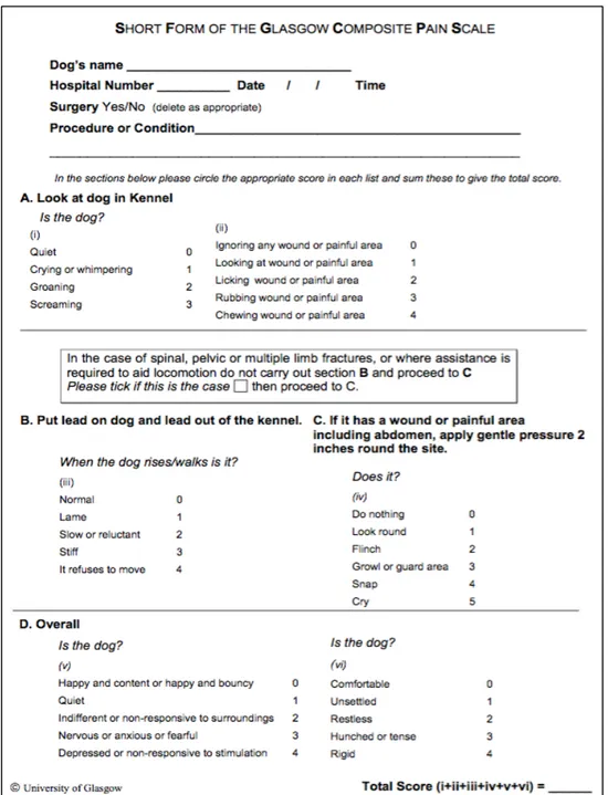
The short form of Glasgow Composite Measure Pain Scale (CMPS-SF), a previously validated decision-making tool is increasingly used in practice for the assessment of pain in dogs. However, few studies have considered the application of a pain scoring system in patients presenting in emergency situations.
This study aimed to evaluate if pain scoring with the Glasgow CMPS-SF was effective in identifying pain in patients in shock.
A prospective study (November 2014 to January 2015), within a first opinion and specialty intensive care service was developed. A total of 31 client-owned dogs (16 females and 15 males) that presented as primary emergencies or transfers. When admitted to the study, all dogs were examined by a veterinarian or registered veterinary nurse. Patients were classified and grouped as Shock (S) or Not Shock (NS) on basis of their shock index (SI). The shock status was defined a priori if the SI was higher than 1.0. Regardless of group, all patients had their pain assessed with the Glasgow CMPS-SF and by a Board Eligible Internist or a Emergency and Critical Care specialist, blinded to both pain score and SI values.
Dogs in shock numbered 18/31 dogs within the not shock group numbered 13/31. Mean age of dogs in the S group was 4.6 years (0.2 – 10) and in the NS group was 8.3 years (1 – 16); a significant difference existed in age between groups (p-value<0.05). Median pain score of the S group was 5 (0 – 17) and on the NS group was 3 (1 – 15). There was no significant difference on pain scores between the groups (p-value>0.05) and between the clinicians’ perception of pain between groups (p-value>0.05). A significant difference was present between pain scores and the clinicians’ opinion on pain (p-value=0.014), including within the shock group (p-value=0.0021). Cohen’s kappa statistic within the shock group was 0.47, which can be interpreted as weak to moderate agreement between the Glasgow CMPS-SF and the clinician opinion on pain. Within the NS group the differences between the pain scores and the clinician’ opinion on pain were not statistically significant (p-value>0.05). These results do not support an acceptable agreement between the Glasgow CMPS-SF and an experienced veterinarians evaluation of pain in patients presenting in shock. Therefore, further investigation into the relevance of the used pain assessment tool in emergency and shock patients is recommended before use in the objective monitoring of this subset of patients.
USO DA FORMA ABREVIADA DA ESCALA COMPOSTA DA DOR DE GLASGOW EM CÃES COM APRESENTAÇÃO DE CHOQUE
Resumo
O reconhecimento e avaliação de dor em doentes veterinários pode ser desafiante, especialmente nos que se encontram em estado crítico. A já validada forma abreviada da Escala Composta da Dor de Glasgow (CMPS-SF) é, cada vez, mais utilizada em ambiente clínico na avaliação da dor aguda em cães. Contudo, poucos estudos têm considerado a aplicação de um sistema de avaliação de dor em doentes que se apresentam em situação de emergência. Foi desenvolvido um estudo experimental com o propósito de avaliar se a pontuação obtida com a CMPS-SF seria capaz de identificar dor em doentes que se apresentassem em choque.
O estudo prospetivo desenvolveu-se numa clínica de primeira opinião e com serviço de cuidados intensivos (entre Novembro de 2014 e Janeiro de 2015). Foram incluídos no estudo 31 cães, admitidos em situação de emergência ou como referências, tendo sido examinados por um médico veterinário ou uma enfermeira veterinária. Os doentes foram classificados como estando em choque (S) ou não (NS) com base no seu índice de choque (IC). O estado de choque foi definido quando IC>1.0. Todos os doentes foram avaliados quanto à dor pela utilização da CMPS-SF e através de um exame físico realizado por um candidato a Internista ou um especialista em Emergências e Cuidados Intensivos, desconhecedores da pontuação obtida com a escala de CMPS-SF e do IC. O nível de significância estabelecido foi de 0.05.
O grupo de cães em choque incluiu 18 cães e o grupo de não choque incluiu 13. A idade média dos animais no grupo S foi de 4.6 anos e no grupo NS foi de 8.3. As diferenças de idade observadas entre os grupos foram consideradas estatisticamente significativas (p-value<0.05). A pontuação média de dor no grupo S foi 5 e no grupo N.S. foi 3. As diferenças observadas na pontuação de dor entre os dois grupos grupos não foi considerada significativa (p-value>0.05). A perceção da dor pelos médicos veterinários nos dois grupos também não foi considerada significativa (p-value>0.05). Considerou-se significativa a diferença observada entre as pontuações de dor e a perceção de dor dos médicos veterinários (p-value=0.014), incluíndo no grupo S (p-value=0.0021). No grupo S, a concordância entre métodos foi de 0.47, interpretada como fraca a moderada.
Face aos resultados obtidos, sugerem-se mais estudos relativos à precisão da utilização de escalas de dor em doentes que se apresentem em emergência e em condições de choque, antes que estas escalas possam ser recomendadas neste tipo de doentes.
Table of Contents
LIST OF FIGURES ... VIII LIST OF TABLES ... IX LIST OF ABBREVIATIONS ... XI LIST OF SYMBOLS ... XIII GLOSSARY ... XIV
PART I - EXTERNSHIP REPORT ... 1
PART II – LITERATURE REVIEW ... 4
2.1INTRODUCTION ... 4 2.2PAIN ... 5 2.2.1DEFINITION ... 5 2.2.2PAIN CLASSIFICATION ... 5 2.2.2.1 Physiologic pain ... 6 2.2.2.2 Pathologic pain ... 6 2.2.2.3 Acute pain ... 6 2.2.2.4 Chronic pain ... 6 2.2.2.5 Neuropathic pain ... 6 2.2.2.6 Inflammatory pain ... 7 2.2.2.7 Visceral Pain ... 7 2.2.2.8 Phantom pain ... 7 2.2.2.9 Cancer pain ... 7 2.2.3PAIN MECHANISMS ... 7 2.2.3.1 Nociception vs Pain... 8 2.2.3.2 Nociceptive processing ... 8 2.2.3.2.1 Transduction ... 9 2.2.3.2.2 Transmission ... 10 2.2.3.2.3 Projection ... 10 2.2.3.2.4 Perception ... 11 2.2.3.2.4.1 Antinociceptive pathways ... 11
2.2.3.3NEUROPLASTICITY AND MEMORY OF PAIN ... 12
2.2.3.3.1 Peripheral sensitization ... 12
2.2.3.3.2 Central sensitization ... 13
2.2.4CONSEQUENCES OF PAIN ... 14
2.2.4.1 Stress response in pain ... 14
2.2.4.1.2 Neuroendocrine axis ... 15
2.2.4.1.3 Effects on metabolism ... 15
2.2.4.1.4 Effects on the immune system ... 15
2.2.5PAIN RECOGNITION AND ASSESSMENT ... 16
2.2.5.1 Physiologic signs ... 17
2.2.5.2 Pain behaviour ... 17
2.2.5.3 Pain Assessment Tools (PATs) ... 18
2.2.5.3.1 Subjective unidimensional scales ... 19
2.2.5.3.1.1 Simple Descriptive Scale (SDS) ... 19
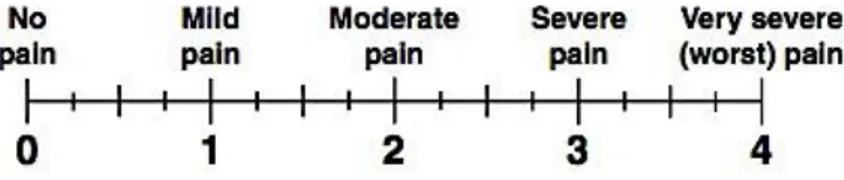
2.2.5.3.1.2 Numerical Rating Scale (NRS) ... 19
2.2.5.3.1.3 Visual Analogue Scale (VAS) ... 20
2.2.5.3.2 Multidimensional pain scales ... 21
2.2.5.3.2.1 University of Melbourne Pain Scale (UMPS) ... 21
2.2.5.3.2.3 Glasgow Composite Measures Pain Scale ... 21
2.2.5.3.2.4 Short Form of the Glasgow Composite Measure Pain Scale ... 22
2.2.5.3.2.5 Further pain assessment tools... 23
2.3PAIN MANAGEMENT ... 24 2.3.1MULTIMODAL ANALGESIA ... 24 2.3.2TREATMENT STRATEGY ... 24 2.3.3DRUG THERAPY ... 25 2.3.3.1OPIOIDS ... 25 2.3.3.1.2 Mechanism of action ... 26 2.3.3.1.3 Tramadol ... 27
2.3.3.1.4 Contraindications and side effects ... 27
2.3.3.2NONSTEROIDAL ANTI-INFLAMMATORY DRUGS ... 28
2.3.2.1 Mechanism of action ... 28
2.3.2.2 Contraindications and adverse effects ... 29
2.3.3 Α2-ADRENERGIC AGONISTS ... 30
2.3.3.1 Mechanism of action ... 30
2.3.3.2 Side effects and contraindications ... 31
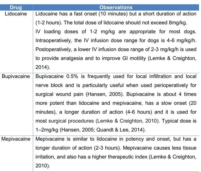
2.3.4LOCAL ANAESTHETICS ... 31
2.3.4.1 Mechanism of action ... 31
2.3.4.2 Local anaesthetics in veterinary medicine ... 32
2.3.4.3 Contraindications and adverse effects ... 32
2.3.5ADJUNCTIVE DRUGS ... 33
2.3.5.1 Anticonvulsants ... 33
2.3.5.2 N-Methyl-D-Aspartate Receptor Antagonists ... 33
2.3.5.3 Tricyclic antidepressants (TCAs) ... 34
2.3.5.4 Glucocorticoids... 34
PART III - SHOCK ... 36
3.1DEFINING SHOCK ... 36
3.1.1GENERAL CONSIDERATIONS IN SHOCK PATHOPHYSIOLOGY ... 36
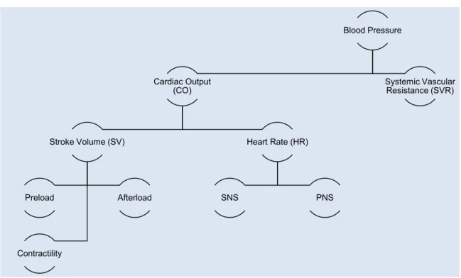
3.1.1.1 Cardiovascular considerations ... 36 3.1.1.2 Cellular injury ... 38 3.1.2SHOCK CLASSIFICATION... 39 3.1.2.1 Hypovolaemic shock ... 40 3.1.2.2 Cardiogenic shock ... 41 3.1.2.3 Distributive Shock ... 41 3.1.2.4 Obstructive shock ... 42 3.1.2.5 Hypoxemic shock ... 42 3.1.2.6 Metabolic shock ... 43 3.1.3COMPENSATORY MECHANISMS ... 43
3.1.4STAGES AND CLINICAL SIGNS OF SHOCK ... 44
3.1.5RECOGNITION AND CLINICAL ASSESSMENT OF SHOCK ... 45
3.1.5.1 Physical examination ... 46
3.1.5.2 Assessing tissue perfusion ... 48
3.1.5.2.1 Arterial Blood pressure ... 48
3.1.5.2.2 Pulse oximetry ... 49
3.1.5.2.3. Blood lactate ... 50
3.1.6SHOCK INDEX ... 51
PART IV – EXPERIMENTAL STUDY ... 53
4.1INTRODUCTION AND OBJECTIVES ... 53
4.2MATERIAL AND METHODS ... 54
4.2.1STUDY POPULATION ... 54
4.2.5ANALGESIA ... 55
4.3RESULTS ... 57
4.3.2PAIN ASSESSMENTS BY THE VETERINARIANS AND WITH THE GLASGOW CMPS-SF ... 59
4.3.2.1 Not Shock (N.S.) group ... 61
4.3.2.1.1 Pain assessment within the not shock group ... 61
4.3.2.2 Shock Group (S) ... 62
4.3.2.2.1 Pain assessment within the shock group ... 62
4.3.3STATISTICAL ANALYSIS ... 63
4.3.3.1 Spearman rank correlation test results ... 63
4.3.3.2 Testing differences in age between groups ... 64
4.3.3.3 Testing the patients’ gender and Glasgow CMPS-SF pain scores ... 64
4.3.3.4 Comparing patients Group and Glasgow CMPS-SF scores ... 65
4.3.3.5 Testing the Veterinarians’ perception of pain (“Vetpain”) between the two groups ... 66
4.3.3.6 Testing the Glasgow CMPS-SF pain score readings (“Scalepain”) in the two groups ... 66
4.3.3.7 Testing the Glasgow CMPS-SF pain scores and the Veterinarian’s perception of pain (“Vetpain”) ... 67
4.3.3.8 Agreement between Glasgow CMPS-SF score readings (“Scalepain”) and the Veterinarians perception of pain (“Vetpain”) ... 68
4.3.3.9 Shock Group ... 68
4.3.3.9.1 Testing the Glasgow CMPS-SF pain scores and the Veterinarians perception of pain (“Vetpain”) within the shock group ... 68
4.3.3.9.2 Testing the agreement between the Glasgow CMPS-SF score readings (“Scale Pain”) and the Veterinarians perception of pain (“Vetpain”) within the shock group ... 69
4.3.3.10 Not Shock group ... 69
4.3.2.10.1 Testing the the Glasgow CMPS-SF pain scores and the Veterinarians perception of pain (“Vet pain”) within the not shock group ... 69
4.3.3.10.2 Testing agreement between the Glasgow CMPS-SF score readings (“Scale Pain”) and the Veterinarians perception of pain (“Vetpain”) within the not shock group ... 70
4.4DISCUSSION ... 70
4.4.1 Study limitations ... 73
5.CONCLUSIONS AND FUTURE PERSPECTIVES ... 74
6. BIBLIOGRAPHY ... 76
ANNEXES ... 83
ANNEX I–EXTERNSHIP FEATURES ... 83
ANNEX II–EVECCS ABSTRACT ... 94
ANNEX III–ABSTRACT IN THE JVECC ... 85
ANNEX IV–UNIVERSITY OF MELBOURNE PAIN SCALE ... 86
ANNEX V–COLORADO STATE UNIVERSITY CANINE ACUTE PAIN SCALE ... 87
ANNEX VI–4A-VET PAIN SCALE ... 88
ANNEX VII–COMMON OPIOID AGENTS USED IN VETERINARY MEDICINE AND RESPECTIVE DOSING ... 89
ANNEX VIII-COMMON NSAIDS USED IN VETERINARY MEDICINE AND RESPECTIVE DOSING ... 90
ANNEX IX-RECOMMENDED DOSES OF SELECTED Α2-AGONISTS FOR ROUTINE SEDATION AND ANALGESIA ... 91
ANNEX X–FORM DEVELOPED FOR THE STUDY ... 92
ANNEX XI–PATIENTS’INDIVIDUAL GLASGOW CMPS-SFSCORES ... 94
List of figures
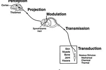
Figure 1 – Pathway and physiologic processes involved in pain sensation ... 8
Figure 2 – Representation of a simple descriptive scale ... 19
Figure 3 – Representation of a numerical rating scale ... 20
Figure 4 – Different representation of a NRS ... 20
Figure 5 – Representation of the visual analogue scale ... 20
Figure 6 – Glasgow Short-Form Composite Measure Pain Scale for assessment of acute pain in canine patients ... 23
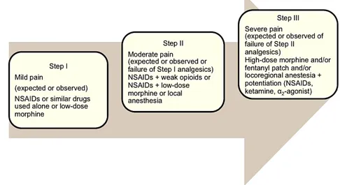
Figure 7 – WHO ladder for pain management approach ... 25
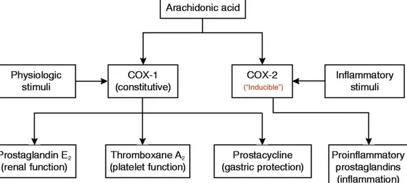
Figure 8 – COX-1 and COX-2 enzymes and NSAID action ... 28
Figure 9 – Schematic representation of parameters involved in Cardiac Output and Blood Pressure maintenance ... 37
Figure 10 – Representation of RAAS as compensatory mechanism in shock ... 44
Figure 11 – Physical examination and complementary tests for diagnostic and monitoring of shock and perfusion ... 46
Figure 12 – Shock index ... 51
Figure 13 – Doppler Ultrasonography equipment used for the measurement of systolic blood pressure ... 56
List of tables
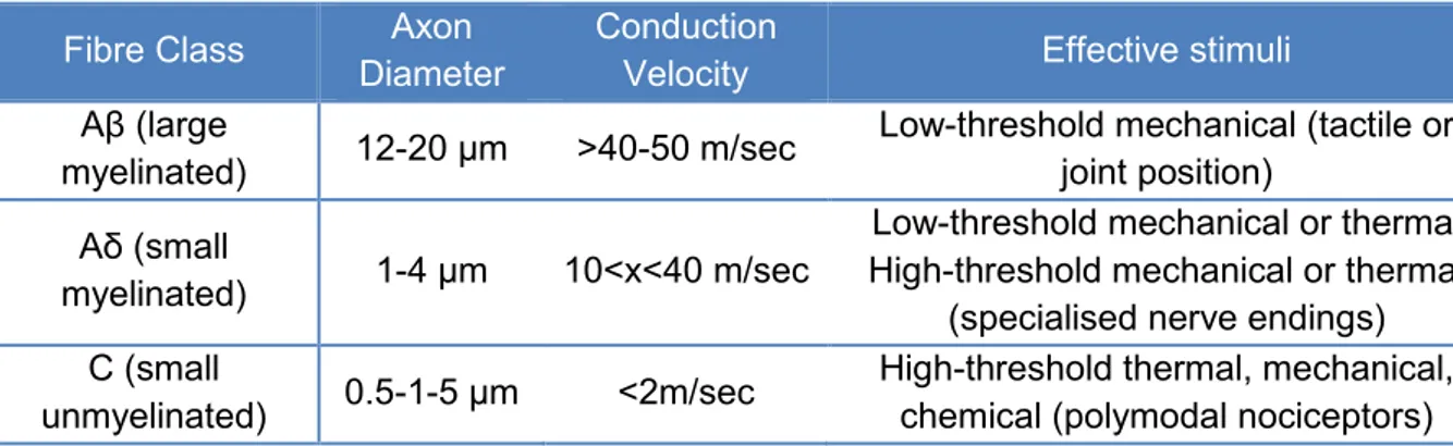
Table 1 – Primary afferent fibres ... 9
Table 2 – Pathophysiologic consequences of pain. ... 14
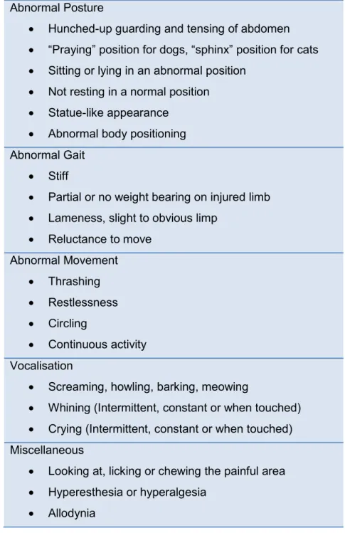
Table 3 – Behavioural and physiologic signs associated with pain in dogs and cats ... 18
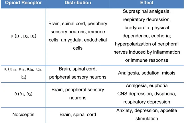
Table 4 – Opioid receptors, distribution and effects. ... 26
Table 5 – General characteristics of most commonly used LAs in veterinary medicine ... 32
Table 6 – Functional Classification and causes of shock ... 40
Table 7 – Clinical parameters at different levels of hypovolemia ... 41
Table 8 – Perfusion parameters during compensatory and decompensatory stages of shock ... 45
Table 9 – Haemodynamic parameters in different types of shock ... 47
Table 10 – Arterial blood pressure ranges for dogs and cats ... 49
Table 11 – Types and causes of hyperlactatemia ... 51
Table 12 – Comparison between Shock Index findings in veterinary medicine studies ... 52
Table 13 –Interpretation of kappa values ... 57
Table 14 – Sample summary characteristics ... 57
Table 15 – Patients admission motives ... 58
Table 16 – Summary of the Glasgow CMPS-SF selected descriptors ... 60
Table 17 – Not Shock group summary characteristics... 61
Table 18 – Shock group summary characteristics ... 62
Table 19 – Results of the Spearman rank correlation testing SI with Age and Glasgow CMPS-SF score ... 63
Table 20 - Pain evaluation with the Glasgow CMPS-SF and by the Veterinarians (“Vetpain”) ... 68
Table 21 – Pain evaluation with the Glasgow CMPS-SF and by the Veterinarians within the shock group ... 69
Table 22 - Pain assessment with the Glasgow CMPS-SF and by the veterinarians within the Not Shock group ... 70
List of charts
Chart 1 – Percentage of time in different clinical area ... 1
Chart 2 – Distribution of hours in each clinical area ... 2
Chart 3 – Dog breeds included in the study ... 58
Chart 4 - Pain perception in the sample with the CMPS-SF (Scale Pain) and the clinicians’ perception of pain (Vet Pain) ... 59
Chart 5 – Pain assessments within the not shock group with the CMPS-SF pain scale and by the veterinarians ... 62
Chart 6 – Pain assessment within the shock group with the CMPS-SF pain scale and by the veterinarians ... 63
Chart 7 – Comparison of patients' age between groups ... 64
Chart 8 – Comparison of gender and Glasgow CMPS-SF scores ... 65
Chart 9 – Comparison of CMPS-SF scores between the two groups ... 65
Chart 10 – Comparison of the veterinarians’ perception of pain in the two groups ... 66
Chart 11 – Comparison of CMPS-SF score readings between the two groups ... 67
Chart 12 – Comparison of CMPS-SF scores and the veterinarians’ perception of pain ... 67
Chart 13 – Comparison of CMPS-SF scores and the veterinarians’ perception of pain within the shock group ... 68
Chart 14 – Comparison of the CMPS-SF scores and the veterinarians’ assessment of pain within the Not Shock group ... 69
List of abbreviations
ABP – Arterial blood pressure
ACE – Angiotensin converting enzyme AcH – Acetylcholine
ACTH – Adrenocorticotrophic hormone ADH – Antidiuretic hormone
AMPA - α-amino-3-hydroxy-5-methyl-4-isoxazolepropionic acid ATP – Adenosine triphosphate
BDNF – Brain-derived neurotrophic factor Bid – Administer two times a day
BP – Blood Pressure Ca2+ – Calcium
CaCO2 – Total arterial oxygen content cAMP – Cyclic adenosine monophosphate CGRP – Calcitonin Gene Related Peptide Cl- – Chloride
CMPS-SF – Short Form Composite Measure Pain Scale CNS – Central nervous system
CO – Cardiac output COX – Cyclooxygenase
CRF – Corticotrophin releasing factor CRI – Constant Rate Infusion
CRT – Capillary refill time CTZ – Chemo-trigger zone CVP – Central Venous Pressure DO2 – Oxygen delivery to tissues E – Epinephrine
ECC – Emergency and critical care ECG - Electrocardiogram
ER – Emergency room
GABA – Gamma-aminobutyric acid
GCMPS – Glasgow Composite Measure Pain Scale GH – Growth hormone
GI - Gastrointestinal H+ - Hydrogen
HPA – Hypothalamic-pituitary-adrenal HR – Heart rate
IASP – International Association for the Study of Pain ICU – Intensive Care Unit
IL-1– Interleukin-1 IL-6 – Interleukin-6 IL-8 – Interleukin-8 IM – Intramuscular IR – Ischemia-reperfusion IV – Intravenous/intravascular K+– Potassium LA – Local anaesthetic LC – Locus ceruleus LOX – Lipoxygenase
MAP – Median Arterial Pressure NS – Not Shock
NE – Norepinephrine
NMDA – N-methyl-D-aspartate NO – Nitric oxide
NY – Neuropeptide Y O2 – Oxygen
p – statistical significance value pKa – acid dissociation constant PAC – Pulmonary Artery Catheter PAG – Periaqueductal Gray PaO2 – Partial pressure of oxygen PAT – Pain assessment tool PGE2 – Prostaglandin E2
PGI2 – Prostaglandin I2/Prostacyclin PGs - Prostaglandins
PO – Per os q - every
QID – Administer four times a day QOL – Quality of life
RAAS – Renin-Angiotensin-Aldosterone-System RAS – Reticular Activating System
S – Shock
SaO2 – Oxygen saturation of hemoglobin SBP – Systolic blood pressure
SC – Sub cutaneous
SDS – Simple Descriptive System Se – Sensitivity
SI – Shock index
Sid – Administer one time a day Sp – Specificity
SV – Stroke volume
TCA – Tricyclic Antidepressant TID – Administer three times a day TNF-α – Tumour necrosis factor alpha TSH – Thyroid stimulating hormone
UMPS – University of Melbourne Pain Scale VAS – Visual Analogue System
List of symbols % percentage α alpha β beta γ gamma δ delta μ mu μg microgram mg milligram kg kilogram ml millilitre < less than > more than = equal to ± more or less
Glossary
Allodynia – pain caused by a stimulus that normally does not cause pain. Analgesia – loss of sensitivity to a stimulus that would normally produce pain
Distress – condition in which stress negatively affects biologic functions critical to the animal's well-being. Distress also means to cause pain or suffering or to make miserable. Hyperalgesia – an increased response to a stimulation that is normally painful (a heightened sense of pain) at the site of injury or in surrounding undamaged tissue.
Primary hyperalgesia – increased sensitivity to a stimulus that is normally painful at the site localized to the area of tissue damage or inflammation.
Secondary Hyperalgesia – increased sensitivity to a stimulus that is normally painful in uninjured or inflamed tissues in areas around and beyond the site of primary site of tissue injury.
Hyperesthesia – increased sensitivity to touch.
Nociception – the physiologic process that leads to the perception of pain.
Noxious stimulus – A stimulus that is damaging or threatens damage to normal tissues. Hypoalgesia – decreased sensitivity to a noxious stimulus.
Pain Threshold – the least amount of pain that an animal can recognize.
Sensitization – an increase in the excitability of neurons, leading to greater sensitivity to stimuli or sensory input.
Suffering – a state of emotional distress associated with events that threaten the biologic and/or psychosocial integrity of the individual.
Wind-Up – sensitization of nociceptors and peripheral and central pain pathways in response to a barrage of afferent nociceptive impulses resulting in expanded receptive fields and an increased rate of discharge.
Chart II – Distribution (in percentage) of time in different clinical areas. Part I - Externship Report
As part of the 6th year of the Integrated Masters in Veterinary Medicine, and held under the Erasmus+ Program, I performed a 4 month training period, starting on October 6th until January 16th, at Village Vet Hampstead (Vet24 London), London, United Kingdom.
My training period was supported under the supervision of Adam Mugford BVetMed MVetMed DACVECC MRCVS (Village Vet Hampstead) and co-supervised by Prof. Dr. Berta São Braz (FMV-UL).
All the procedures and activities I enrolled were performed under the supervision of one of the veterinarians, veterinary nurses or patient care assistants and by the end of this time period, I carried out approximately 700 hours (698h) of training.
The majority of the hours were spent in wards and intensive care unit (Chart I and II); here, I was able to follow and monitor the patients admitted to the practice and many of emergency and critical care cases I came across in this training period. Further information is displayed on annex I.
53%
29% 9%
6% 3%
Wards Medicine Surgery/Minors Imaging Others Chart 1 - Percentage of time in different clinical areas
Chart 2 – Distribution of hours in each clinical area
The routine in wards would start with morning rounds, where all the clinical information about the in-patients was transmitted and discussed by the night-shift veterinarian to the veterinarians on the day rota. After distribution of patients through veterinarians, my role was to assist them and veterinary nurses with their tasks, including: physical examinations, patient restraining, intravenous (IV) catheter checks, IV catheterisation and removal, placing and changing bandages, monitoring of critical care patients, taking blood samples, preparation and administration of medications, set fluid therapy systems and bags, perform scheduled brief major body system examinations (Temperature, Pulse and Respiratory rate - TPR) and non-invasive blood pressure (NIBP) monitoring, pain assessment and general patient welfare, feeding the patients, changing bed and cleaning kennels, and Tender Loving and Care.
In Medicine I had the opportunity to assist consultations, and by doing so I was able to improve my knowledge on how to conduct a conversation with clients in order to get a complete clinical history. I was also present on morning and late ward rounds, where all in-accommodation cases were discussed regarding clinical history, patient condition and medical or surgical management.
My part in Surgery included all aspects of the perioperative period (pre, intra and post-operative). Pre-operatively I helped with patient’s admission, pre-medication preparation and administration, and with the patient’s preparation to surgery (IV placements, clipping and cleaning). In intraoperative care, I helped with the patient’s anaesthesia and vital signs monitoring. In the postoperative phase, I would help with monitoring the patient’s recovery. In some occasions I was allowed to scrub-in with the surgeon and watch the procedures and
Wards Medicine Surgery/Mino
rs Imaging Others Total hours 370 202 63 42 21 0 50 100 150 200 250 300 350 400 Ho u rs
assist in surgery (e.g. perform feline castrations and participate on female spays, watch and assist arthroscopies, splenectomies or mammary gland removals, among others). I was also able to watch and assist veterinarians and veterinary nurses with procedures such as endoscopies (upper and lower tract), central catheterization and urinary catheter placements, naso- and oesophageal feeding tube placements, observe and assist with pleural effusion aspiration, among others.
The Imaging area consisted of radiography and ultrasound, and I was able to participate in both, where I helped the veterinarians and veterinary nurses in the positioning of patients and could discuss the imaging findings with the clinicians.
Regarding other tasks, I was able to help in the laboratory with procedures such as packed cell volume reading, blood film examinations and cytology, urine and urine sediment analysis; whenever possible I would read books or scientific papers about several medical subjects of interest or regarding cases present at the hospital.
During this training period, I was able to perform a prospective study project for my masters’ degree dissertation, regarding the assessment of pain and shock in dogs presenting in emergencies, while using the Glasgow’s short form Composite Measure Pain Scale and the shock index, respectively. The interest on the topic for this dissertation was supported by a small number of data on the evaluation of pain in veterinary patients upon arrival to the clinical environment in emergency situations, with a validated pain scoring system.
As result of this study, an abstract entitled “A Prospective Study Of The Utility Of The Short Form Glasgow Composite Measure Pain Scale (CMPS-SF) In Canine Patients In Shock” (Annex II) was accepted and presented as an oral communication at the 14th Congress of the European Veterinary Emergency and Critical Care Society (EVECCS) held in Lyon and published on the Journal of Veterinary Emergency and Critical Care (JVECC), volume 25, issue S1 (Annex III).
PART II – Literature review
2.1 Introduction
As stated by Colin Allen (1998), “scientific attitudes are, in large measure, a product of education” and in Veterinary Medicine the attitudes towards pain in animals have changed dramatically over the past two decades (Weary, Niel, Flower & Fraser, 2006; Reid, Scott, Nolan & Wiseman-Orr, 2013). For many years the subject of pain had been a neglected area in the veterinary field, since it was common belief that animals would not experience pain as humans do because they do not express it in the same way (Mathews, 2000; Morton, Reid, Scott, Holton & Nolan, 2005; Lockhead, 2010; Mathews et al, 2014). Currently, pain is recognised as a complex sensation that is universally shared by all mammals (Mathews et al, 2014) and it is now well defined in veterinary medicine that the provision of optimal patient care includes the management of pain (Mathews, 2000; Hansen, 2005; Lockhead, 2010; Mathews et al, 2014; Epstein et al, 2015). Regardless of the frequency of situations in which it is encountered, both in human and veterinary medicine, pain is a highly subjective, multidimensional and individual experience. As a consequence, the recognition and assessment of pain in small animal practice is particularly challenging and furthermore complicated by the many limitations in communication that veterinarians experience with their patients, as animals are nonverbal and unable to self-report pain (Muir III & Woolf, 2001; Gogny, 2006; Schnakers, 2012; Sharkey, 2013; Mathews et al, 2014; Perkowski, 2014; Epstein et al, 2015). The consequences of pain, especially when unmanaged, are serious threats to the animal’s well being; it causes both physical and psychological damages; increase the metabolic rate, delays healing, suppresses the immune system and can increase the metastatic rate of some cancers (Downing, 2014). For these reasons, ethical principles of beneficence and nonmaleficence should oblige professionals to provide pain management and comfort to all patients, including those less likely or able to display pain such as critically ill animals (Hansen, 2005; Shaffran et al, 2005; Herr, 2006); those animals in pain may not demonstrate overt signs of distress, and failure to manage pain in these patients can compromise their recovery and contribute to patient mortality (Hansen, 2003; Shaffran et al, 2005; Lockhead, 2010; Mathews et al, 2014). Shock is considered a life-threatening condition often found in the emergency rooms and intensive care units (ICU), frequently associated with pain (Shaffran et al, 2005; Lockhead 2010). In fact, acute pain, in conjunction with blood loss has been shown to be an important factor to increase mortality risk associated with traumatic shock, compared with mortality risk for simple hemorrhagic shock (Wiese, Muir & Wittum, 2004). Although there is no gold standard for pain assessment in veterinary medicine, it is recognized that behavioural signs may be more useful in the identification of abnormal processes as pain and also, the use of a pain scoring system is
beneficial in the assessment and quantification of pain (Lockhead 2010; Sharkey, 2013; Rooney, 2014; Mathews et al, 2014; Epstein et al, 2015). Given that many of the pain assessment tools developed focus on the evaluation of post-operative pain, and that few studies considered the assessment of pain of veterinary patients presenting as emergencies or requiring critical care assistance, it is the purpose of this dissertation to evaluate the use of pain scoring in the identification of pain in canine patients presenting in shock.
2.2 Pain
2.2.1 Definition
Pain is a complex experience and for that reason, defining it can be equally challenging (Sawyer, 1998). Pain is not just a physical impression (“how it feels”); it involves both a physiologic sensation and an emotional component associated to that sensation (“how it makes you feel”) (Lamont, Grimm & Tranquili, 2000; Muir III & Woolf, 2001; Wiese et al, 2004; Mathews et al, 2014), and it is this emotional charge that causes the suffering commonly associated with pain (Mathews et al, 2014). The International Association for the Study of Pain (IASP) (1979) defines it as “an unpleasant sensory and emotional experience, associated with actual or potential tissue damage, or described in terms of such damage”. However, as veterinary patients cannot self-report their experiences verbally, Molony, Kent, Hosie and Graham (1997) suggest an alternative definition: “an aversive, sensory experience representing awareness by the animal of damage or threat to the integrity of its tissues”. Additionally, the IASP also states that the inability to communicate verbally the experience of pain, cannot exclude the possibility that it is present and analgesia is required (Muir III & Woolf, 2001; Meintjes, 2012; Mathews et al, 2014; Epstein et al, 2015). These definitions also recognize the importance of an unpleasant emotional experience, such as fear, in triggering homeostatic responses similar to those induced by noxious stimuli (Muir III & Woolf, 2001).
2.2.2 Pain classification
At simplest, pain can be classified based on its duration as either acute or chronic (Mathews et al, 2014) but there are several proposed classifications. Some authors consider pain either as adaptive (pain that contributes to survival) or as maladaptive (pain as a disease) but despite slight variations on its categorization, it can be classified as physiologic, pathologic, neuropathic, inflammatory or visceral (Lamont et al, 2000; Hellyer et al, 2007; Gaynor & Muir III, 2014; Mathews et al, 2014; Epstein et al, 2015).
2.2.2.1 Physiologic pain
Physiologic pain (also termed nociceptive pain) is considered an adaptive, protective and survival-oriented response quite distinct from the one resulting from overt damage to tissues or nerves (Lamont et al, 2000; Muir III, Wiese & Wittum, 2004). This type of pain occurs after most types of noxious stimulation and initiates self-protective escape and avoidance behaviors and activates a variety of hierarchical homeostatic autonomic responses (stress responses) designed to maintain and restore normal body functions (Muir III et al, 2004). Physiologic pain is only elicited when intense noxious stimuli threaten to injure tissue, and is characterized by a high stimulus threshold, well localized and transient, and demonstrates a stimulus-response relationship similar to those of the other somatic sensations (Lamont et al, 2000).
2.2.2.2 Pathologic pain
Pathologic pain can also be termed as maladaptive or clinical (Lamont et al, 2000; Epstein et al, 2015). It is usually associated with tissue injury incurred at the time of surgery or trauma (Lamont et al, 2000; Gaynor & Muir III, 2014) but can also occur when pain is uncoupled from the noxious stimulus or healing process (Muir III et al, 2004). It is pain as disease and it has been attributed to a variety of pathologic processes as hyperalgesia, allodynia, expansion of the painful field beyond its original boundaries and pain protracted beyond the expected time of inflammation and healing (Muir III, et al 2004; Gaynor & Muir III, 2014a; Epstein et al, 2015).
2.2.2.3 Acute pain
Acute pain has been defined as the one that exists following injury and during the expected time of inflammation and healing. It’s usually self-limiting and should resolve in less than 3 months (Gaynor & Muir III, 2014a; Epstein et al, 2015).
2.2.2.4 Chronic pain
It can be described as pain that lasts beyond the expected time of healing (more than 3 months) (Meintjes, 2012; Gaynor & Muir III, 2014a; Epstein et al, 2015) or as persistent pain caused by conditions where healing did not occur or has recurred (Mathews et al, 2014). 2.2.2.5 Neuropathic pain
Defined as the pain arising from injury or involvement of the peripheral or central nervous system and is possibly associated with motor, sensory or autonomic deficits (Gaynor & Muir III, 2014a). After nerve injury, some changes occur in the sensory transmission of pain, including modifications in the expression of neurotransmitters, neuromodulators, receptors, ion channels and structural proteins. Examples of neuropathic pain in veterinary patients include the one induced by lumbosacral lesions, intervertebral disc herniation and other
spinal cord injuries and discospondylitis, among others (Meintjes, 2012). 2.2.2.6 Inflammatory pain
Inflammatory pain is the most common type (Lemke, 2004). This type of pain is a result of tissue damage and activation of the immune system and release of inflammatory mediators, such as prostaglandins, hydrogen ions and histamine. Usually, inflammatory pain decreases along with the reduction of inflammation (Gaynor & Muir III, 2014a; Epstein et al, 2015). Although it may be seen as pathological by some authors (Lamont et al, 2000), it can also be described as adaptive, in the sense that it contributes to survival by limiting or preventing contact or movement of the affected part, until healing is complete (Gaynor & Muir III, 2014a).
2.2.2.7 Visceral Pain
As the name suggests, visceral pain arises from distension or inflammation of the viscera. The noxious input arising from internal organs is processed by the same nociceptive fibres (Aδ and C fibers) that accompanies sympathetic and parasympathetic pathways (Gaynor & Muir III, 2014a), and therefore the pain emerging from the viscera can be accompanied by typical signs of sympathetic stimulation (tachycardia and tachypnea) and typical behavioural signs. It is usually described as deep, cramping, aching or gnawing and diffuse (without a good localization) (Lamont et al, 2000; Gaynor & Muir III, 2014a).
2.2.2.8 Phantom pain
Phantom pain is described as perceived sensations related to a limb or organ than is not physically part of the body and may be associated with the development of neuromas in amputated limbs (Meintjes, 2012).
2.2.2.9 Cancer pain
Cancer pain can be acute, chronic or intermittent and may be related to the disease itself or to the treatment (Gaynor & Muir III, 2014a).
2.2.3 Pain mechanisms
Pain is the perception of the sensory experience induced by a noxious stimulus (Muir III & Woolf, 2001). Although pain responses are singular to each individual, the pain mechanism and sensory components are similar in all mammals (despite some individual variation in pain sensitivity and response to analgesics within species) (Lamont et al, 2000; Lemke, 2004; Viñuela-Fernandez, et al, 2007). Pain is a sensory event that involves the peripheral and central nervous systems, arising from and reciprocally affecting processes of higher consciousness (Lamont, 2008).
2.2.3.1 Nociception vs Pain
Nociception is the unconscious sensory component of pain - the recognition and neural processing of high intensity noxious stimuli captured by specialized peripheral nerve endings called nociceptors (Lamont et al, 2000; Lemke, 2004; Epstein et al, 2015). As mentioned, this process plays an integral adaptive role as part of the body’s normal defence mechanisms, warning of contact with potentially damaging environmental insults and initiating behavioural and reflex avoidance strategies (Lamont et al, 2000; Meintjes, 2012; Valtolina & Goggs, 2012;). Pain is the endpoint of nociceptive input and processing at higher neural structures and can only occur in a conscious animal (Epstein et al, 2015).
2.2.3.2 Nociceptive processing
Nociception comprises a sequence of processes: transduction, transmission, modulation and perception of the neural signs generated in response to external noxious stimuli (Figure 1). Exemplifying this process as a chain, a first neuron (1st order neuron), originating in the peripheral tissues, is the primary afferent neuron responsible for the recognition, transduction – the transformation of various environmental stimuli into electrical signs (action potentials) – and transmission of the signals from its origin to the dorsal horn of the spinal cord. A second neuron (2nd order neuron), also named projection neuron, receives information from the primary afferent neuron, encodes it (modulation) and projects it to neurons in the medulla, pons, midbrain, thalamus and hypothalamus. Finally, a third neuron in chain (3rd order neuron), also named supraspinal neuron, integrates signals from the lower order neurons and projects them to the subcortical and cortical areas, where pain is finally perceived (Lamont et al, 2000; Lemke, 2004; Wiese & Yaksh, 2014). Though it is possible to describe it through a three-neuron chain, the pain pathway involves much more complexity. Accompanying the ascending pain pathway, from stimulus to its cognitive perception, there is also a descending path – descending inhibitory neurons from the midbrain that modulates afferent transmission of painful stimuli (Lamont et al, 2000; Meintjes, 2012).
2.2.3.2.1 Transduction
Nociceptors respond to high-intensity stimuli that have potential to cause cell damage, such as heat, pressure, vibration and chemicals. Once the nociceptor is triggered, the process of transduction is mediated by membrane bound receptors activated by these stimuli and lead to an influx of sodium and calcium ions along a diffusion gradient, which results in depolarisation of the plasma membrane and generation of an action potential (Meintjes, 2012). These nociceptors are the free endings of afferent nerve fibers that are composed of different populations of axons, including large myelinated, small myelinated and unmyelinated axons. Accordingly to their associated afferent nerve fibers and stimulus sensitivities, pain receptors can be generally classified as A- or C-fibers nociceptors, and as unimodal (sensitive to only one type of stimulus) or polymodal (reactive to several different types of painful stimuli), mechanical, thermal or silent (activated by chemicals and inflammatory mediators) (table 1) (Lemke, 2004; Lamont, 2008; Meintje, 2012; Wiese & Yaksh, 2014).
Table 1 – Primary afferent fibres (Adapted from Handbook of Veterinary Pain Management, Gaynor & Muir III, 3rd Edition, 2014)
The large myelinated and rapidly conducting Aβ fibers respond to nonnoxious low-intensity mechanical stimuli (touch, pressure) but not to noxious stimuli directly. The small myelinated Aδ fibres respond to low and high-intensity thermal or mechanical stimuli and carry the nociceptive input responsible for the fast, sharp pain (first pain) that occurs immediately after injury. The small unmyelinated C fibres typically respond to high-intensity thermal, mechanical and chemical products and are responsible for the prolonged, dull pain (second pain) that occurs after injury (Lemke, 2004; Wiese & Yaksh, 2014). Silent nociceptors are activated by chemical stimuli (inflammatory mediators) and respond to mechanical and thermal stimuli only after they have been activated. These nociceptors also have small, unmyelinated C fibers that conduct impulses at a velocity of less than 3 m/s (Lemke, 2004).
Fibre Class Axon Diameter
Conduction
Velocity Effective stimuli Aβ (large
myelinated) 12-20 μm >40-50 m/sec
Low-threshold mechanical (tactile or joint position)
Aδ (small
myelinated) 1-4 μm 10<x<40 m/sec
Low-threshold mechanical or thermal High-threshold mechanical or thermal
(specialised nerve endings) C (small
unmyelinated) 0.5-1-5 μm <2m/sec
High-threshold thermal, mechanical, chemical (polymodal nociceptors)
2.2.3.2.2 Transmission
The cell bodies of both types of afferent nociceptive nerve fibers are contained in the dorsal root ganglia and extend axons to synapse with dorsal horn neurons (2nd order neurons) within the grey matter of the spinal cord (Lamont et al, 2000). The primary synaptic transmitter present in all types of primary afferents is glutamate, although these fibres corelease other neuropetptides (substance P, neurokinin A, calcitonin gene related peptide) that bind to receptors on dorsal horn neurons (Lemke, 2004; Lamont, 2008). With normal afferent input, glutamate bind to alpha-amino-3-hydroxy-5-methyl-4-isoxazolepropionic acid (AMPA) receptors and neuropeptides bind to neurokinin receptors. The activation of AMPA receptors is responsible for the generation of fast postsynaptic potentials that last a few miliseconds, while the activation of neurokinin receptors is responsible for generating slow synaptic potentials that last several seconds, and reinforce the effects of AMPA receptor activation. In addition to having a prolonged duration of action, neuropeptides diffuse away from the synapse and activate neurons outside of the immediate area, and with intense afferent input, prolonged activation of AMPA and neurokinin receptors lead to progressive cellular depolarization and activation of additional types of glutamate receptors (N-methyl-D-aspartate [NMDA]) on dorsal horn neurons (Lemke, 2004). The dorsal horn represents the first relay point for somatic sensory information. Primary afferent axons may form direct or indirect connections with one of three functional populations of dorsal horn neurons: 1) interneurons, excitatory or inhibitory, 2) propriospinal neurons, which extend over multiple spinal segments and are involved in segmental reflex activity and interactions among stimuli acting at separate sources; 3) projection neurons, which participate in rostral transmission by extending axons beyond the spinal cord to terminate in supraspinal centers such as the midbrain and the cortex.
2.2.3.2.3 Projection
Output from the spinal cord arises from the superficial dorsal horn and this information is projected by the second-order neurons to supraspinal sites (Wiese & Yanksh, 2014). The most prominent ascending nociceptive pathway is the spinothalamic tract and it is divided into two components: medial and lateral. The medial component projects to medial thalamic nuclei and then (via 3rd order neurons) to the limbic system and is involved with the affective-motivational aspect of pain. The lateral component projects to the lateral thalamic nuclei and then to the somatosensory cortex and is responsible for transmission of nociceptive input involved with the sensory-discriminative aspect of pain. The spinoreticular pathway projects to the reticular formation (essential to the integration of nociceptive input) in the medulla and pons, to the thalamic nuclei, and then to the somatosensory cortex. The spinomesecephalic tract projects to the reticular formation and to the periaqueductal grey matter (PAG), which has a central role in the integration and modulation of nociceptive input at supraspinal level
(Lamont et al, 2000; Lemke, 2004). These pathways reflect the underlying dichotomy that pain may be considered to have. In fact, these supraspinal systems can be also be described as somatosensory or affective-motivacional pathways. The somatosensory pathway projecting through the somatosensory thalamus to the somatosensory cortex serves to encode stimulus localisation and intensity (sensory-discriminative), whereas the affective-motivational pathway projects to more medial aspects of the thalamus and others regions such as the anterior cingulate and insula, which are classically associated with emotions and affects (Wiese & Yaksh, 2014).
2.2.3.2.4 Perception
The recognition and processing of sensory information (perception) occurs in multiple specific areas of the brain, which communicate via interneurons to produce an integrated response that reflects the coordinated contributions of arousal, somatosensory input, and autonomic and motor output. The reticular activating system (RAS), located in the brainstem, mediates motor, autonomic and endocrine responses and is a critical center for the integration of these sensory experiences and the subsequent affective and emotional aspects of pain through projection to the medial thalamus and limbic system. Nociceptive information arriving from the dorsal horn is processed in regions such as the pons and medulla, midbrain, PAG and thalamus. The PAG and thalamus serve as relay centers for sensory information transfer; the PAG transfers information to the thalamus and hypothalamus whereas the thalamus transfers information to the cerebral cortex (Wiese & Yaksh, 2014). The thalamus relays information to the somatosensory cortex, which projects information to other cortical regions involved with input association, such as the limbic system. In turn, the limbic system includes several regions such as the cingulate gyrus (responsible for behavior and emotion), amygdala (conditioned fear, anxiety), hippocampus (memory), hypothalamus (sympathetic autonomic activity) and locus ceruleus (arousal, vigilance, behavior). The caudal extension of the limbic system, the PAG, receives descending information from the cortex, amygdala and hypothalamus and descending projections from the medulla, medullary reticular formation (including locus ceruleus) and spinal cord, and is of great importance in the antinociceptive pathway (Valtolina & Gobbs, 2012; Wiese & Yaksh, 2014).
2.2.3.2.4.1 Antinociceptive pathways
Accompanying the ascending nociceptive pathways there are descending antinociceptive pathways that modulate the nociceptive input (Lamont, 2008; Lemke & Creighton, 2010; Valtolina & Gobbs, 2012; Wiese & Yaksh, 2014). These antinociceptive pathways refer to the pain-inhibitory mechanisms that can either reduce the probability of nociceptive stimuli to be perceived as painful or reduce the perceived intensity of pain (Argoff, 2011). The descending
antinociceptive pathways originate at supraspinal level and project to neurons in the dorsal horn of the spinal cord (Lemke & Creighton, 2010). Structures such as the PAG, locus ceruleus and medulla are all particularly important in the modulation of nociceptive input (Lamont, 2008; Lemke & Creighton, 2010; Argoff, 2011). Especially, the PAG is considered to be one of the most important relay points for descending facilitative and inhibitory modulation of nociceptive input (Wiese & Yaksh, 2014). Endogenous opioids (β-endorphins, enkephalins, dynorphins), serotonin and norepinephrine are the main transmitters in the descending antinociceptive pathway (Lamont et al, 2000; Lamont, 2008; Lemke & Creighton, 2010; Argoff, 2011).
2.2.3.3 Neuroplasticity and memory of pain
Neuroplasticity refers to the capacity of the nervous system to modify or adapt its biochemical and physiologic functions in response to different environmental (internal and external) stimuli (Lemke & Creighton, 2010; Wiese & Yaksh, 2014). Neuroplasticity implies that multiple minor sensory events or a single major one can change the stimulus-response characteristics of the nervous system. Several aspects, including the animal’s behaviour pattern, the environment, the expectation of pain and the intensity of previous painful events form the memory of pain, but it is the peak intensity of pain that is the single most important factor in determining the real memory. Additionally, animals that have an inherent memory of pain or of a significant painful event are more difficult to manage, and those in which pain has been persistent (days to weeks) are less responsive to treatment (Wiese & Yaksh, 2014). As result of the neuroplasticity, peripheral and central sensitization occur in response to the barrage of nociceptive input that accompanies tissue trauma and play a central role in the development of pathological pain (Lemke, 2004). This phenomenon occurs as a result of severely altered nervous system function, with dynamic changes both peripherally (peripheral sensitization) and centrally (central sensitization) (Lamont et al, 2000; Lamont, 2008; Argoff, 2011).
2.2.3.3.1 Peripheral sensitization
Peripheral sensitization occurs as a direct consequence of tissue trauma and inflammation and generally reverts after treatment and healing (Viñuela-Férnandez et al, 2007; Lamont, 2008; Lemke & Creighton, 2010; Argoff, 2011; Toth, 2013). Tissue trauma leads to the release of inflammatory mediators such as hydrogen ions (H+), potassium (K+), bradykinin, serotonin, histamine and cytokines (Lemke & Creighton, 2010; Argoff, 2011; Moffat & Rae, 2011; Toth, 2013). The damage to cell membranes also activates the arachidonic acid pathway, which leads to the production of prostaglandins and leukotrienes. Some inflammatory mediators activate nociceptors directly (bradykinin), while others sensitize nociceptors (prostaglandins). Stimulation of nociceptors also leads to antidromal activation of
nociceptive nerve terminals and release of substance P and calcitonin gene related peptide, which causes mast cell degranulation, vasodilation and edema, leading to further sensitization and activation of nociceptors (neurogenic inflammation). Additionally, sympathetic nerve terminals can also contribute to the activation and sensitization of nociceptors by releasing norepinephrine and prostaglandins. The final result is what is commonly named, a “sensitizing soup” of chemical mediators that act synergistically to lower the nociceptors threshold (Lemke & Creighton, 2010; Toth, 2013).
2.2.3.3.2 Central sensitization
In the same way that the peripheral nociceptors can become sensitized, dorsal horn nociceptive neurons can also exhibit increased excitability (Lamont et al, 2000; Lamont, 2008). Central sensitization emphasizes the large plasticity of the somatosensory nervous system in response to activity, inflammation and neural injury. Its occurrence is characterized by a functional improvement of neural circuits in nociceptive pathways, which result from an increase in membrane excitability and a decreased synaptic inhibition. This central sensitization leads to a state of facilitation, potentiation, augmentation or amplification of action potentials (Latremoliere & Woolf, 2009) and therefore it manifests itself as pain hypersensitivity – particularly allodynia, secondary hyperalgesia, aftersensations and an heightened temporal summation (Woolf, 2011). Incessant stimulation of peripheral nociceptors leads to sustained release of glutamate and neuropeptides from afferent nerve fibres. As consequence, continuous activation of AMPA and neurokinin receptors on dorsal horn projection neurons lead to progressive cellular depolarization and further activation of other types of glutamate receptors (NMDA) (Lemke, 2004).
In general, many of the alterations underlying central sensitization are similar to those that produce peripheral sensitization. Numerous intracellular signaling pathways are activated in the dorsal horn by the neurotransmitter glutamate in addition to other neuromodulators (such as substance P and brain derived neutropic factor [BDNF]) (Lamont, 2008). Furthermore, there has been some evidence that glial cells are key players in the formation and maintenance of pathologic pain states. Glial cells (Schuman cells, microglia, astrocytes and oligodendrocytes) are now known to have an important role in the initiation and facilitation of the development of central sensitization (Lamont, 2008; Lemke & Creighton, 2010; Argoff, 2011). Microglia and astrocytes normally are also activated by glutamate and neuropeptides released from primary afferent fibers and are capable of releasing several nociceptive sensitizing agents such as adenosine triphosphate (ATP), nitric oxide (NO), TNF-α, IL-1 and other cytokynes, which directly increase nerve excitability, indicating a role for these cells in the initiation and maintenance of enhanced pain states, including neuropathic pain (Lamont, 2008; Lemke & Creighton, 2010; Argoff, 2011).
2.2.4 Consequences of pain
Animal welfare, well-being and contentment are all key-aspects of animal quality of life (QOL), which has been defined as “a multidimensional, experiential continuum” and should include the “five freedoms”: 1) freedom from thirst, hunger and malnutrition; 2) freedom from discomfort; 3) freedom to express normal behaviour; 4) freedom from fear and distress; 5) freedom from pain, injury and disease. Acute and chronic pain states produce stress and activate defensive biologic responses that result in serious physiologic and behavioural alterations. Generally, the pain phenotype clinically observed results from one or more clinical pathologies and produces multiple neuroendocrine responses (table 2) (Wiese & Yaksh, 2014). Furthermore, stress can change the whole pain experience by causing changes in the brain chemistry, affecting the level of alertness, learning performance and memory, which lead to behavioural adjustments in addition to interdependent autonomic, endocrine and immune alterations (Muir III, 2014a).
Table 2 – Pathophysiologic consequences of pain (Adapted from Handbook of Veterinary Pain Management, 3rd Edition, Gaynor & Muir III, 2014)
Source of pain Symptoms
Cardiovascular Tachycardia, hypertension, vasoconstriction, increased cardiac work and oxygen consumption
Pulmonary Hypoxia, hypercarbia, atelectasis, decreased cough,
ventilation/perfusion mismatch, predisposition to pulmonary infection
Gastrointestinal Nausea, vomiting, ileus
Renal Oliguria, urine retention
Extremities Skeletal muscle pain, limited mobility, thromboembolism Endocrine Vagal inhibition, increased adrenergic activity, increased metabolism,
increased oxygen consumption Central nervous
system Anxiety, fear, sedation, fatigue, depression
Immunologic Impairment, “sickness syndrome”
2.2.4.1 Stress response in pain
Stress triggers a response that prepares the animal for emergency situations (the “fight or flight” response). Regardless of cause, the pain response induces activation of the sympathetic nervous system, secretion of glucocorticoids, hypermetabolism, sodium (Na+) and water retention and altered carbohydrate and protein metabolism (Muir III, 2014a). If untreated, the consequences of pain may extend well beyond unnecessary suffering (Hansen, 2005). In fact, severe or persistent stress can stimulate self-sustained neuroendocrine and immune cascades that degrade homeostatic mechanisms, leading to self-mutilation, immune-incompetence and a “sickness syndrome”. This “sickness syndrome” occurs when animals are intermittently or constantly exposed to factors that activate the immune-inflammatory response and the animals generally demonstrate clinical signs of
hyperalgesia, depression, inappetence and somnolence, or may actually display signs of hyper-vigilance such as anxiety, restlessness and an over-sensitivity, that interfere with the animal’s ability to rest or sleep (Muir III, 2014a).
2.2.4.1.2 Neuroendocrine axis
External stimuli (auditory, visual and somatosensory information) are transmitted to the thalamus or directly to the amygdala, activating the hypothalamic-pituitary-adrenal (HPA) system. This will further stimulate the secretion of CRF and vasoactive intestinal peptide (VIP), which in turn stimulates the pituitary gland to release ACTH, melanocortin, prolactin, vasopressin and thyroid-stimulating hormone (TSH), and growth hormone (GH). The metabolic consequences of these hormonal changes are increased catabolism, the mobilization of substrates to provide energy for tissue repair, and salt and water retention to maintain fluid volume and cardiovascular homeostatis. Additionally, acetylcholine (ACh) released from preganglionic descending sympathetic nerves during the stress response triggers secretion of NE, epinephrine (E) and neuropeptide Y (NPY, a vasoconstrictor) into systemic circulation. E and NE bind to adrenergic receptors, producing a general systemic arousal and prepares the animal for “fight or flight”, increase heart rate and breathing, activate muscles, or dilate blood vessels (muscle, brain, lungs, heart), and increase blood supply to organs involved in fight or flight. The release of CRF in the brain is one of the major components of the stress response, if not the most important. CRF acts synergistically with vasopressin to stimulate the production of ACTH and β-endorfins, thereby enhancing survival and producing analgesic effects, respectively. CRF also stimulates the adrenomedullary release of ACTH and catecholamines. CRF is an excitatory neurotransmitter in the brain, producing increased cortical NE release and excitation (Muir III, 2014a).
2.2.4.1.3 Effects on metabolism
As result of this neuroendocrine response, there is increased secretion of catabolic hormones and thus, a catabolic state succeeds. Generally, pain states are responsible for hyperglycaemia, lipolysis and proteolysis. Hyperglycaemia is due to stress-induced production of glucagon and cortisol (and insulin resistance), and is associated with higher incidence of wound infection, morbidity and mortality. Cortisol, catecholamines and GH stimulate the lipolytic activity, and ultimately, the resultant glycerol is a source for additional gluconeogenesis in the liver. Likewise, protein catabolism is also increased and the resultant amino acids from its breakdown can be used to form new proteins, glucose and other substrates (Muir III, 2014a).
2.2.4.1.4 Effects on the immune system
The immune system can also be perceived as a sensory organ communicating injury-related information to the brain, where the messengers are cytokines (such as interleukin 1 [IL-1],
interleukin 6 [IL-6] and tumour necrosis factor alpha [TNF-α]). In chronic pain states sustained increases of cortisol, norepinephrine, epinephrine and glucagon can suppress the humoral and cellular immune responses. Additionally, the systemic release of endogenous opioids, such as endorphins and encephalin, may contribute to immunosuppression (Muir III, 2014a).
2.2.5 Pain recognition and assessment
Pain assessment presents many challenges. In veterinary medicine there is a struggle to effectively recognise, measure and manage pain as part of ongoing efforts to address the issue of quality of life in companion animals. Pain is a subjective and an individual experience. Considering that and even humans (who can self-report pain) struggle to accurately describe their discomfort and pain quality, this issue is amplified in veterinary medicine, as veterinary patients are unable to verbally express themselves with their caretakers. Therefore, the burden of pain assumption, recognition and assessment rests within veterinary professionals (Morton et al, 2005; Lockhead, 2010; Sharkey, 2013; Perkowski, 2014; Epstein et al, 2015;). In this regard, assessing pain in dogs can be similar to the neonatal and paediatric fields of human medicine, and with the cognitively impaired or nonverbal human patients, where pain assessment depends upon an observer interpretation (Sharkey, 2013).
There are several factors that can affect the observer’s assessment, including both observer and patient related factors. Observers’ age, gender, personal health and clinical experience can introduce bias (Perkowski, 2014). Environmental factors, such as hospital setting and confinement, may alter the likelihood of an animal to display characteristic pain behaviours, and thereby confounding the evaluator’s assessment. Breed and temperament influence the display of pain behaviour as well; some breeds are more stoic and less prone to exhibit pain related behaviour, whereas small toy breeds tend to be more open to showing pain (Lockhead, 2010; Wiese, 2014). Age may also play a role in pain perception. According to Lockhead (2010), paediatric and neonatal patients tend to be more vocal and possibly more communicative about their pain or discomfort, while adults or geriatric patients have a tendency toward stoicism and therefore may tolerate painful states without much complaint. Yet, this subject is still a matter of debate since there have been studies with mixed results, reporting increased, decreased or no change in pain sensitivity with increasing age (Wiese, 2014). Also influencing the recognition of pain in clinical practice are temporal restrictions, as often the staff is unable to perform frequent and whole evaluations of each patient (Lockhead, 2010). Nevertheless, an effort should be made to have all patients evaluated for pain on admission and at regular intervals throughout the hospitalization period (Shaffran et al, 2005).
2.2.5.1 Physiologic signs
Physiologic manifestations of pain are largely related to activation of the sympathetic nervous system. As result, the animal may show increased serum cortisol and catecholamine concentrations, hyperventilation or tachypnea, tachycardia, hypertension, hyperthermia, pale mucous membranes, salivation and pupil dilation. Despite their potential value as indicators of pain, physiologic parameters should not be the only method used to identify or assess pain because the sympathetic nervous system and the stress response can be triggered by non-painful conditions such as disease, fear and anxiety (Lockhead, 2010; Crompton, 2014; Wiese, 2014; Perkowski, 2014).
2.2.5.2 Pain behaviour
As mentioned, it is currently accepted that observation of pain related behaviour is preferred when assessing pain in animals (Sharkey 2013; Wiese, 2014; Mathews et al, 2014; Epstein et al, 2015). Becoming familiarized with normal behaviour in a particular animal and species is the first step in learning to assess abnormal or painful behaviour, as behaviour modification is one of the most import signs of pain in veterinary species (Lockhead, 2010). History taking and the owner’s judgement can also be a valuable aid. Common behaviours displayed by animals in pain can include anxiety, depression, inappetence, reluctance to move and changes in body posture, reclusion and aggression. Further common behavioural changes associated with pain in dogs and cats are briefly described in table 3, but as mentioned the key-factor in pain awareness is the ability of the observer to recognize a change in the behavioural pattern of the animal in pain (Wiese, 2014).
Inappropriately, many people tend to focus on vocalisation and agitation as signs of pain but these behaviours are the least specific (Lockhead, 2010; Wiese, 2014). When the caretaker requires animals to show dramatic signs of pain, the patients are forced to prove to that they are in pain. However, many animals may instinctively resist to a change in behaviour, for reasons such as 1) a way to mask injury (as they would in the wild), 2) because they may be too ill or injured to commit to behavioural change 3) it may be a learned response in some. In regard to critical illness and debilitating disease, these conditions limit the options for coping and the pain related behaviours that an animal would normally show; some patients may not move, stand, shift position, withdraw, vocalise, or show other recognizable responses to pain. They may lose their ability and motivation to care for themselves and may not groom, eat, drink, or ask to be let out and urinate and defecate. Therefore, when assessing an animal for pain a range of factors should be considered, including the type, anatomical location and duration of surgery, medical problem or extent of injury and it should be assumed that pain is present in animals whose condition puts them at risk (Hansen, 2005; Shaffran et al, 2005; Perkowski, 2014).