AR
TIGO ORIGINAL / ORIGINAL AR
TICLE
INTRODUCTION
Celiac disease (CD) is a life-long inlammatory autoimmune disease (AID) of the gastrointestinal tract affecting genetically susceptible individuals(12). This enteropathy shows a well-known familiar predis-position and a prevalence of 16% among irst-degree relatives(1, 6). The risk is even greater in families with affected siblings carrying the human leukocyte antigen (HLA)-DQ2. Multiple cases in the same family are not rare and the risk of CD in second degree relatives is still signiicant(11, 19). Evidence for a familial risk in CD has been accumulated from many sources, including biopsy and serological studies in families with known CD, HLA-genotyping studies, genome-wide expres-sion and linkage studies(14, 18).
Several AID are more prevalent among CD patients and their close relatives compared to the general population(21). The coexistence of CD with other AID reinforces the involvement of common immune mechanisms and genetic factors in the phi-siopathology of these disorders(10). It is possible that chronic lymphocyte stimulation in the intestine of CD patients could result in increased autoantibody
pro-AUTOANTIBODIES IN RELATIVES OF
CELIAC DISEASE PATIENTS:
a follow-up of 6-10 years
Flávia Raphaela NASS
1, Lorete Maria KOTZE
2, Renato M. NISIHARA
1,
Iara Taborda de MESSIAS-REASON
1and Shirley R. da Rosa UTIYAMA
1ABSTRACT – Context - Autoimmune diseases are 3 to 10 times more frequently in patients with celiac disease and their relatives than in the general population. Objective - To investigate a broad spectrum of autoantibodies in celiac disease relatives from Southern Brazil, in a serological follow-up of 6-10 years, aiming to associate with other autoimmune diseases, degree of parentage, demographic and clinical data. Methods - Serum samples of 233 relatives were analyzed in two different phases: n = 186 in phase I (1997-2000) and n = 138 (being 91 = follow-up group and 47 = newly tested) in phase II (2006-2007). As controls, 100 unrelated individuals were evaluated. Autoantibodies to smooth muscle, mitochondrial, liver-kidney microssome, parietal cell and thyroid microssome were tested by indirect immunoluorescence. Results - A signiicant increase of autoantibodies, in both phases, was observed in the rela-tives when compared to the non-relarela-tives (P = 0.0064), speciically to anti-thyroid microssome and anti-parietal cell. In both phases, the female/male proportion of autoantibodies was of 4:1 to 3:1 (P≤0.041). The frequency of autoantibodies amongst 1st and 2nd degree relatives was 11.8% and 9.68% in phase I and 4% and 6.67% in phase II. Conclusion - Celiac disease relatives presented other autoantibodies and serological screening is a useful instrument for identifying autoimmune diseases along the years.
HEADINGS – Autoantibodies. Celiac disease. Autoimmune diseases. Family.
Declared conflict of interest of all authors: none. Financial support: none.
1 Laboratory of Immunopathology, Clinical Hospital, Federal University of Paraná; 2 Gastroenterology Service, Cajuru Hospital, Catholic University of Paraná, Curitiba, PR, Brazil.
Correspondence: Prof. Shirley Ramos da Rosa Utiyama - Rua Padre Camargo, 280 - 80060-240 - Curitiba, PR, Brazil. E-mail: [email protected]
duction and therefore, stimulate the development of other AID. The most frequently reported conditions associated with CD are type 1 diabetes mellitus and autoimmune thyroiditis(4, 11).
Considering that the delay in the diagnosis of CD can be associated to complications such as osteoporo-sis, anemia, infertility, malignancy, and more recently related to other AID(15), the serological screening for autoantibodies (AAB) in relatives of celiac patients represents an important instrument for early detection of these diseases in these individuals. In this study, a broad spectrum of AAB was investigated in relatives of celiacs from southern Brazil, in a serological follow-up of 6-10 years, aiming to associate the data with occur-rence of other AID, the degree of parentage, as well as demographic and clinical data of the individuals.
METHODS
characteristics of these individuals.
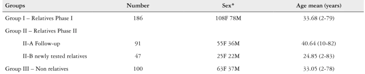
Serum samples of 233 relatives (100♂, 133♀; 2-83 years) were analyzed, being 186 collected in phase I of the study (years 1997 to 2000) and 138 in phase II (years 2006 to 2007). Amongst the last one, 91 relatives were recovery from the phase I, and constitute the follow-up group; while 47 were relatives not evaluated in phase I (newly tested relatives). As control group, we studied 100 unrelated vo-lunteers from the same geographical area, which reported no familial CD cases.
AAB to smooth muscle (SMA), mitochondrial (AMA), liver-kidney microssome (LKM), parietal cell (PCA) and thyroid microssome (ATM) were tested by indirect immuno-luorescence (IFI), as previously described(7, 20), using FITC polyclonal anti-human globulin conjugate (Dako, Denmark). The substrate used in the tests were cryostatic tissue sections of mouse stomach (SMA, PCA), liver (LKM) and kidney (AMA, LKM), and human thyroid (ATM). The sample was considered positive if luorescence was seen at dilution of 1:40 to SMA, 1:10 to ATM, and 1:20 to all the other AAB. All positive samples were tittered up to the end point. Positive and negative controls were used for each bath.
All positive serum for ATM was tested for anti-peroxidase
antibody (anti-TPO) by quimioluminescence (kitDPC -
Dia gnostic Products Corporation, Los Angeles, CA, USA).
The data were compiled in frequency and contingency tables and statistical analyses were performed with Epi-Info, using Chi squared, Fisher and Proportion tests when indi-cated. The signiicance level was set at 0.05.
RESULTS
Signiicant increase of AAB was observed in relatives (10.3%; 24/233) compared to the non-relatives (1%; 1/100;
P = 0.0064), being raised and constant so in phase I (10.75%; 20/186) as in phase II of research (8.7%; 12/138). Amongst the evaluated AAB, an increased frequency for the anti-ATM (6.45%; 12/186; P≤0.0050 and 5.8%; 8/138; P≤0.0117) and anti-PCA (3.76%; 7/186; P≤0.0117 and 3.62%; 5/138;
P≤0.0635) was detected in the two phases, respectively. AMA and anti-LKM were not detected. SMA was positive in two relatives in phase I (1.08%; 2/186) and in one subject in the control group (Table 2).
The female/male proportion of AAB was of 4:1 (16♂; 4♀; P<0.0001) to 3:1 (9♂; 3♀; P = 0.041) in each phase. The frequency of AAB in 1st and 2nddegree relatives was 11.8% (19/161) and 4% (1/25) in phase I and 9.68% (9/93) and 6.67% (3/45) in phase II, without signiicant difference in both. Among different groups of relatives, it was observed increased and constant frequency of positive AAB in siblings
TABLE 1. Demographic data of the relatives of celiac disease patients and control group
Groups Number Sex* Age mean (years)
Group I – Relatives Phase I 186 108F 78M 33.68 (2-79)
Group II – Relatives Phase II
II-A Follow-up 91 55F 36M 40.64 (10-82)
II-B newly tested relatives 47 25F 22M 24.85 (2-83)
Group III – Non relatives 100 63F 37M 33.05 (2-78)
*F=Females; M=Males
TABLE 2. Frequency of autoantibodies in relatives of celiac and non-relatives
Autoantibodies
Phase I Phase II
Non-relatives P* Relatives phase I x Non relatives P* Relatives phase II x Non relatives P* Relatives phase I x Relatives phase II
Follow-up Newly tested
(n = 186) (n = 91) (n = 47) (n=100)
n(%) ATM PCA SMA LKM AMA TOTAL 12 (6.45) 7 (3.76) 2 (1.08) 0 (0) 0 (0) 20 (10.75) 6 (6.59) 3 (3.30) 0 (0) 0 (0) 0 (0) 8 (8.79) 2 (4.26) 2 (4.26) 0 (0) 0 (0) 0 (0) 4 (8.51) 0 (0) 1 (1) 0 (0) 0 (0) 1 (1) 0.0050 0.0473 NS -0.0055 0.0117 0.0635 NS -0.0220 NS NS NS -NS
ATM = anti-thyroid microssome PCA = anti-parietal cell SMA = anti-smooth muscle LKM = anti-liver-kidney microssome
AMA = anti-mitochondrial NS = non signiicant Fisher
in both phases (9.43%; 6.25%, respectively) (Figure 1). No statistical difference was reached between AAB distribution by age, with trend to the signiicance (P = 0.083) in relatives older than 60 years compared to younger individuals in phase II (Figure 2).
The serological follow-up in 91 relatives showed posi-tivity of 7.69% to AAB in phase I, being 2.2% (2/91) to anti-ATM, 4.4% (4/91) to anti-PCA and 1.1% (1/91) to anti-SMA. In a second occasion, after 6-10 years, the total positivity to AAB in the same ones was 8.79% (8/91), with 6.59% (6/91) to anti-ATM and 3.3% (3/91) to anti-PCA, with one relative positive concomitantly to these both antibodies in phase II. After 6-10 years, 12.09% (11/91) of the family members reevaluated showed changes in their serological proile, or remained positive for the same AAB. At this, amongst the seven positive relatives in irst testing, four remained positive (three to anti-PCA; one to anti-ATM) and three became negative, whereas four pre-viously negative individuals became positive to anti-ATM and one relative positive to anti-PCA since phase I demon-strated seroconversion for anti-ATM either (P = NS). Until moment, clinical diagnosis of auto-immune gastritis was conirmed in one followed-up relative.
DISCUSSION
Even though untreated celiac patients, as well as their relatives, present high prevalence of AAB, it has been not yet clariied if CD is an inlammatory disease with secondary auto-immune reactions, or if it is a primary AID induced by known environmental factors. The present study shows an increased prevalence of AAB in relatives of celiac patients, which was signiicantly higher in relation to non-relatives (P = 0.0064; 10.3% and 1% respectively). These data are in concordance with previous reports(10).
This is a pioneer follow-up for AAB in relatives of celiac patients, and the percentage of individuals recovered during 6-10 years was relevant (48.9%; 91/186) compared to previous
studies, where the maximum of recovery samples has been 30.3% (40/132)(5).
Our indings suggest that one time testing is insuficient to identify all AAB positive individuals, and reinforce the need to re-evaluate family members of celiac patients who have been negative on a irst serological screening. In addition, a persistent risk of developing AAB was seen in adults and children in both, irst and second degree relatives. These data suggest that CD relatives should be screened not only for CD but also for other AID, even in the absence of symptoms.
Although the association of thyroid’s AID and CD has been shown to be frequent, the reports on thyroid’s AID in CD relatives are scarce. In both phases of this study a high prevalence to anti-ATM in relatives (P = 0.0050; P = 0.0117), followed for anti-PCA (P = 0.0473; P = 0.0635) was observed in CD relatives when compared with the control group. Ansaldi et al.(2) showed that 26% of Italian CD patients had positive autoimmune thyroid serology compared to 10% in the control subjects. Ventura el al.(24) demonstrated high titers of serum TPO antibodies present in 14.4% of CD patients from Italy. Similar results for parietal cell auto-antibodies were obtained by Utiyama et al.(10) in a Brazilian study that found 3.6% of positivity in CD patients and 3.4% in irst degree relatives.
The serological follow-up of 6-10 years in 91 relatives showed that AAB proile is dynamic, being important to perform more than one screening. This inding is corro-borated by the fact that four previously negative relatives have become positive for anti-ATM in phase II and three relatives remained positive for anti-PCA in the both phases. At present, the clinical-laboratorial evaluation of positive relatives for ATM suggested sub-clinical forms of the disease, requiring a criterious follow-up, considering that celiac rela-tives when in use of gluten can present thyroid disfunction throughout the time(22).
The concurrently occurrence of CD and pernicious ane-mia and/or atrophic gastritis is rare(20). However, in this study,
(1)Group > 60 years phase II x 0-18 years and 19-60 years phase II: P = 0.083
Chi-square with Yates’ correction
FIGURE 2. Positivity to autoantibodies in relatives of phases I and II
in relation to age
(1)Parents phase II x siblings phase II: P = 0.0356 (2)Parents phase II x offsprings phase II: P = 0.002
Fisher
FIGURE 1. Positivity of autoantibodies in relatives of phases I and II in
relation to parentage
P = 0.0356(1)
one female 52 years old, that was amongst the ive relatives positive to anti-PCA in phase II (Table 2), had clinical and endoscopic diagnosis of auto-immune gastritis, while the others relatives still await for the results.
A highly signiicant positivity for AAB was seen in the women when compared to males in both phases (P<0.0001;
P = 0.041, respectively). These results corroborate with the higher predisposition of female including celiac relatives to the development of different AID(8, 9).
The positivity of AAB between relatives > 60 years (17.65%) demonstrated a trend to the significance (P = 0.083) in relation to other individuals, suggesting that the persistent consumption of gluten can represent a trigger to these positivity in genetically susceptible individuals, such as celiac relatives. Although CD occurs at any age and with a great variety of manifestations, there is a marked increase in the incidence rates of CD among adults > 60 years(16). It is important to systematically include serological screening tests for CD and other AID in the evaluation of these susceptible
adults. Atypical manifestations and low suspicion can delay diagnosis even during years(13, 17).
The indings of parentage were interesting. The value of this serological study between siblings of celiac patients was emphasized, since AAB positivity were similar after 6-10 years, in contrast to the frequency of AAB in the other relatives. This issue highlights the increased risk to the development of other AID in these individuals (Figure 1), similarly to the higher risk to develop CD(3, 14).
CONCLUSION
In conclusion, our data suggest that irst and second degree relatives of celiac patients are a group of individuals that shows positive serological test results for AID, hence, this population need to be evaluated for the presence of AAB more than once, irrespective to the presence of symptoms. Serological screening is a useful instrument for identifying these affections along the years.
Nass FR, Kotze LM, Nisihara RM, Messias-Reason IT, Utiyama SRR. Autoanticorpos em familiares de pacientes celíacos: seguimento por 6 a 10 anos. Arq Gastroenterol. 2012;49(3):199-203.
RESUMO – Contexto - Doenças autoimunes são 3 a 10 vezes mais frequentes em pacientes com doença celíaca e em seus familiares que na população em geral. Objetivos - Realizar amplo peril de autoanticorpos em familiares de celíacos do sul do Brasil, em seguimento sorológico de 6-10 anos, visando associá-lo com outras doenças autoimunes, grau de parentesco, dados demográicos e clínicos desses indivíduos. Métodos - Foram analisadas amostras de 233 familiares em duas etapas diferentes: n = 186 na etapa I (1997-2000) e n = 138 (91 recoleta e 47 novos familiares testados) na etapa II (2006-2007). Como controle foram avaliadas amostras de 100 não-familiares. Anticorpos antimúsculo liso, antimitocondrial, anticélula gástrica parietal, antimicrossomal de fígado e rim e antimicrossomal tireoidiano foram testados por imunoluorescência indireta. Resultados - Foi observado um aumento signiicativo de positividade para os autoanticorpos em familiares de celíacos, quando comparados aos não-familiares (P = 0,0064), especiicamente para o antimicrossomal tireoidiano e anticélula gástrica parietal. Entre os indivíduos com autoanticorpos positivos, a proporção do sexo feminino para o masculino foi de 4:1 e 3:1 em ambas as etapas (P≤0,041). A frequência de autoanticorpos detectada entre familiares de primeiro e segundo graus foi de 11,8% e 9,68% na etapa I e 4% e 6,67% na etapa II. Conclusão - Familiares de pacientes celíacos apresentam autoanticorpos positivos e o acompanhamento sorológico desses indivíduos é utilizado como instrumento na identiicação de doenças autoimunes ao longo dos anos.
REFERENCES
1. Almeida PL, Gandoli L, Modelli IC, Martins R de C, Almeida RC, Pratesi R.
Prevalence of celiac disease among irst degree relatives of Brazilian celiac patients. Arq Gastroenterol. 2008;45:69-72.
2. Ansaldi N, Palmas T, Corrias A, Barbato M, D’Altiglia MR, Campanozzi A,
Baldassarre M, Rea F, Pluvio R, Bonamico M, Lazzari R, Corrao G. Autoim-mune thyroid disease and celiac disease in children. J Pediatr Gastroenterol Nutr. 2003;37:63-6.
3. Armstorng MJ, Robins GG, Howdle PD. Recent advances in celiac disease. Curr
Opin Gastroenterol. 2009;25:100-9.
4. Baptista ML, Koda YK, Mitsuneri R, Nisihara RM, Ioshii SO. Prevalence of
celiac disease in Brazilian children and adolescents with type 1 diabetes mellitus. J Pediatr Gastroenterol Nutr. 2005; 41: 621-4.
5. Biagi F, Campanella J, Zanellati G. Incidence of celiac disease in irst negree
relatives [abstract]. In: XII International Celiac Disease Sympsium, New York; 2006. poster D-259 p.86.
6. Biagi F, Corazza GR. First-degree relatives of celiac patients: are they at an
increased risk of developing celiac disease? J Clin Gastroenterol. 2009;43:3-4.
7. Bigazzi PE, Rose NR. Pruebas para anticuerpos contra antígenos tissulares
es-pecíicos. In: Rose NR, Friedman H, ed. El laboratorio en immunologia clínica. Buenos Aires: Pan Americana; 1984. p.968-79.
8. Bonaci-Nikolic B, Andrejevic S, Rodlovic N, Davidovic I, Sofronic L, Spuran
M, Micev M, Nikolic MM. Serological and clinical comparison of children and adults with anti-endomysial antibodies. J Clin Immunol. 2007;27:163-71.
9. Bonamico M, Ferri M, Mariani P, Nenna R, Thanasi E, Luparia RP, Picarelli A,
Magliocca FM, Mora B, Bardella MT, Verrienti A, Fiore B, Uccini S, Megiorni F, Mazzilli MC, Tiberti C. Serologic and genetic markers of celiac disease: a sequential study in the screening of irst degree relatives. J Pediatr Gastroenterol Nutr. 2006;42:150-4.
10. da Rosa Utiyama SR, da Silva Kotze LM, Nisihara RM, Carvalho RF, de Car-valho EG, de Sena MG, de Messias Reason IJ. Spectrum of autoantibodies in celiac patients and relatives. Dig Dis Sci. 2001;46:2624-30.
11. da Rosa Utiyama SR, Nass FR, Kotze LM, Nisihara RM, Ambrosio AR, Messias-Reason IT. Serological screening of relatives of celiac disease patients:
antiendomysium antibodies, anti tissue transglutaminase or both? Arq Gastro-enterol. 2007;44:156-61.
12. da Rosa Utiyama SR. Doença celíaca: aspectos genéticos. In: Barbieri D, Kotze LMS, Rodrigues M, Romaldini CC, ed. Atualização em doenças diarréicas da criança e do adolescente. São Paulo, SP: Atheneu;2010:329-48.
13. Fernández A, González L, de-la-Fuente J. Coeliac disease: clinical features in adult populations. Rev Esp Enferm Dig. 2010;102:466-71.
14. Freeman HJ. Risk factors in familial forms of celiac disease. World J Gastroenterol. 2010;16:1828-31.
15. Gudjónsdóttir AH, Nilsson S, Ek J, Kristiansson B, Ascher H. The risk of celiac disease in 107 families with at least two affected siblings. J Pediatr Gastroenterol Nutr. 2004;38:338-42.
16. Hawkes ND, Swift GL, Smith PM, Jenkins HR. Incidence and presentation of coeliac disease in South Glamorgan. Eur J Gastroenterol Hepatol 2000;12:345-9. 17. Jones S, D’Souza C, Haboubi NY. Patterns of clinical presentation of adult coeliac
disease in a rural setting. Nutr J. 2006;Sep 14;5:24.
18. Kotze LMS. Celiac disease in Brazilian patients: associations, complications and causes of death. Forty years of clinical experience. Arq Gastroenterol 2009;46:261-9. 19. Nass FR, Kotze LM, Nisihara RM, de Messias-Reason IJ, da Rosa Utiyama
SR. Serological and clinical follow-up of relatives of celiac disease patients from Southern Brazil. Digestion. 2011;83:89-95.
20. Rizzetto M, Swana G, Doniach D. Microssomal antibodies in active chronic hepatitis and other disorders. Clin Exp Immunol. 1973;15:331-44.
21. Rostom A, Murray JA, Kagnoff MF. American Gastroenterological Association (AGA) Institute technical review on the diagnosis and management of celiac disease. Gastroenterology. 2006;131:1981-2002.
22. Sategna-Guidetti C, Soleiro E, Scaglione N, Aimo G, Mengozzi G. Duration of gluten exposure in adult coeliac disease does not correlate with the risk for autoimmune disorders. Gut. 2001;49:502-5.
23. Sheehan NJ, Stanton-King K. Polyautoimmunity in a young woman. Br J Rheu-matol. 1993;32:254-6.
24. Ventura A, Neri E, Ughi C, Leopaldi A, Città A, Not T. Gluten-dependent dia-betes-related and thyroid-related autoantibodies in patients with celiac disease. J Pediatr. 2000;137:263-5.