Prevalence of celiac disease in
Brazilian children of short stature
1Departamento de Endocrinologia, Hospital Brigadeiro, São Paulo, SP, Brasil 2Unidade de Diabetes, LIM 25, Divisão de Endocrinologia, and
3Departamento de Gastroenterologia and LIM 06,
Faculdade de Medicina, Universidade de São Paulo, São Paulo, SP, Brasil
M.S. Queiroz1,2,
M. Nery2,
E.L. Cançado3,
D. Gianella-Neto2
and B. Liberman1
Abstract
The aim of the present study was to determine the prevalence of celiac disease in children of short stature and to assess whether some of the routine laboratory examinations performed to determine the cause of short stature could suggest the presence of celiac disease. A total of 106 children of short stature and no gastrointestinal symptoms were studied. An extensive endocrine work-up had been negative for all of them and an additional investigation was performed by measuring the concentration of antiendomysial antibody. Patients who were positive for antiendomysial antibody (≥1:10) or who exhibited IgA deficiency (less than 5 mg/dl) were referred for an endoscopic intestinal biopsy. We detected a pathological titer of antiendomysial IgA in six of these patients. Five of them showed histological abnormalities compatible with celiac disease and one had normal histology and was considered to have potential celiac disease. The prevalence of celiac disease in the population studied was 4.7% (with another 0.9% of the subjects being considered to have potential celiac disease). The children with celiac disease did not differ in any of the parameters tested when compared to those without celiac disease, though they showed an improvement in growth velocity after treatment with a gluten-free diet. We conclude that it is important to test all children with short stature for celiac disease by measuring antiendomysial IgA.
Correspondence
M.S. Queiroz Rua Jaci, 3310
15015-810 São José do Rio Preto, SP Brasil
E-mail: marcia.s.queiroz@uol.com.br Research supported by FAPESP (No. 99/00840-0).
Received February 5, 2003 Accepted September 17, 2003
Key words
•Celiac disease •Short stature
•Antiendomysial antibody •Growth failure
•Celiac sprue
Introduction
Celiac disease is characterized by malab-sorption resulting from inflammatory injury of the mucosa of the small intestine after ingestion of wheat gluten or related rye and barley proteins. The highest reported preva-lence has been observed among western Eu-ropeans and in countries to which EuEu-ropeans emigrated, notably North America and
ap-parently healthy blood donors, while an-other study (3) carried out on patients diag-nosed as having celiac disease described a spectrum of presentation similar to that ob-served in other developing countries.
Celiac disease has a wide spectrum of gastrointestinal and extraintestinal manifes-tations, with many patients showing atypical symptoms or none at all. Classically, infants with celiac disease present impaired growth, diarrhea and abdominal distention between the ages of 4 and 24 months. Atypical dis-ease is usually seen in older children or adolescents, who often have no overt fea-tures of malabsorption. In addition to recur-rent abdominal pain, aphthous stomatitis, arthralgia, defects in dental enamel, short stature, and delayed puberty, affected chil-dren may show behavioral disturbances such as depression and irritability, and may per-form poorly in school (4).
Although celiac disease is a known cause of short stature in children, its diagnosis is often difficult because of the presence of few symptoms and of biochemical param-eters that fall within the normal range. Some-times short stature could be the principal or only finding (5) and the rate of diagnosis depends on the level of suspicion for the disease. The diagnosis is based on clinical symptoms, positive antibodies, and an intes-tinal biopsy, which is considered to be the Gold Standard (6,7). Histological evidence of celiac disease among patients consuming a regular (gluten-containing) diet includes small-bowel mucosal villous atrophy, crypt hyperplasia and increased numbers of intra-epithelial lymphocytes, with clinical im-provement and complete remission of symp-toms occurring after the introduction of a gluten-free diet. Furthermore, the presence of specific antibodies at the time of diagno-sis and their disappearance after treatment with a gluten-free diet has been considered to be a helpful diagnostic criterion.
Serum IgA-class antireticulin, antigliadin, and antiendomysial antibodies are widely used for the screening for celiac disease. However,
and as recommended by the European Society of Paediatric Gastroenterology and Nutrition and by the American Gastroenterological As-sociation, the diagnosis requires histological evidence through a small bowel biopsy (6,7). Prospective studies (8-13) in patients with positive autoantibodies and no abnormalities in the intestinal biopsy have revealed that 28 to 100% of these patients show histological evi-dence of celiac disease within 4 months to 5 years. These findings suggest that positive antibodies could be a marker for gluten sensi-tivity even in the absence of typical histologi-cal abnormalities.
The purpose of the present study was to determine the prevalence of celiac disease among Brazilian children with short stature who have no gastrointestinal symptoms, and to assess whether some of the routine labora-tory examinations performed during investi-gation of the cause of short stature could suggest the diagnosis of celiac disease.
Patients and Methods
A total of 106 children, 34 girls and 72 boys with height less than the 3rd percentile adjusted for age and sex (14), were enrolled in the study. Age ranged from 1.3 to 16.4 years (mean = 9.6 years, SD = 3.6 years). The patients and their parents answered a structured questionnaire, and gastrointesti-nal symptoms were not a major complaint for these patients. The research protocol was reviewed and approved by the Medical Eth-ics Committee of the University Hospital, Faculty of Medicine, University of São Paulo, where the laboratory investigation was per-formed, and of the Brigadeiro Hospital, where the patients enrolled in this study were under endocrinological investigation. Written in-formed consent was obtained from the children’s parents.
total proteins and albumin, determination of immunoglobulin A (IgA), assessment of liver and renal function (determined by standard methods), and hormonal evolution through the measurement of thyroid-stimulating hor-mone, free-thyroxin, growth hormone (GH) (Immulite Diagnostic Products Corporation, Los Angeles, CA, USA), and concentrations of IGF1 and IGF-binding protein-3 (IRMA, Diagnostic Systems Lab. Inc., Webster, CA, USA). Routine GH stimulation testing using either clonidine or insulin-induced hypogly-cemia as secretagogues was performed (oral clonidine, 150 µg/m2; insulin, 0.1 U/kg of weight, iv). Patients were considered not to be GH deficient when the peak GH value during the stimulation test was equal to or higher than 5 ng/ml. All etiologic factors known to produce growth failure had also been excluded, e.g., diabetes mellitus, he-matological and liver disease, renal failure, fetal growth failure, disease of bone metabo-lism, and chromosomal abnormalities.
When no cause of the short stature was found, additional investigation was per-formed by measuring the concentrations of antiendomysial antibody (immunofluores-cence in umbilical cord). Patients who either had positive results for antiendomysial anti-body (titer ≥1:10) or exhibited IgA defi-ciency (IgA less than 5 mg/dl) were referred for an endoscopic intestinal biopsy and four to seven biopsy specimens were taken from the distal part of the duodenum. The slides were examined by the routine anatomy and pathology service of the Hospital, and the results were confirmed by a pathologist ex-perienced in celiac disease. The histological results were scored according to Marsh cri-teria (15). Bone age was determined using the Greulich and Pyle atlas (16). Pubertal stages were evaluated according to Tanner (17).
The results are reported as means ± SD. Statistical analysis was performed by the unpaired Student t-test (GraphPad Prism Software Incorporated), with the level of significance set at P < 0.05.
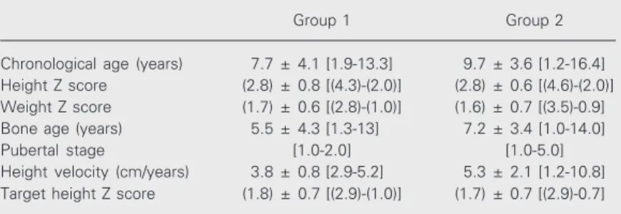
Table 1. Clinical data of patients with short stature with active or potential celiac disease (group 1) and patients with short stature of undetermined cause (group 2).
Group 1 Group 2
Chronological age (years) 7.7 ± 4.1 [1.9-13.3] 9.7 ± 3.6 [1.2-16.4] Height Z score (2.8) ± 0.8 [(4.3)-(2.0)] (2.8) ± 0.6 [(4.6)-(2.0)] Weight Z score (1.7) ± 0.6 [(2.8)-(1.0)] (1.6) ± 0.7 [(3.5)-0.9] Bone age (years) 5.5 ± 4.3 [1.3-13] 7.2 ± 3.4 [1.0-14.0] Pubertal stage [1.0-2.0] [1.0-5.0] Height velocity (cm/years) 3.8 ± 0.8 [2.9-5.2] 5.3 ± 2.1 [1.2-10.8] Target height Z score (1.8) ± 0.7 [(2.9)-(1.0)] (1.7) ± 0.7 [(2.9)-0.7]
The results are reported as means ± SD. The numbers in brackets are range values and the numbers in parentheses are negative values. Height Z score was calculated as patient height minus average height of the population of the same age and sex/ average SD of height for age and sex. Weight Z score was calculated as patient weight minus average weight of the population of the same age and sex/average SD of weight for age and sex. Target height Z score was calculated as patient height minus final height of population of the same sex/average SD of final height of the population of the same sex. P > 0.05 for all parameters (unpaired Student t-test).
Results
Six of the 106 patients had positive serol-ogy for antiendomysial antibody, and under-went an endoscopic intestinal biopsy (group 1, N = 6). Children with a negative antiendo-mysial antibody were included in group 2 (N = 100).
The following histological results were obtained: a) three of six patients (patients 2, 4 and 6) had complete mucosal villous atro-phy with a villus:crypt ratio of 1:1; b) two patients (patients 3 and 5) had subtotal mu-cosal villous atrophy with a villus:crypt ratio of 3:1 associated with a dense inflammatory infiltrate in the lamina propria and an intra-epithelial lymphocyte infiltrate; c) one pa-tient (papa-tient 1) had a normal mucosa. There-fore, the prevalence of well-diagnosed ce-liac disease among children with short stat-ure in this study was 4.7% (5 of 106 patients) and one patient (0.9%) was diagnosed as having potential celiac disease.
disease received a gluten-free diet. The chil-dren under treatment were followed up for 3 months (patient 5), 6 months (patient 6), and 1.5 year (patients 2, 3 and 4); unfortunately patient 1 was lost to follow-up. We observed improvement of growth rate in all patients and patient 4 had complete catch-up of growth after one year on a gluten-free diet (Table 4).
Discussion
The present data show an expressive num-ber of celiac disease (4.7%) children in a group of short stature, thereby justifying the search for this disease in all children with short stature. Previous studies on children with growth failure but without gastrointes-tinal symptoms have shown a variable inci-dence of celiac disease (0 to 59.0%) depend-ing on the region where the study was per-formed (18-25). All patients with a diagnosis of celiac disease showed an improvement in growth velocity after the introduction of a gluten-free diet, indicating that this param-eter would be useful to confirm the correct diagnosis, as also reported by others (19, 23,26). Unfortunately, the patient diagnosed as having potential celiac disease (13,27) because of a positive result in the antiendo-mysial antibody test and no histological ab-normalities was lost to follow-up. These find-ings might be explained by patched altera-tion of celiac disease (28), or by silent/sub-clinical disease (15,26).
Antiendomysial antibody has been shown to have a high sensitivity and specificity for the diagnosis of celiac disease and correlates well with villous atrophy in untreated pa-tients, but false-negative results have been obtained for patients with IgA deficiency, justifying its measurement (29-32). How-ever, an intestinal biopsy continues to be the Gold Standard for the diagnosis of celiac disease (6,7). Our children were also tested for IgA deficiency and all were found to have normal IgA values. Hence, the negative result of the antiendomysial antibody test cannot be attributed to IgA deficiency.
Table 2. Hormonal measurements of patients with short stature and active or poten-tial celiac disease (group 1) and patients with short stature of undetermined cause (group 2).
Group 1 Group 2
T4F (ng/dl) 1.4 ± 0.3 [1.0-1.8] 1.5 ± 0.4 [0.6-2.4] TSH (µIU/ml) 2.2 ± 1.2 [0.6-4.7] 2.2 ± 1.3 [0.2-5.0] GH peak in the stimulatory 11.8 ± 4.8 [7-19.2] 17.1 ± 12.6 [5.5-77.3]
test (ng/ml)
IGF1 Z score (1.2) ± 0.4 [(1.7)-(0.7)] (0.7) ± 0.9 [(2.4)-1.4] IGFBP3 Z score (0.1) ± 0.7 [(0.8)-0.9] (0.3) ± 0.8 [(1.8)-1.4]
The results are reported as means ± SD. The numbers in brackets are range values and the numbers in parentheses are negative values. GH = growth hormone; T4F = free thyroxin; TSH = thyroid-stimulating hormone. IGF1Z score was calculated as patient IGF1 value minus average IGF1 value for the population of the same puberty stage/average SD of the IGF1 value for puberty stage. IGFBP3 Z score was calculated as patient IGFBP3 value minus average IGFBP3 value for the population of same puberty stage/average SD of the IGFBP3 value for puberty stage. P > 0.05 for all parameters (unpaired Student t-test).
Table 3. Correlation between histological results and measurements of plasma antien-domysial antibody titer among group 1 patients.
Patient EmA titer Histological results (IgA)
Patient 1 1/320 Without abnormalities
Patient 2 1/10 Complete mucosal villous atrophy in areas of variable atrophy; villus:crypt ratio, 1:1 to 3:1
Patient 3 1/40 Subtotal mucosal villous atrophy with hypercellular mucosa; villus:crypt ratio, 3:1
Patient 4 1/320 Complete mucosal villous atrophy; villus:crypt ratio, 1:1
Patient 5 1/10 Subtotal mucosal villous atrophy with heavy lympho-plasmocyte infiltrate in the lamina propria; villus:crypt ratio, 3:1
Patient 6 1/320 Complete mucosal villous atrophy; villus:crypt ratio, 1:1
EmA = antiendomysial antibody.
results of laboratory (data not shown) and hormonal assessment were also not signifi-cantly different (P > 0.05, Table 2). None of the patients had cutaneous lesions or dental anomalies that could be associated with a diagnosis of celiac disease.
Table 3 shows the relationship between positive antiendomysial antibody and histo-logical evidence of celiac disease.
A review of the literature (5,18,19,21,33-36) led us to conclude that there is no single parameter suggesting the presence of celiac disease in children of short stature. Our data support the view that there is no single test or measurement that can identify all subjects with celiac disease, with the occurrence of even a few false-positive results. None of measurements (clinical, laboratory and hor-monal) were positive in all of our patients with documented duodenal villous atrophy, nor did they differ significantly between pa-tients diagnosed as having celiac disease and others of short stature of unknown etiol-ogy.
Currently, very little is known about the pathogenesis of growth failure in children with celiac disease. There are reasons to believe that nutritional deficiencies can re-sult in growth failure associated with changes in hormonal status such as poor GH release in a stimulatory test (18,19) and low levels of IGF1 (5). Eichler et al. (37) found a strong relationship between the duration of gluten exposure and reduced IGF1 levels and con-cluded that reduction in IGF1 levels occurs only after prolonged exposure to gluten. The children enrolled in the present study had no gastrointestinal symptoms or significant malnutrition (Z score for weight = -1.6 ± 0.7, and Z score for height = -2.8 ± 0.6) or abnormal GH secretion and a reduction in
Table 4. Clinical data before and after the introduction of a gluten-free diet for subjects with duodenal abnormalities (group 1).
Patient
1 2 3 4 5 6
Before the diet
CA (years) 8.66 9.83 5.91 1.91 13.3 5.41 Height Z score (2.0) (3.2) (3.1) (2.4) (4.3) (2.9) GV (cm/years) 5.1 2.9 3.7 3.6 5.2 3.3 Weight Z score (1.5) (1.3) (1.7) (1.8) (2.5) (2.5)
After the diet
Height Z score - (3.2) (3.2) (1.2) (4.2) (1.5) GV (cm/years) - 5.9 5.2 10.0 6.5 15.3 Weight Z score - (2.2) (1.7) 0 (2.2) (1.2)
The results are reported as means ± SD. The numbers in brackets are range values and the numbers in parentheses are negative values. CA = chronological age; GV = growth velocity. Height Z score was calculated as patient height minus average height of the population of the same age and sex/average SD of height for age and sex. Weight Z score was calculated as patient weight minus average weight of the population of the same age and sex/average SD of weight for age and sex. Patient 1 was lost to follow-up.
IGF1 levels (Z indices for IGF1 = -0.8 ± 0.9) that could explain the delayed growth.
The prevalence of celiac disease amongst children of short stature was 4.7% (with another 0.9% with potential celiac disease). The children affected by celiac disease did not differ from those without celiac disease in any of the parameters tested. Hence, it is important to search for celiac disease in all children with short stature.
References
1. Trier JS (1991). Celiac sprue. New England Journal of Medicine, 325: 1709-1719.
2. Gandolfi L, Pratesi R, Cordoba JC, Tauil PL, Gasparin M & Catassi C (2000). Prevalence of celiac disease among blood donors in Brazil.
American Journal of Gastroenterology, 95: 689-692.
3. de Freitas IN, Sipahi AM, Damiao AO, de Brito T, Cancado EL, Leser PG & Laudanna AA (2002). Celiac disease in Brazilian adults. Journal of Clinical Gastroenterology, 34: 430-434.
4. Farrell RJ & Kelly CP (2002). Celiac sprue. New England Journal of Medicine, 346: 180-188.
5. Verkasalo M, Kuitunen P, Leisti S & Perheentupa J (1978). Growth failure from symptomless celiac disease. A study of 14 patients.
Helvetica Paediatrica Acta, 33: 489-495.
6. Anonymous (2001). American Gastroenterological Association
medi-cal position statement: celiac sprue. Gastroenterology, 120: 1522-1525.
7. Anonymous (1990). Revised criteria for diagnosis of coeliac dis-ease. Report of Working Group of European Society of Paediatric Gastroenterology and Nutrition. Archives of Disease in Childhood, 65: 909-911.
8. Collin P, Helin H, Maki M, Hallstrom O & Karvonen AL (1993). Follow-up of patients positive in reticulin and gliadin antibody tests with normal small-bowel biopsy findings. Scandinavian Journal of Gastroenterology, 28: 595-598.
9. Kaukinen K, Maki M, Partanen J, Sievanen H & Collin P (2001). Celiac disease without villous atrophy: revision of criteria called for.
Digestive Diseases and Sciences, 46: 879-887.
(1998). Small-bowel mucosal inflammation in reticulin or gliadin antibody-positive patients without villous atrophy. Scandinavian Journal of Gastroenterology, 33: 944-949.
11. Maki M, Holm K, Koskimies S, Hallstrom O & Visakorpi JK (1990). Normal small bowel biopsy followed by coeliac disease. Archives of Disease in Childhood, 65: 1137-1141.
12. O’Farrelly C, Graeme-Cook F, Hourihane DO, Feighery C & Weir DG (1987). Histological changes associated with wheat protein anti-bodies in the absence of villous atrophy. Journal of Clinical Patholo-gy, 40: 1228-1230.
13. Troncone R, Greco L, Mayer M, Paparo F, Caputo N, Micillo M, Mugione P & Auricchio S (1996). Latent and potential coeliac dis-ease. Acta Paediatrica. Supplement, 412: 10-14.
14. Marques RM, Marcondes E, Berquo E, Prandi R & Yunes J (1982).
Crescimento e Desenvolvimento Pubertário em Crianças e Adoles-centes Brasileiros. II. Altura e Peso. Editora Brasileira de Ciências, São Paulo, SP, Brazil.
15. Oberhuber G, Granditsch G & Vogelsang H (1999). The histopathol-ogy of coeliac disease: time for a standardized report scheme for pathologists. European Journal of Gastroenterology and Hepatolo-gy, 11: 1185-1194.
16. Greulich WW & Pyle SI (1959). Radiographic Atlas of Skeletal Devel-opment of the Hand and Wrist. 2nd edn. Stanford University Press, Stanford, CA, USA.
17. Tanner JM (1962). Growth at Adolescence with a General Consider-ation of the Effects of Hereditary and Environmental Factors upon Growth and Maturation from Birth to Maturity. 2nd edn. Blackwell Scientific Publications, Oxford, England.
18. Bonamico M, Scire G, Mariani P, Pasquino AM, Triglione P, Scaccia S, Ballati G & Boscherini B (1992). Short stature as the primary manifestation of monosymptomatic celiac disease. Journal of Pedi-atric Gastroenterology and Nutrition, 14: 12-16.
19. Cacciari E, Salardi S, Lazzari R, Cicognani A, Collina A, Pirazzoli P, Tassoni P, Biasco G, Corazza GR & Cassio A (1983). Short stature and celiac disease: a relationship to consider even in patients with no gastrointestinal tract symptoms. Journal of Pediatrics, 103: 708-711.
20. de Lecea A, Ribes-Koninckx C, Polanco I & Calvete JF (1996). Serological screening (antigliadin and antiendomysium antibodies) for non-overt coeliac disease in children of short stature. Acta Paediatrica. Supplement, 412: 54-55.
21. Groll A, Candy DC, Preece MA, Tanner JM & Harries JT (1980). Short stature as the primary manifestation of coeliac disease. Lan-cet, 2: 1097-1099.
22. Oliveira MCLA, Reis FJC, Chagas AJ, Brasileiro-Filho G, Bahia M, Silva LD & Penna FJ (1998). Estudo de doenças de má absorção intestinal como causa de baixa estatura monossintomática. Pala-vras-chave. Jornal de Pediatria, 74: 213-216.
23. Rosenbach Y, Dinari G, Zahavi I & Nitzan M (1986). Short stature as
the major manifestation of celiac disease in older children. Clinical Pediatrics, 25: 13-16.
24. Rossi TM, Albini CH & Kumar V (1993). Incidence of celiac disease identified by the presence of serum endomysial antibodies in chil-dren with chronic diarrhea, short stature, or insulin-dependent dia-betes mellitus. Journal of Pediatrics, 123: 262-264.
25. Stenhammar L, Fallstrom SP, Jansson G, Jansson U & Lindberg T (1986). Coeliac disease in children of short stature without gas-trointestinal symptoms. European Journal of Paediatrics, 145: 185-186.
26. Ciclitira PJ (2001). AGA technical review on celiac sprue. American Gastroenterological Association. Gastroenterology, 120: 1526-1540. 27. Troncone R (1995). Latent coeliac disease in Italy. The SIGEP Work-ing Group on Latent Coeliac Disease. Italian Society for Paediatric Gastroenterology and Hepatology. Acta Paediatrica, 84: 1252-1257. 28. Marsh MN (1993). Clinical and pathological spectrum of coeliac
disease. Gut, 34: 1740.
29. Ferreira M, Davies SL, Butler M, Scott D, Clark M & Kumar P (1992). Endomysial antibody: is it the best screening test for coeliac dis-ease? Gut, 33: 1633-1637.
30. Hin H, Bird G, Fisher P, Mahy N & Jewell D (1999). Coeliac disease in primary care: case finding study. Bristish Medical Journal, 318: 164-167.
31. Johnston SD, Watson RG, McMillan SA, McMaster D & Evans A (1996). Preliminary results from follow-up of a large-scale popula-tion survey of antibodies to gliadin, reticulin and endomysium. Acta Paediatrica. Supplement, 412: 61-64.
32. McMillan SA, Haughton DJ, Biggart JD, Edgar JD, Porter KG & McNeill TA (1991). Predictive value for coeliac disease of antibodies to gliadin, endomysium, and jejunum in patients attending for jeju-nal biopsy. Bristish Medical Journal, 303: 1163-1165.
33. Knudtzon J, Fluge G & Aksnes L (1991). Routine measurements of gluten antibodies in children of short stature. Journal of Pediatric Gastroenterology and Nutrition, 12: 190-194.
34. Corazza GR, Frisoni M, Treggiari EA, Valentini RA, Filipponi C, Volta U & Gasbarrini G (1993). Subclinical celiac sprue. Increasing occur-rence and clues to its diagnosis. Journal of Clinical Gastroenterol-ogy, 16: 16-21.
35. Bottaro G, Cataldo F, Rotolo N, Spina M & Corazza GR (1999). The clinical pattern of subclinical/silent celiac disease: an analysis on 1026 consecutive cases. American Journal of Gastroenterology, 94: 691-696.
36. Lejarraga H, Caino S, Salvador A & De Rosa S (2000). Normal growth velocity before diagnosis of celiac disease. Journal of Pedi-atric Gastroenterology and Nutrition, 30: 552-556.