• Nenhum resultado encontrado

Arq. Bras. Cardiol. vol.98 número4 en v98n4a14

N/A
N/A
Protected

Academic year: 2018

Share "Arq. Bras. Cardiol. vol.98 número4 en v98n4a14"

Copied!
4
0
0

Texto

(1)

Case Report

Introduction

Antiphospholipid antibody syndrome (APS) is a clinical entity characterized by two fundamental components: first, the presence of serum antibodies against phospholipids or phospholipid-binding proteins, called antiphospholipid antibodies, of which the best known are lupus anticoagulant, anticardiolipin and anti-β2-glycoprotein I; second, at least one of the several clinical manifestations, of which the most common are venous and arterial thromboses, gestational morbidity and thrombocytopenia1.

This syndrome can be present as the primary condition, but is also secondary to other diseases, particularly systemic lupus erythematosus (SLE)2. Although the thrombosis more often affects deep venous segments of the lower limbs, arterial thrombosis may also occur. Brain vessels are the most common site of arterial thrombosis, which, more rarely, can affect the coronary arteries2.

Case Report

We report on a 37-year-old female patient, diagnosed with SLE 14 years before with the following criteria of diagnostic classification by the American College of Rheumatology3:

arthritis, serositis, photosensitivity and positive serology for antinuclear antibodies (ANA). The patient is undergoing outpatient follow-up, receiving prednisone, chloroquine, calcium carbonate and vitamin D. She had positive antiphospholipid antibodies in plasma (anticardiolipin antibody and lupus inhibitor) at the time of diagnosis of SLE, but as she had no clinical manifestations of APS at that time, this diagnosis was not confirmed. Primary prophylaxis was started with acetylsalicylic acid (ASA). She had an episode of atypical chest pain about two months before the current hospitalization, at the time investigated through myocardial scintigraphy with 99mTc-sestamibi associated to pharmacological stress testing with intravenous dipyridamole and normal perfusion was observed.

She was admitted with a history of precordial pain of moderate intensity, with chest tightness, irradiating to the back for about 18 hours, with no other associated symptoms. Initially she had sought a basic health unit, where she was treated with painkillers, with partial pain improvement, and was released to go home. Approximately 12 hours after the first assessment, she once again sought medical care in the same unit due to chest pain persistence, and at this time she was referred for hospital evaluation, arriving at our service 18 hours after symptom onset. She denied a history of systemic arterial hypertension, dyslipidemia, diabetes mellitus, smoking and family history of coronary artery disease. At the clinical examination, she had only tachycardia (HR: 110 bpm) without any other significant alterations.

The electrocardiography showed sinus tachycardia, electrically inactive area in the antero-septal wall, ST-segment elevation in the high lateral wall (DI and aVL) and ST segment depression in the inferior wall (Figure 1).

During the clinical evolution, elevated serum troponin I levels were observed (4.10 mcg/L, 99th percentile: 0.01) and high values in the CK-MB time curve (186, 121, 110, 28 U/L; at admission and after 6, 12 and 48 hours, respectively).

The diagnosis of acute myocardial infarction (AMI) was confirmed. As the time of symptom evolution was > 12 hours, no myocardial reperfusion therapy was performed. She received dual antiplatelet aggregation therapy (aspirin and clopidogrel), low molecular weight heparin and captopril.

On the second day of hospitalization, she underwent an echocardiogram, which showed moderately depressed global left ventricular systolic performance, with ejection fraction of 36%, and akinesis of segments: basal anterior

Keywords

Lupus erythematosus, systemic; antiphospholipid syndrome / complications; coronary thrombosis; myocardial infarction.

The antiphospholipid syndrome (APS) is an autoimmune thrombophilia, characterized by the presence of plasma antibodies against phospholipids, associated with recurrent episodes of venous and/or arterial thrombosis and gestational morbidity (especially recurrent miscarriage).

We report the case of a young female patient diagnosed with systemic lupus erythematosus (SLE) associated with the presence of antiphospholipid antibodies for a long time, presenting with acute myocardial infarction (AMI) due to proximal thrombosis of the anterior descending artery as the first clinical complication of APS.

Coronary Thrombosis as the First Complication of Antiphospholipid

Syndrome

Carlos Henrique Miranda

1

, Luís Gustavo Gali

1

, José Antônio Marin-Neto

2

, Paulo Louzada-Júnior

2

, Antônio Pazin-Filho

2

Unidade Coronariana da Unidade de Emergência do Hospital das Clínicas da Faculdade de Medicina de Ribeirão Preto da Universidade de São Paulo1, Ribeirão Preto, SP; Divisão de Cardiologia - Divisão de Imunologia e Divisão de Emergências Clínicas da Faculdade de Medicina de

Ribeirão Preto da Universidade de São Paulo2, Ribeirão Preto, SP, Brazil

Mailing address: Carlos Henrique Miranda •

Rua Bernardino de Campos, 1000 - Centro - 14015-130 - Ribeirão Preto, SP, Brazil

E-mail: chmirand@cardiol.br, chmirand@yahoo.com.br

Manuscript received February 12, 2011; revised manuscript received July 01, 2011; accepted July 04, 2011.

(2)

Case Report

Miranda et al AMI and antiphospholipid antibody syndrome

Arq Bras Cardiol 2012;98(4):e66-e69

descending artery (DA), presence of inter and intracoronary collateral circulation, left ventricular anterior wall regional contraction disorder and moderately depressed global function, as well as an image suggestive of mural thrombus (Figure 2).

Myocardial scintigraphy with thallium-201 at rest was performed to evaluate myocardial viability on the 7th day septal, mid-anterior septal, apical septal, basal anterior,

mid-anterior, antero-apical and hypokinesis of the segments: mid-inferior septal and lateral-apical. The pericardium was normal, without effusions.

She underwent a coronary angiography on the fourth day of hospitalization, which disclosed a one-vessel obstruction, with proximal thrombotic occlusion of the anterior

Figure 1 - 12-lead electrocardiogram at rest showing sinus tachycardia, electrically inactive area in the antero-septal wall, ST-segment elevation in the high lateral wall (DI and aVL) and ST-segment depression in inferior wall. Reprinted with authorized consent from project CEP / HCFMRP-USP-11738/2004.

Figure 2 – Coronary angiography showing proximal thrombotic occlusion of the anterior descending artery and presence of inter and intracoronary collateral circulation.

AD- anterior descending artery; CX - circumlex artery, RC - right coronary artery; CC collateral circulation; * - site of acute thrombotic occlusion. Reprinted with authorized

consent from project CEP / HCFMRP-USP-11738/2004.

(3)

Case Report

Miranda et al

AMI and antiphospholipid antibody syndrome

Arq Bras Cardiol 2012;98(4):e66-e69

of hospitalization, demonstrating viability associated with non-transmural fibrosis in a large extension of the AD artery territory. She was submitted to percutaneous transluminal coronary angioplasty on the following day, with non-pharmacological stent implant in the AD artery, resulting in arterial recanalization without complications.

The anticardiolipin antibody (ACA) measurement was repeated with IgG of 33.8 GPL/mL (positive> 14 GPL / mL) and IgM of 3.7 MPL/mL (negative < 7 MPL/ ml) and anti-β2-Glycoprotein I (anti-β2GP I) with IgG of 38.22 (positive > 8 U/mL) and IgM of 3.17 (negative < 5.0 U/ mL). Thus, the presence of arterial thrombosis associated with antiphospholipid antibodies (ACA and anti-β2GP I) defined the diagnosis of APS, according to the international consensus of 20061.

Pa t i e n t e v o l u t i o n w a s a s y m p t o m a t i c , w i t h o u t hemodynamic instability or signs of pulmonary congestion (Killip I). She was discharged after 14 days of hospitalization, with prescriptions for three antithrombotic agents (aspirin, clopidogrel and warfarin) and anti-remodeling medications (captopril, and beta-blocker). These three antithrombotic agents were scheduled to be taken for a month and after that, clopidogrel would be discontinued, while maintaining aspirin and warfarin.

Discussion

SLE may cause chest pain due to pleuritis, pericarditis, pneumonia, pulmonary thromboembolism, arthritis and heart disease. Studies have shown that patients with SLE have a 2 to 50-fold higher risk of atherosclerotic heart disease, which is a major cause of premature mortality in this disease4.

SLE patients are, in most cases, young women who are usually considered at low risk for coronary atherosclerosis. Furthermore, the chest pain associated with obstructive coronary artery disease in these patients may be atypical. These characteristics may delay the correct diagnosis of AMI as a cause of chest pain in these individuals, which may increase morbimortality due to loss of temporal windows of opportunity for appropriate treatment, as observed in our case.

The mechanism responsible for the accelerated atherosclerosis process in SLE patients is multifactorial and not fully understood. There is high prevalence of traditional risk factors for atherosclerosis such as diabetes, dyslipidemia, hypertension, which may be partly secondary to adverse effects of the prolonged use of corticosteroids, and the intrinsic inflammatory process of the disease must play a central role in this process5,6.

In addition to the aforementioned factors, there is an association of SLE with APS. There is high prevalence of antiphospholipid antibodies in SLE patients: approximately 12% to 30% have anticardiolipin antibodies and 15% to 34%, lupus anticoagulant antibodies. About 50% to 70% of these patients with SLE and antiphospholipid antibodies develop APS with clear clinical manifestations, during a follow-up period of 20 years2. Additionally, when APS develops in the context of SLE, there is high prevalence of thrombosis, especially deep venous thrombosis of the lower limbs, which occurs in 29% to 55% of patients during a six-year follow-up7.

Arterial thrombosis can also occur in these patients, 50% of the time in brain vessels, presenting as stroke or transient ischemic attack; in 23% of the cases, coronary arteries are involved, presenting as AMI or angina, and the remaining 27 % correspond to other diverse arterial beds, such as renal, subclavian, retinal arteries, etc2.

The analysis of 59 patients followed in our hospital with APS (27 primary APS and 32 with APS secondary to SLE) showed that arterial thrombosis is more prevalent in primary APS and that there were no cases of coronary involvement in this series8.

Although acute in situ thrombosis is the most likely mechanism of coronary occlusion causing AMI in this patient, we cannot rule out the possibility of embolism from noninfectious thrombotic vegetations that can affect these patients (thrombotic non-infectious Libman Sacks endocarditis). However, this type of embolization occurs preferentially in brain vessels, and the echocardiography did not detect any sign of this abnormality.

Regarding the chronic approach of these patients with APS, oral anticoagulation with warfarin associated with aspirin use is recommended in cases of arterial thrombosis, and most specialists advise the extended and indefinite use due to the high recurrence rate of thrombotic events (93% for arterial thrombosis and 76% for venous thrombosis). In our case, the indication for oral anticoagulation was reinforced by the presence of the intraventricular mural thrombus9.

There is no scientific evidence for the use of clopidogrel as secondary prophylaxis in these patients and its use was justified only in our patient due to the performance of percutaneous transluminal coronary angioplasty with stent implant.

In patients with SLE and antiphospholipid antibodies, but no clinical manifestations of APS, one should consider the use of aspirin as the primary prophylaxis9.

Conclusion

The definitive diagnosis of the cause of chest pain in patients with SLE is always a challenge for the physician, but the possibility of a coronary artery disease should always be considered among the possible diagnoses. APS is an autoimmune disease associated with an increased risk of thrombosis, especially in deep venous territories, but it also can affect the coronary arteries.

Potential Conflict of Interest

No potential conflict of interest relevant to this article was reported.

Sources of Funding

There were no external funding sources for this study.

Study Association

This study is not associated with any post-graduation program.

(4)

Case Report

References

1. Miyakis S, Lockshin MD, Atsumi T, Branch DW, Brey RL, Cervera R, et al. International consensus statement on an update of the classification criteria for definite antiphospholipid syndrome (APS). J Thromb Haemost. 2006;4(2):295-306.

2. Levine JS, Branch DW, Rauch J. The antiphospholipid syndrome. N Engl J Med. 2002;346(10):752-63.

3. Hochberg MC. Updating the American College of Rheumatology revised criteria for the classification of systemic lupus erythematosus. Arthritis Rheum. 1997;40(9):1725.

4. Manzi S, Meilahn EN, Rairie JE, Conte CG, Medsger TA Jr, Jansen-McWilliams L, et al. Age-specific incidence rates of myocardial infarction and angina in women with systemic lupus erythematosus: comparison with the Framingham Study. Am J Epidemiol. 1997;145(5):408-15. 5. Petri M, Perez-Gutthann S, Spence D, Hochberg MC. Risk factors for

coronary artery disease in patients with systemic lupus erythematosus. Am J Med. 1992;93(5):513-9.

6. Bruce IN, Gladman DD, Urowitz MB. Premature atherosclerosis in systemic lupus erythematosus. Rheum Dis Clin North Am. 2000;26(2):257-78.

7. Cervera R, Piette JC, Font J, Khamashta MA, Shoenfeld Y, Camps MT, et al.; Euro-Phospholipid Project Group. Antiphospholipid syndrome: clinical and immunologic manifestations and patterns of disease expression in a cohort of 1,000 patients. Arthritis Rheum. 2002;46(4):1019-27.

8. Freitas MVC, Silva LM, Petean FC, Carvalho IF, Franco RF, Donadi EA, et al. Síndrome do anticorpo antifosfolípide: estudo comparativo entre a forma primária e secundária ao lúpus eritematoso sistêmico. Rev Bras Reumatol. 2003;43(3):153-9.

9. Khamashta MA, Cuadrado MJ, Mujic F, Taub NA, Hunt BJ, Hughes GR. The management of thrombosis in the antiphospholipid-antibody syndrome. N Engl J Med. 1995;332(15):993-7.

Miranda et al AMI and antiphospholipid antibody syndrome

Imagem

Figure 2 –  Coronary angiography showing proximal thrombotic occlusion of the anterior descending artery and presence of inter and intracoronary collateral circulation

Referências

Documentos relacionados

Antiphospholipid antibodies (aPL) are a heterogeneous group of antibodies that are detected in the serum of patients with a variety of conditions, including autoimmune (systemic

In our study, the presence of antiphospholipid antibodies in premenopausal patients with SLE was associated with lower circulating estradiol levels, but not with prolactin levels..

Objective: To determine the frequency of antiparvovírus B19 (B19) antibodies in patients with rheumatoid arthritis (RA) and systemic lupus erythematosus (SLE), and the possible

Herein, we report a case with neovascular glaucoma secondary to ischemic central retinal vein occlusion associated to antiphospholipid antibodies in a young patient..

We report a case study describing the diagnosis and management of limbic encephalitis in a patient with active Systemic Lupus Erythematosus disease (SLE) and past medical history

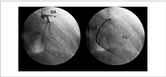
W e describe a patient with systemic lupus erythematosus associated with antiphospholipid antibodies who developed chorea, which has been considered a sign of lupus activity..

Unilateral central retinal artery occlusion as the sole presenting sign of Susac syndrome in a young man: case report.. Arq

De acordo com WOITCHUNAS et al (2010), a mordida cruzada é classificada de três formas: 1) Más posições dentárias: A mordida cruzada dentária envolve somente uma