Autoimmune limbic encephalitis
A manifestation of systemic lupus erythematosus
in the central nervous system
Débora Bartzen Moraes Angst, Nathália Stela Visoná de Figueiredo, Valmir Passarelli, Meire Argentoni Baldocchi, Maria Sheila Guimarães Rocha, Sonia Maria Dozzi Brucki
ABSTRACT. Autoimmune limbic encephalitis (ALE) associated with systemic lupus erythematosus (SLE) is a rare entity with few reports in the literature to date. In general, ALE associated with SLE has a satisfactory response to immunosuppressive treatment (RIT), but the pathogenesis of this association is poorly understood and may include an autoimmunity component. We report a case study describing the diagnosis and management of limbic encephalitis in a patient with active Systemic Lupus Erythematosus disease (SLE) and past medical history of cancer (endometrial adenocarcinoma in 2004 and papillary urothelial carcinoma in 2011 with curative treatment), followed over a one-year period. We discuss the possible association between limbic encephalitis and all past neoplastic and immune-mediated conditions of this patient. In this particularly case, autoimmunity was the most relevant factor associated with limbic encephalitis given negative neoplastic screening. Moreover, a good response was observed to immunotherapy, not seen with paraneoplastic limbic encephalitis, which is associated with poor response. In this case, the association of ALE with SLE is possible, since laboratory testing disclosed lupic activity and the patient had involvement of other systems (such as hematologic) during the period. However, the presence of other surface membrane antibodies are possible in the search for alternative etiologies.
Key words: limbic encephalitis, lupus erythematosus systemic, neoplasms.
ENCEFALITE LÍMBICA AUTO-IMUNE: MANIFESTAÇÃO DO LÚPUS ERITEMATOSO SISTÉMICO NO SISTEMA NERVOSO CENTRAL RESUMO. Encefalite Límbica Autoimune (EL) associada a lúpus eritematoso sistêmico (LES) é uma entidade rara, com poucos relatos na literatura até o momento. Em geral, EL associada com LES tem uma resposta satisfatória ao tratamento imunossupressor, mas a patogênese desta associação é pouco compreendida e pode incluir um componente de autoimunidade. Descrevemos em um estudo de caso o diagnóstico e o tratamento empregado na encefalite límbica ocorrida no contexto de uma paciente com LES ativo e história pregressa de doenças neoplásicas (adenocarcinoma endometrial em 2004 e carcinoma papilar urotelial em 2011 ambos com o tratamento curativo), a qual foi seguida durante um ano. Discutimos uma possível associação de encefalite límbica e todos os antecedentes neoplásicos e imunomediados desta paciente. Neste caso em particular, a autoimunidade é o fator mais relevante relacionado com a encefalite límbica devido a uma triagem neoplásica negativa. Além disso, houve uma grande resposta com a imunossupressão, o que não é visto na encefalite límbica paraneoplásica, mais relacionada com uma má resposta. Neste caso, a associação de EL com LES é possível, uma vez que testes laboratoriais confirmaram a atividade lúpica, bem como a paciente apresentava envolvimento de outros sistemas (como hematológico) neste interim. No entanto, a presença de outros anticorpos de superfície da membrana é possível em busca de diferentes etiologias.
Palavras-chave: encefalite límbica, lúpus eritematoso sistêmico, neoplasias.
INTRODUCTION
L
imbic encephalitis (LE) is a rare neurologi-cal syndrome that selectively afects the structures of the limbic system.1 he mainclinical manifestations of limbic encephalitis
are seizures associated with episodic memory impairment and behavioral changes. In addi-tion, there may be diferent degrees of involve-ment in extra-limbic-system tissues such as the cerebellum, brainstem and thalamus.1,2
Department of Neurology, Hospital Santa Marcelina, São Paulo SP, Brazil.
Sonia Maria Dozzi Brucki. Rua Rio Grande, 180 / apto 61 – 04018-000 São Paulo SP – Brazil. Email: [email protected] Disclosure: The authors report no conflicts of interest.
Received February 17, 2015. Accepted in final form April 29, 2015.
In 1960, Brierley et al. irst referred to the entity which afects the limbic areas as ‘subacute encepha-litis’.3,4 he disease was given its inal name of ‘limbic
encephalitis’ in1968 by Corsellis et al.5,6 Initial reports
of this disease were accompanied by a positive history of cancer in the clinical context.7 Subsequent
investiga-tions conirmed this initially reported association and, based on substantial evidence, it was referred to as a classical paraneoplastic syndrome.8
Consequently, up until the mid‐1990s, most cases of LE were considered to be paraneoplastic.9,10 However,
there is a growing number of reports of patients whose clinical, radiological and CSF indings suggest a clinical picture of limbic encephalitis, with both diagnostic tests and follow‐up excluding an underlying cancer.11 For this
reason, the concept of limbic encephalitis has now been expanded. Although it is still considered a classical para-neoplastic syndrome, its association with autoimmune disease has been extensively studied.12,13
he discovery of these autoimmune disorders has changed the diagnostic approach to clinical problems as diverse as catatonia, subacute memory disturbance, as well as limbic encephalitis. For instance, some patients previously thought to have viral encephalitis will be found to have a treatable autoimmune disease.13 he
in-cidence of these disorders related with an autoimmune mechanism is unknown, but collectively they are at least 5 times more frequent than all encephalitis cases associ-ated with classic paraneoplastic antibodies.13
he association of autoimmune limbic encephalitis (ALE) and Systemic Lupus Erythematosus (SLE) has been recently highlighted.14-17 However, few articles
have described this feature. herefore, there is a lack of understanding on the frequency and power of this association.16
We report a case study, followed up for a one-year pe-riod, of a patient with limbic encephalitis with active Sys-temic Lupus Erythematosus Disease (SLE), who showed a good response to immunosuppressors and whose diagnostic tests excluded underlying active cancer.
CASE REPORT
A right-handed 44-year-old female patient with 15 years of schooling was admitted in early February/2014 to our service with a history of asthenia and myalgia which started 7-10 days prior to admission. hese symptoms were followed by anterograde amnesia and temporal disorientation initiated 3 days before the hospitaliza-tion. Clinical and neurologic examination was normal except for temporal disorientation, low scores on the Mini-Mental State Examination and episodic memory impairment (Table 1).
he patient reported previous diagnosis of SLE as well as endometrial adenocarcinoma in 2004 and papil-lary urothelial carcinoma in 2011, with a curative treat-ment in the past, and no complaints related to these diseases.
Her Magnetic Resonance Image (MRI) disclosed bi-lateral hippocampi hyperintense signal on T2 and Flair with restriction in difusion and absence of abnormali-ties in ADC at admission (Figure 1). Moreover, CSF had mild lymphocytic pleocytosis (5 cells), 36.3 mg/dL of protein and 56 glucose. Her electroencephalography re-vealed a TIRDA pattern in the left temporal region with
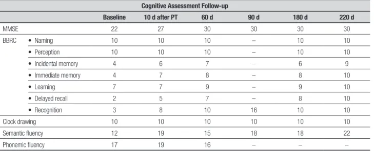
Table 1. Cognitive performance on baseline and follow-up.
Cognitive Assessment Follow-up
Baseline 10 d after PT 60 d 90 d 180 d 220 d
MMSE 22 27 30 30 30 30
BBRC • Naming 10 10 10 – 10 10
• Perception 10 10 10 – 10 10
• Incidental memory 4 6 7 – 6 9
• Immediate memory 4 7 8 – 8 10
• Learning 7 7 9 – 9 10
• Delayed recall 2 5 7 – 8 10
• Recognition 3 8 10 16 10 10
Clock drawing 10 10 10 10 10 10
Semantic fluency 12 19 15 18 18 22
Phonemic fluency 17 19 16 – – –
an electrographic seizure in the right temporal region on the same exam (Figure 2).
Extensive laboratory work-up was performed and serum assays showed low C3 and C4 complement frac-tion, presence of anti-P ribosomal, positive anti SSA (276ua/ml) and ANA (1/160) as well as lymphopenia and thrombocytopenia, clear signs of active SLE. A di-agnosis of limbic encephalitis and active SLE was then reached.
Additionally, during the hospital stay, a search for tu-mors was performed. A laboratory study with biomark-ers showed carcinoembryonic antigen, CA15-3, CA125 and CA19/9 at normal levels. Computed Tomographic imaging of the thorax, abdomen and pelvis, breast and transvaginal ultrasound were normal. MRI of the tho-rax, abdomen and pelvis was also performed and were normal. In addition, a Positron Emission Tomography (PET-CT) scan of the whole body was negative for any occult neoplastic focus.
Immunosuppressive treatment (IT) with methyl-prednisolone (1 g/d for 4 days) and cyclophosphamide 1g - single dose) was indicated. Video-electroencepha-lography was performed on the third day of immuno-suppressive treatment, while using oxcarbazepine, and showed normal background activity without epileptic discharges. After this irst session, at the end of
Febru-Figure 1. [A, B] Coronal flair, [C] Axial T2WI. [A, B] Bilateral hipocampi hyperintense sign at clinical onset of symptoms. [C] The same signal is already present on axial T2 sequence. [D, E] Coronal flair, [F] Axial T2Wi. [D, E] Later in the follow up (220 days af-ter), MRI has an improvement. [F] The patient’s total recovery is showed.
ary/2014, the patient improved her scores on learning, delayed recall and recognition. Her semantic and pho-nemic luency were substantially increased. As clini-cal response was efective, it was decided to maintain the patient under this treatment regimen for at least 6 months to consolidate immunosuppression, with monthly infusions administered in the hospital setting. She was treated from February to October/2014.
During the follow-up, after two cycles of immu-nosuppression, a repeat MRI (April/2014) revealed a marked improvement in hippocampal signal on T2 and Flair compared to that observed in the irst exam. An-other MRI exam was performed in October/2014 (9 months from irst admission) with normal signal in the hippocampi. A follow-up EEG remained normal, there-fore the antiepileptic drug was discontinued. A fully and persistent recovery of her cognitive abilities was observed (Table 1) up until her last evaluation. Current-ly, she has a subjective complaint of slow thinking and normal instrumental activities of daily living. After con-cluding 6 months of immunosuppression treatment, she returned to work.
DISCUSSION
he classical manifestation of LE includes episodic memory impairment9, seizures10, confusion16, sleeping
problems18, and psychiatric symptoms.19 he most
char-acteristic clinical feature is short-term memory loss, but associated symptoms such as confusion and seizures might limit the memory assessment.12
Gultekin et al. proposed diagnostic criteria that in-cludes a pathological demonstration of LE or all of the following four: [1] short-memory loss, seizures or psy-chiatric symptoms suggestive of limbic system involve-ment; [2] less than 4 years between the neurologic symp-toms and cancer diagnosis; [3] exclusion of metastasis, infection, metabolic and nutritional deicits, stroke and side-efects of therapy that can cause LE; [4] at least one out of: CSF with inlammatory indings; or hyperinten-sity of temporal lobes bilaterally on magnetic resonance image (MRI) T2/FLAIR sequences; or EEG with epileptic / slow activity involving focally temporal lobes35. Other
authors, Graus and Saiz, revised the criteria, changing some accepted clinical characteristics for diagnosis (un-der this criteria, the patient has to have all four items): [1] subacute onset (less than 12 weeks) of the clinical signals and symptoms cited above; [2] neuropathologic or radiologic (MRI, or single photon-emission computer tomography (SPECT); positron-emission computed to-mography (PET-CT) evidence of limbic involvement; [3] exclusion of other possible etiologies; [4] demonstration
of cancer within 5 years of the neurologic symptoms or the evidence of well-characterized paraneoplastic anti-bodies associated with this clinical picture36.
All patients with LE should undergo a neuroimag-ing evaluation of the medial temporal area.1 In patients
with predominant anterograde amnesia, MRI usually dis-closes FLAIR or T2 abnormalities in this area.1 In
indi-viduals with a wide range of symptoms, the MRI shows more extensive abnormalities in the temporal lobes or beyond the limbic system.1,20 EEG often demonstrates
unilateral or bilateral temporal lobe epileptic discharges or slow background activity.12 However, LE can present
as an unexplained subacute onset of neurological symp-toms, with normal MRI and no cerebrospinal luid (CSF) evidence of inlammation.11
It is crucial to rule out any underlying malignancy as LE is commonly related to neoplasm, as a paraneo-plastic manifestation. herefore, the most frequent as-sociated tumors are lung (particularly small cell lung cancer - SCLC), breast, ovarian, testicular, and prostate cancer and can be associated with thymoma, neuroen-docrine tumors or Hodgkin´s disease.12-14 To our
knowl-edge, only a single study has identiied a case of limbic encephalitis, with positive VGKC antibodies associated with endometrial adenocarcinoma, whereas no reports of papillary urothelial carcinoma and limbic encephalitis were found.21
hus, Fluorodeoxyglucose-PET is useful for detect-ing many occult malignancies but has limited utility for ovarian teratomas.11 For this type of tumor, MRI of
the abdomen and pelvis is the test of choice, followed by CT and abdominal or transvaginal ultrasound (if age-appropriate).11,13 It is also important to order tests
of tumor markers such as CA125, human chorionic go-nadotropin, and alpha-fetoprotein.11,13
On the other hand, a few reports had previously de-scribed a possible association between LE and SLE. In this context, SLE for instance can mimic or be associ-ated with limbic encephalitis.15,22,23 As lupus can
pres-ent in many forms, it has been called ‘the disease with a thousand faces’.11,17,23-25 he disturbances in
neuropsy-chiatric SLE are wide-ranging and include cerebrovas-cular disease, seizures, myelopathy, aseptic meningitis, movement disorders, demyelinating syndrome as well as moderate or severe cognitive dysfunction, psychosis, acute confusional state and depression.15
encephali-tis with active SLE, although with a poorly understood pathophysiology, could also share some similarities and diferences in this picture. It is believed that a conver-gence point between the three conditions involves an autoimmunity component shared by all of them.16
In general, several studies show that ive features characterize autoimmune physiopathology.13,18,26
First-ly, the epitopes are extracellular and the antibody bind-ing is visible in cells transfected with the target antigen. Secondly, the antibodies alter the structure or function of the corresponding neuronal antigen. hirdly, the ef-fects of the antibodies are often reversible. Lastly, the clinical picture resembles that of pharmacologic or ge-netic models in which the antigen is disrupted.13,18,26
Speciically in the context of ALE and PLE, some researchers propose that a logical way to diferentiate these two conditions is to identify whether the target antigen is intracellular, synaptic or on the cell surface and whether the immune response is primarily medi-ated by cellular or humoral mechanisms.11 Many
stud-ies indicate that the disorders associated with antibod-ies against intracellular antigens are mediated by T-cell mechanisms, which represent markers of an associated cancer but have not been shown to be pathogenic.12,18,27,28
hese typically afect older individuals. Moreover, they are paraneoplastic and largely resistant to immunother-apy, even after tumor removal.16,29,30
In contrast, autoimmune limbic encephalitis is as-sociated with antibodies to synaptic or cell surface antigens. hese are likely to contribute directly to the pathology of the condition.10,11 hey afect younger
indi-viduals as well as children and are often not associated with tumors. his condition appears to be antibody me-diated, and is often highly responsive to treatment.11,12
Some immune-mediated cases appear to have a mono-phasic course, but others may relapse.11 he main
anti-bodies related to nonparaneoplastic autoimmune LE are against NMDA receptors and VGKC complex.31
herefore, with regard to ALE and PLE, there are many types of antibodies against extra or intracellular struc-tures, such as: α-amino-3-hydroxy-5-methyl-4- isoxa-zolepropionic acid receptors (AMPARs), γ-aminobutyric acid-B receptors (GABABRs), glutamic acid decarboxyl-ase (GAD), N-methyl-D-aspartate receptor (NMDAR) and voltage-gated potassium channel (VGKC) com-plex antigens: leucine rich glioma inactivated protein 1 (LGI1), and contactin-associated protein-2 (CASPR2). Additionally, there are onconeural antibodies, particu-larly anti-Hu, anti-Ma 1/2, CV-2, and amphiphysin.11,32-34
Speciically in SLE, some autoantibodies such as anti-phospholipids (β2-glycoprotein 1 and cardiolipin),
anti-ribosomal P protein, anti-NMDA, speciically subtype Glun2 or NR2, and anti-microtubule-associated protein 2 (MAP-2) are found but with variable frequency in neu-ropsychiatric SLE.15 Besides all these, anti-glutamate
re-ceptor may be found to link these two diseases.14
Recent data suggests that neuropsychiatric events occur in 6–12% of patients with newly diagnosed SLE during the irst year of the illness. he most common neuropsychiatric syndromes attributed to SLE are sei-zure disorders, cerebrovascular disease, acute confu-sional states and neuropathies . 37
However, general SLE-related disease activity, pre-vious or concurrent neuropsychiatric symptoms, and persistent positivity for antiphospholipid antibodies at moderate-to-high titers have been shown to be the most informa tive indicators of neuropsychiatric events attributed to SLE.15
For instance, the presence of anti-NMDA, specii-cally the subtype Glun2 or NR2, in patients with SLE is estimated at 14 to 37% and especially SLE patients with neuropsychiatric manifestations this igure can reach up to 80%.14 his association is a recent inding in the
liter-ature, and many authors are focusing on this previously unknown association. However, more elevated levels of anti-NMDA subtype Glun1 or NR1 are more frequently associated with Anti-NMDA receptor Encephalitis and seem to play a more evident pathological role in LE and may be dose-dependent.14, 15
Consequently, irrespective of age, previous medical history and main presentation, ideally all such patients should be tested for these types of antibodies. Unfor-tunately, these antibodies in our patient could not be tested due to technical laboratory limitations.
In the management of ALE, it is important to high-light the beneits of early treatment11. General
con-cepts about treatment of classical paraneoplastic CNS syndromes do not apply in these cases. For example, whereas classical paraneoplastic syndromes do not re-spond to immunotherapy unless the tumor is success-fully treated12, when it then has a limited response11,16,21,
ALE may respond to immunotherapy regardless of tu-mor removal.12,13,26
contribute by reporting a possible case of LE with active SLE, adding more data to this discussion.
he main limitation of this article is the absence of neuronal antibody tests. However, the good evolution of the patient and excellent, rapid response to immuno-therapy make it reasonable to assume an underlying au-toimmune LE, as this has been discussed previously in the literature.37 By contrast, paraneoplastic LE usually
has a poor response. he association of ALE with SLE is also reasonable, since laboratory testing showed SLE activity besides involvement of other systems (hemato-logic) concomitant to CNS involvement.
he presence of other surface membrane antibodies is possible, since there are associations among diferent types of these as mentioned above. his case highlighted a need for rapid treatment when an autoimmune cause is suspected.
To sum up, further studies are necessary to
deter-mine the true association between limbic encephalitis and autoimmune diseases, especially Systemic Lupus Erythematosus. Eforts should be made to establish whether this association is pathologic. Future studies should explore which antibodies are related to neuro-psychiatric lupus and the pathologic mechanism trig-gered by them. To this end, determining the role of autoantibodies will be essential in order to conirm the true relationship.
Authors contributions. Débora Bartzen Moraes Angst: drafting/revising the manuscript, study concept, and analysis of data. Nathália Stela Visoná de Figueiredo: drafting/revising the manuscript, study concept, and analysis of data. Valmir Passarelli: study concept, and analysis of data. Meire Argentoni Baldocchi: study con-cept, and analysis of data. Maria Sheila Guimarães Ro-cha: study concept, and analysis of data.
REFERENCES
1. Urbach H, Soeder BM, Jeub M, Klockgether T, Meyer B, Bien CG. Serial MRI of limbic encephalitis. Neuroradiology 2006;48:380-386. 2. Kalkman PH, Allan S, Birchall IW. Magnetic resonance imaging of limbic
encephalitis. Can Assoc Radiol J 1993;44:121-124.
3. Bierley JB, Correllis JAN, Hierons R, et al. Subacute encephalitis of late adult life mainly affecting the limbic areas. Brain 1960;83:357-368. 4. Alamowitch S, Graus F, Uchuya M, Rene R, Bescansa E, Delattre JY.
Limbic encephalitis and small cell lung cancer Clinical and imunological features. Brain 1997;120:923-928.
5. Corsellis JA, Goldberg GJ, Norton AR. “Limbic encephalitis” and its association with carcinoma. Brain 1968;91:481-496.
6. Bowyer S, Webb S, Millward M, Jasas K, Blacker D, Nowa A. Small cell lung cancer presenting with paraneoplastic limbic encephalitis. Asia Pac J Clin Oncol 2011;7:180-184.
7. Rosenfeld MR, Dalmau J. Paraneoplastic limbic encephalitis associated with small-cell lung cancer. Comm Oncol 2007;4:491-494.
8. White D, Beringer T. Paraneoplastic limbic encephalitis in an elderly patient with small cell lung carcinoma. Ulster Med J 2010;79:22-24. 9. McKeon A, Lennon VA, Pittock SJ. Immunotherapy-responsive
demen-tias and encephalopathies. Continuum (Minneap Minn).2010;16:80-101. 10. Machado S, Pinto AN, Sarosh IR. What should you know about limbic
encephalitis? Arq Neuropsiquiatr 2012;70:817-822.
11. Lee R, Buckley C, Irani SR, Vincent A. Autoantibody testing in encepha-lopathies. Pract Neurology 2012;12:4-13.
12. Rosenfeld M, Dalmau J. Paraneoplastic Disorders of the CNS and Auto-immune Synaptic Encephalitis. Continuum (Minneap Minn 2012;18: 366-383.
13. Lancaster E, Martinez-Hernandez E, Dalmau J. Encephalitis and anti-bodies to synaptic and neuronal cell surface proteins. Neurology 2011; 77:179-89.
14. Levite M. Glutamate receptor antibodies in neurological diseases: anti-AMPA-GluR3 antibodies, NR1 antibodies, anti-NMDA-NR2A/B antibodies, anti-mGluR1 antibodies or anti-mGluR5 antibodies are present in subpopulations of patients with either: epilepsy, encepha-litis, cerebellar ataxia, systemic lupus erythematosus (SLE) and neuro-psychiatric SLE, Sjogren’s syndrome, schizophrenia, mania or stroke. These autoimmune anti-glutamate receptor antibodies can bind neurons in few brain regions, activate glutamate receptors, decrease glutamate receptor’s expression, impair glutamate-induced signaling and function, activate blood brain barrier endothelial cells, kill neurons, damage the brain, induce behavioral/psychiatric/cognitive abnormali-ties and ataxia in animal models, and can be removed or silenced in some patients by immunotherapy. J Neural Transm 2014;121:1029-75.
15. Jeltsch-David H, Muller S. Neuropsychiatric systemic lupus erythe-matosus: pathogenesis and biomarkers. Nat Rev Neurol 2014;10: 579-596.
16. Kano O, Arasaki K, Ikeda K, et al. Limbic encephalitis associated with systemic lupus erythematosus. Lupus 2009;18:1316-9.
17. Senécal JL. Treating lupus with medication. In: Lupus Canada, Lupus: the disease with a thousand faces, 2nd edition. Quebec: Lupus Canada;
1990:357-358.
18. Anderson NE, Barber PA. Limbic encephalitis – a review. J Clin Neurosci 2008;15:961-971.
19. Porto FH, Coutinho AM, Lucato LT, et al. Paraneoplastic limbic enceph-alitis with prominent neuropsychiatric apathy. J Neurol Sci 2014;337: 224-227.
20. Dalmau J, Graus F, Villarejo A, et al. Clinical analysis of anti-Ma2-asso-ciated encephalitis. Brain 2004;127:1831-1844.
21. Irani SR, Alexander S, Waters P, et al. Antibodies to Kv1 potassium channel-complex proteins leucine-rich, glioma inactivated 1 protein and contactin-associated protein-2 in limbic encephalitis, Morvan’s syndrome and acquired neuromyotonia. Brain 2010;133:2734-2748. 22. Yamaguchi Y1, Wada M, Kurita K, Takahashi Y, Kato T. Case of acute
limbic encephalitis associated with SLE accompanied with anti-gluta-mate receptor antibodies. Rinsho Shinkeigaku 2012;52:545-50. 23. Stübgen JP. Nervous system lupus mimics limbic encephalitis. Lupus
1998;7:557-60.
24. Scolding NJ, Joseph FG. The neuropathology and pathogenesis of systemic lupus erythematosus. Neuropathol Appl Neurobiol 2002;28: 173-189.
25. Johnson RT, Richardson EP. The neurological manifestations of systemic lupus erythematosus. Medicine 1968;47:337-69.
26. Dalmau J, Tuzun E, Wu HY, et al. Paraneoplastic anti-Nmethyl- D-aspar-tate receptor encephalitis associated with ovarian teratoma. Ann Neurol 2007;61:25-36.
27. Said S, Cooper CJ, Reyna E, Alkhateeb H, Diaz J, Nahleh Z. Paraneo-plastic limbic encephalitis, an uncommon presentation of a common cancer: Case report and discussion. Am J Case Rep 2013;14:391- 394.
28. Dreessen J, Jeanjean AP, Sindic CJ. Paraneoplastic limbic encepha-litis: Diagnostic relevance of CSF analysis and total body PET scanning. Acta Neurol Belg 2004;104:57-63.
29. Ryu JY, Lee SH, Lee EJ, et al. A case of paraneoplastic limbic encepha-litis associated with small cell lung cancer. Tuberc Respir Dis 2012;73: 273-277.
para-neoplastic limbic and brain-stem encephalitis in patients with testicular cancer. N Engl J Med 1999;340:1788-1795.
31. Sarkis RA, Nehme R, Chemali ZN. Neuropsychiatric and seizure outcomes in nonparaneoplastic autoimune limbic encephalitis. Epilepsy Behav 2014;39: 21-25.
32. Iizuka T, Sakai F, Ide T, et al. Anti-NMDA receptor encephalitis in Japan: long-term outcome without tumor removal. Neurology 2008;70: 504-511.
33. Lai M, Hughes EG, Peng X, et al. AMPA receptor antibodies in limbic encephalitis alter synaptic receptor location. Ann Neurol 2009;65: 424-34.
34. Yu Z, Kryzer TJ, Griesmann GE, Kim K, Benarroch EE, Lennon VA. CRMP-5 neuronal autoantibody: marker of lung cancer and thymoma-related autoimmunity. Ann Neurol 2001;49:146-154.
35. Gultekin SH, Rosenfeld MR, Voltz R, et al. “Paraneoplastic limbic encephalitis: neurological symptoms, immunological findings and tumour association in 50 patients”. Brain 2000;123(7):1481-1494. 36. Graus F, Saiz A. “Limbic encephalitis: a probably under-recognized
syndrome”. Neurologia 2005;20(1):24-30.