phosphate nanoparticles
Mary Anne S. Melo,
1,2Lei Cheng,
1,3Michael D. Weir,
1Ru-Ching Hsia,
4Lidiany K. A. Rodrigues,
2Hockin H. K. Xu
1,5,6,71Biomaterials and Tissue Engineering Division, Department of Endodontics, Prosthodontics and Operative Dentistry,
University of Maryland School of Dentistry, Baltimore, Maryland 21201
2Faculty of Pharmacy, Dentistry and Nursing, Federal University of Ceara, Fortaleza, CE, Brazil
3State Key Laboratory of Oral Diseases, West China School of Stomatology, Sichuan University, Chengdu, China 4Electron Microscopy Core Facility, University of Maryland School of Dentistry, Baltimore, Maryland 21201 5Center for Stem Cell Biology and Regenerative Medicine, University of Maryland School of Medicine,
Baltimore, Maryland 21201
6Marlene and Stewart Greenebaum Cancer Center, University of Maryland School of Medicine, Baltimore, Maryland 21201 7Department of Mechanical Engineering, University of Maryland, Baltimore County, Maryland 21250
Received 15 July 2012; revised 11 October 2012; accepted 11 November 2012
Published online 20 December 2012 in Wiley Online Library (wileyonlinelibrary.com). DOI: 10.1002/jbm.b.32864
Abstract:Secondary caries remains the main reason for den-tal restoration failure. Replacement of failed restorations accounts for 50%–70% of all restorations performed. Antibac-terial adhesives could inhibit biofilm acids at tooth-restoration margins, and calcium phosphate (CaP) ions could remineralize tooth lesions. The objectives of this study were to: (1) incorpo-rate nanoparticles of silver (NAg), quaternary ammonium dimethacrylate (QADM), and nanoparticles of amorphous calcium phosphate (NACP) into bonding agent; and (2) investi-gate their effects on dentin bonding and microcosm biofilms. An experimental primer was made with pyromellitic glycerol dimethacrylate (PMGDM) and 2-hydroxyethyl methacrylate (HEMA). An adhesive was made with bisphenol-A-glycerolate dimethacrylate (BisGMA) and triethylene glycol dimethacry-late (TEGDMA). NAg was incorporated into primer at 0.1 wt %. The adhesive contained 0.1% NAg and 10% QADM, and 0%– 40% NACP. Incorporating NAg into primer and NAg-QADM-NACP into adhesive did not adversely affect dentin bond strength (p > 0.1). Scanning electron microscopy showed
numerous resin tags, and transmission electron microscopy revealed NAg and NACP in dentinal tubules. Viability of human saliva microcosm biofilms on primer/adhesive/com-posite disks was substantially reduced via NAg and QADM. Metabolic activity, lactic acid, and colony-forming units of biofilms were much lower on the new bonding agents than control (p<0.05). In conclusion, novel dental bonding agents containing NAg, QADM, and NACP were developed with the potential to kill residual bacteria in the tooth cavity and inhibit the invading bacteria along tooth-restoration margins, with NACP to remineralize tooth lesions. The novel method of combining antibacterial agents (NAg and QADM) with remi-neralizing agent (NACP) may have wide applicability to other adhesives for caries inhibition. VC 2012 Wiley Periodicals, Inc. J Biomed Mater Res Part B: Appl Biomater 101B: 620–629, 2013.
Key Words:antibacterial primer and adhesive, dentin bond strength, silver nanoparticles, calcium phosphate nanopar-ticles, human saliva microcosm biofilm, caries inhibition
How to cite this article:Melo Mary Anne S., Cheng L, Weir MD, Hsia R-C, Rodrigues Lidiany K. A., Xu Hockin H. K.. 2013. Novel dental adhesive containing antibacterial agents and calcium phosphate nanoparticles. J Biomed Mater Res Part B 2013:101B:620– 629.
INTRODUCTION
Resin composites are being increasingly used as esthetic dental restorations.1–3 Their popularity is a result of
signifi-cant improvements in properties and performance.4–7 None-theless, composites tend to accumulate more biofilms in vivo,8,9 which could produce acids and cause dental caries.10,11 Recurrent caries is the main reason for
restora-tion failure, and replacement of the failed restorarestora-tions accounts for 50%–70% of all operative work.12–14To reduce
secondary (recurrent) caries, novel antibacterial resins containing quaternary ammonium salts (QAS) were devel-oped.15–20Resins containing 12-methacryloyloxydodecylpyri-dinium bromide (MDPB) significantly reduced the bacteria growth.15,16,21Other resins used antibacterial agents such as
Correspondence to:H. H. K. Xu; e-mail: hxu@umaryland.edu or L. K. A. Rodrigues; e-mail: lidianykarla@yahoo.com
Contract grant sponsor: NIH; contract grant numbers: R01DE17974, R01DE14190
Contract grant sponsor: Coordination for Improvement of Higher Education/Fulbright Doctoral Program; contract grant number: BEX0523/11-9 Contract grant sponsor: National Natural Science Foundation of China; contract grant number: 81100745
methacryloxylethyl cetyl dimethyl ammonium chloride (DMAE-CB) and cetylpyridinium chloride (CPC).17,18,22,23
Another method to combat dental caries involved the development of antibacterial resins by incorporating silver (Ag) filler particles.24–26Nanoparticles of Ag (NAg) were dem-onstrated to be effective for antibacterial applications.20,26,27A different method for caries-inhibition used calcium phosphate (CaP) particle-filled composites, which released Ca and P ions and remineralized tooth lesions.28–30 Recently, CaP nanopar-ticles were synthesized via a spray-drying technique.31 The new nanocomposites containing nanoparticles of amorphous calcium phosphate (NACP) released Ca and P ions similar to traditional CaP composites, while possessing much higher me-chanical properties for load-bearing tooth restorations.32,33
Besides composites, it is also important to develop caries-inhibiting adhesives. Adhesive bonds the composite to the tooth structure and infiltrates into dentinal tubules.34–36 Extensive studies were performed to improve and charac-terize enamel and dentin bonding.37–40 Residual bacteria could exist in the prepared tooth cavity, and microleakage could allow bacteria to invade the tooth-restoration interfa-ces.15,22 Therefore, antibacterial adhesives were developed using MDPB and other antibacterial agents.21–23 It is also beneficial to render the primer antibacterial because it directly contacts tooth structure.41,42 Recently, quaternary ammonium dimethacrylate (QADM) was combined with NAg in a commercial primer (SBMP, 3M).43 However, QADM and NAg have not been incorporated into any other adhesives. It remains to be investigated (1) whether QADM and NAg could be incorporated into another bonding system to also impart potent antibacterial functions; (2) whether NACP (as a remineralizing agent with Ca and P ions) can be incorpo-rated into the antibacterial adhesive without compromising the dentin bond strength; (3) whether the NACP and NAg nanoparticles could flow into the dentinal tubules with the adhesive. The rationale was that while QADM and NAg could eradicate residual bacteria in the tooth cavity and dentinal tubules, NACP could remineralize the remnants of lesions in the tooth cavity, thus yielding a unique combina-tion of antibacterial/remineralizing capabilities.
Accordingly, the objectives of this study were to incorpo-rate NAg, QADM, and NACP into an experimental bonding system and to investigate the effects on anti-biofilm and dentin bonding properties. It was hypothesized that: (1) The new experimental bonding agent containing NAg and QADM would greatly reduce the biofilm viability, metabolic activity, and lactic acid production in a dental plaque micro-cosm biofilm model; (2) Incorporation of NACP into the ex-perimental bonding agent containing NAg and QADM would not decrease the dentin bond strength; (3) NAg and NACP could flow with the bonding agent into the dentinal tubules.
MATERIALS AND METHODS
Synthesis of experimental bonding agent containing NAg, QADM, and NACP
The experimental primer contained pyromellitic glycerol dime-thacrylate (PMGDM) (Hampford, Stratford, CT) and 2-hydrox-yethyl methacrylate (HEMA) (Esstech, Essington, PA) at a mass
ratio 3.3/1, with 50% acetone solvent (all mass fractions).44 The photo-initiator for the primer and adhesive was 1% phenyl bis(2,4,6-trimethylbenzoyl) phosphine oxide (BAPO) (Irga-cure819, Ciba Chemicals, Japan). The experimental adhesive consisted of bisphenol A glycerolate dimethacrylate (BisGMA) and triethylene glycol dimethacrylate (TEGDMA) (Esstech) at
70/30 mass ratio.45
Silver 2-ethylhexanoate salt (Strem, New Buryport, MA) of 0.1 g was dissolved in 1 g of 2-(tert-butylamino)ethyl methacrylate (TBAEMA, Sigma).20,26 TBAEMA was used because it improves the solubility by forming AgAN coordi-nation bonds with Ag ions, thereby facilitating the Ag salt to dissolve in the resin solution. TBAEMA was selected as it contains reactive methacrylate groups and therefore can be chemically incorporated into a dental resin on photopolyme-rization.26 This method produced NAg
in situ in the resin
matrix with particle sizes of less than 10 nm.20,26This study used a silver 2-ethylhexanoate/(primerþsilver 2-ethylhexa-noate) mass fraction of 0.1%, because this imparted a strong antibacterial effect, without adversely affecting dentin bond strength or primer color in preliminary studies. The adhesive contained a silver 2-ethylhexanoate/(adhesive þ
silver 2-ethylhexanoate) of 0.1%.
In addition, QADM and NACP were incorporated into the adhesive. Bis(2-methacryloyloxy-ethyl) dimethylammonium bromide, a quaternary ammonium dimethacrylate (QADM), was recently synthesized.19 The synthesis of QADM was performed using a modified Menschutkin reaction, where a tertiary amine group was reacted with an organo-halide. A benefit of this reaction is that the reaction products are gen-erated at virtually quantitative amounts and require mini-mal purification.19 Briefly, 10 mmol of 2-(
N,N -dimethylami-no)ethyl methacrylate (Sigma-Aldrich, St. Louis, MO) and 10 mmol of 2-bromoethyl methacrylate (Monomer-Polymer Laboratories, Trevose, PA) were combined in ethanol and stirred at 60C for 24 h.19,20This yielded QADM as a clear,
colorless, and viscous liquid, which was mixed with the ad-hesive at QADM/(adad-hesiveþQADM) mass fraction of 10%, following a previous study.43
A spray-drying technique was used to make NACP.31,32,46
Amorphous calcium phosphate (ACP, Ca3[PO4]2) is important
because it is a precursor that can convert to apatite, similar to the minerals in tooth enamel and dentin. A spraying solu-tion was prepared by adding 1.5125 g of acetic acid glacial (J.T. Baker, Phillipsburg, NJ) into 500 mL of distilled water. Then, 0.8 g of calcium carbonate (CaCO3, Fisher, Fair Lawn,
NJ) and 5.094 g of DCPA (Baker) were dissolved into the ace-tic acid solution. The final Ca and PO4 ionic concentrations
were 8 mmol/L and 5.333 mmol/L, respectively. This yielded a Ca/P molar ratio of 1.5, the same as that for ACP. This solu-tion was sprayed through a nozzle into a heated chamber. The water and volatile acid were evaporated into the dry, heated air flow, and expelled into an exhaust-hood. The dried particles were collected by an electrostatic precipitator.32 This method produced NACP with a mean particle size of 112 nm.32 The NACP were mixed with the adhesive at
NACP/(NACPþ adhesive) mass fractions of 0%, 10%, 20%,
30%, and 40%, following previous studies.32,33
1. Experimental primer control and adhesive control (termed ‘‘P control, A control’’).
2. PþNAg, A control (Primer had 0.1% NAg, while adhesive had no NAg).
3. PþNAg, AþNAgþQADM (Both P and A had 0.1% NAg. A also had 10% QADM).
4. PþNAg, AþNAgþQADMþ10NACP (10NACP means 10% NACP by mass).
5. PþNAg, AþNAgþQADMþ20NACP. 6. PþNAg, AþNAgþQADMþ30NACP.
7. PþNAg, AþNAgþQADMþ40NACP.
Dentin bond strength and scanning electron microscopy and transmission electron microscopy examinations
The use of extracted human teeth was approved by the Univer-sity of Maryland. Caries-free third-molars were ground to remove the occlusal enamel.43,47Flat mid-coronal dentin surfa-ces of caries-free molars were prepared by cutting off the tips of crowns with a diamond saw (Isomet, Buehler, Lake Bluff, IL). Each tooth was embedded in a poly-carbonate holder (Bos-worth, Skokie, IL) and ground perpendicularly to the longitudi-nal axis on 320-grit silicon carbide paper until occlusal enamel was removed. Dentin was etched with 37% phosphoric acid for 15 s.43,47A primer was applied, then an adhesive was applied and light-cured for 10 s (Optilux VCL401, Demetron, Danbury, CT). A stainless-steel iris (inner diameter¼4 mm, thickness¼
1.5 mm) was held against the adhesive-treated dentin, and the opening was filled with a composite (TPH, Caulk/Dentsply, Mil-ford, DE) and light-cured for 60 s. The specimens were stored in water at 37C for 24 h.43,47 To measure the dentin shear
bond strength, a chisel was aligned parallel to the composite-dentin interface and loaded (MTS, Eden Prairie, MN) at 0.5 mm/min until the composite-dentin bond failed. Dentin shear bond strengthSD¼4P/(pd2), wherePis load-at-failure, andd
is composite diameter.43,47
To examine the dentin-adhesive interface, the bonded teeth were cut longitudinally. The sections were treated with 50% phosphoric acid and 10% NaOCl,43 then
gold-coated and examined via scanning electron microscopy (SEM, Quanta 200, FEI, Hillsboro, OR). For transmission electron microscopy (TEM), thin sections with an approxi-mate thickness of 120lm were cut and fixed with 2%
para-formaldehyde and 2.5% glutaraldehyde following a previous study.48Samples were embedded in epoxy (Spurr’s, Electron Microscopy Sciences, PA). Ultra-thin sections with approxi-mate thickness of 100 nm were cut using a diamond knife (Diatome, Bienne, Switzerland) with an ultra-microtome (EM-UC7, Leica, Germany). The non-demineralized sections were examined in TEM (Tecnai-T12, FEI).
Dental plaque microcosm biofilm model and live/dead assay
The microcosm biofilm model was approved by the Univer-sity of Maryland. Human saliva was shown to be ideal for growing plaque microcosm biofilms in vitro, with the
biofilm inoculums was collected from a healthy adult donor having natural dentition without active caries or periopa-thology, and without the use of antibiotics within the last 3 months, following a previous study.43 The donor did not brush teeth for 24 h and abstained from food/drink intake for at least 2 h before donating saliva. Stimulated saliva was collected during parafilm chewing and kept on ice. Saliva was diluted in sterile glycerol to a concentration of 70% saliva and 30% glycerol, and stored at80
C.43
Layered disks were fabricated as schematically shown in Figure 3(A), following previous studies.41,43 A polyethylene mold with an inner diameter of 9 mm and a thickness of 2 mm was situated on a glass slide. A primer was first applied into the mold to cover the glass slide. After drying with a stream of air, the adhesive was applied and cured for 20 s with an Optilux curing unit (VCL 401, Demetron Kerr, Danbury, CT). Then, a composite (TPH) was placed on the adhesive to fill the disk mold and light-cured for 1 min. The cured disks were removed from the molds, immersed in sterile water and agitated for 1 h to remove any uncured monomer, following a previous study.41The disks were then dried and sterilized with ethylene oxide (Anprolene AN 74i, Andersen, Haw River, NC).
The saliva-glycerol stock was added, with 1:50 final dilu-tion, to a growth medium as inoculum. The growth medium contained mucin (type II, porcine, gastric) at a concentration of 2.5 g/L; bacteriological peptone, 2.0 g/L; tryptone, 2.0 g/L; yeast extract, 1.0 g/L; NaCl, 0.35 g/L, KCl, 0.2 g/L; CaCl2, 0.2
g/L; cysteine hydrochloride, 0.1 g/L; haemin, 0.001 g/L; and vitamin K1, 0.0002 g/L, at pH 7.50 The inoculum was
cultured at 37
C in an incubator containing 5% CO2for 24 h.
Each disk was placed into a well of 24-well plates, with the primer surface on the top. To each well was added 1.5 mL of inoculum, which was incubated for 8 h. The disks were trans-ferred to new 24-well plates with fresh medium and incu-bated. After 16 h, the disks were transferred to new 24-well plates with fresh medium and incubated for 24 h. This constructed a 2-day incubation, which was shown to form dental plaque microcosm biofilms.43 The disks with 2-day biofilms were washed with phosphate buffered saline (PBS) and stained using a live/dead bacterial viability kit (Molecu-lar Probes, Eugene, OR). Live bacteria were stained with Syto 9 to produce a green fluorescence, and bacteria with compro-mised membranes were stained with propidium iodide to produce a red fluorescence. The stained disks were examined using an epifluorescence microscope (TE2000-S, Nikon, Melville, NY).20
Metabolic activity of microcosm biofilms
The MTT (3-[4,5-Dimethylthiazol-2-yl]-2,5-diphenyltetrazo-lium bromide) assay is a colorimetric assay that measures the enzymatic reduction of MTT, a yellow tetrazole, to form-azan.20 Each disk with the 2-day biofilm was transferred to
a new 24-well plate, then 1 mL of MTT dye (0.5 mg/mL MTT in PBS) was added to each well and incubated at 37
bacteria reduced the MTT to purple formazan. After 1 h, the disks were transferred to a new 24-well plate, 1 mL of di-methyl sulfoxide (DMSO) was added to solubilize the forma-zan crystals, and the plate was incubated for 20 min with gentle mixing at room temperature in the dark. After mixing via pipetting, 200 lL of the DMSO solution from each well
was transferred to a 96-well plate, and the absorbance at 540 nm (optical density OD540) was measured via a
micro-plate reader (SpectraMax M5, Molecular Devices, Sunnvale, CA). A higher absorbance is related to a higher formazan concentration, which indicates a higher metabolic activity in the biofilm on the disk.20
Lactic acid production and colony-forming units of microcosm biofilms
To measure lactic acid production, disks with 2-day biofilms were transferred to 24-well plates containing buffered-pep-tone water (BPW) plus 0.2% sucrose, and incubated for 3 h to allow the biofilms to produce acid. The lactate concentrations in the BPW solutions were determined using an enzymatic (lactate dehydrogenase) method, following previous stud-ies.20,43The microplate reader (SpectraMax M5) was used to measure the absorbance at 340 nm (optical density OD340) for
the collected BPW solutions. Standard curves were prepared using a lactic acid standard (Supelco, Bellefonte, PA).
For colony-forming units (CFUs), disks with 2-day bio-films were transferred into tubes with 2 mL cysteine pep-tone water, and the biofilms were harvested by sonication and vortexing via a vortex mixer (Fisher, Pittsburgh, PA). Four types of agar plates were used. First, tryptic soy blood agar culture plates were used to determine total microor-ganisms.43 Second, mitis salivarius agar (MSA) culture plates, containing 15% sucrose, were used to determine total streptococci.51 This is because MSA contains selective agents crystal violet, potassium tellurite, and trypan blue, which inhibit most negative bacilli and most gram-positive bacteria except streptococci, thus enabling strepto-cocci to grow.51 Third, cariogenic mutans streptococci are known to be resistant to bacitracin, and this property is often used to isolate mutans streptococci from the highly heterogeneous oral microflora. Hence, MSA agar culture plates plus 0.2 units of bacitracin per mL were used to determine mutans streptococci.52 Forth, Rogosa SL culture plates were used to determine lactobacilli. Rogosa SL plates contained high levels of sodium acetate and ammonium citrate at a low pH which would inhibit most microorgan-isms but allow the growth of lactobacilli.53
One-way analysis of variance was performed to detect the significant effects of the variables. Tukey’s multiple com-parison was used to compare the data at apvalue of 0.05.
RESULTS
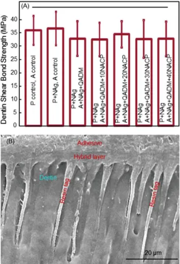
Figure 1 shows the dentin bonding results: (A) Dentin shear bond strengths (mean 6sd;n¼10), and (B) representative SEM image of dentin-adhesive interface. In (A), dentin shear bond strengths were similar between the control and those containing NAg, QADM, and NACP (p > 0.1). Therefore,
adding NAg, QADM, and NACP from 10% to 40% did not
sig-nificantly compromise the dentin bond strength, compared with the control. In (B), SEM examination revealed the for-mation of numerous resin tags from well-filled dentinal tubules. An example is shown for control [Figure 1(B)], while other groups had similar dentin-adhesive interfacial features.
Representative TEM images of the dentin-adhesive inter-face are shown in Figure 2. In (A), an example of a resin tag in a dentinal tubule shows that the NACP were successfully flowed with the adhesive resin into the dentinal tubule. This example was for the bonding system of PþNAg, AþNAgþ
QADMþ30NACP. An even higher magnification in (B) shows that the NAg was also incorporated into the dentinal tubules.
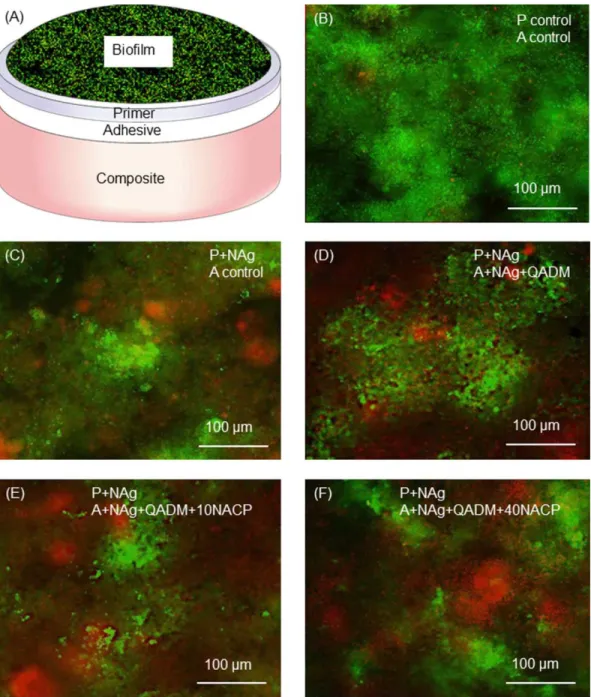
Figure 3 shows the live/dead assay results. The layered disk structure is schematically shown in (A). The 2-day bio-film adherent on the disk surface was stained, with the live
FIGURE 1.Dentin bond testing. (A) Dentin shear bond strength using
extracted human molars. Each value is mean 6sd (n ¼ 10). P¼ primer. A ¼adhesive. PþNAg had 0.1% NAg (by mass) in primer. AþNAgþQADM had 0.1% NAgþ10% QADM in adhesive. The hybrid layer is the demineralized collagen of dentin infiltrated by primer and adhesive (hybrid layer¼collagenþprimerþadhesive). Adding NAg, QADM, and NACP into the bonding agent did not decrease the dentin bond strength. Horizontal line indicates statistically similar values (p
>0.1). (B) SEM examination of dentin-adhesive interfaces revealed numerous resin tags from well-filled dentinal tubules, with an exam-ple shown for the control. Other groups had similar dentin-adhesive interfacial features. [Color figure can be viewed in the online issue, which is available at wileyonlinelibrary.com.]
bacteria staining a green color, and the dead and compro-mised bacteria staining red. The control disks showed pri-marily live bacteria (B). Disks with NAg and NAgþQADM
The MTT assay of biofilms is plotted in Figure 4 (mean 6sd;n¼6). As expected, biofilms on the control disks had a relatively high metabolic activity. Incorporation of QADM and NAg greatly decreased the metabolic activity of the ad-herent biofilms (p < 0.05). Adding 10% and 20% NACP
into the adhesive did not significantly change the metabolic activity, while 30% and 40% NACP significantly lowered the metabolic activity (p<0.05).
Figure 5 plots the lactic acid production of biofilms (mean6 sd;n¼ 6). Biofilms on control disks secreted the most amount of lactic acid. Incorporation of NAg and QADM into the control bonding agent substantially reduced the lac-tic acid production (p <0.05). Further adding NACP to the
adhesive in general did not significantly affect the lactic acid production of the biofilms, except for the 40% NACP in the adhesive which had a significantly lower lactic acid (p <
0.05).
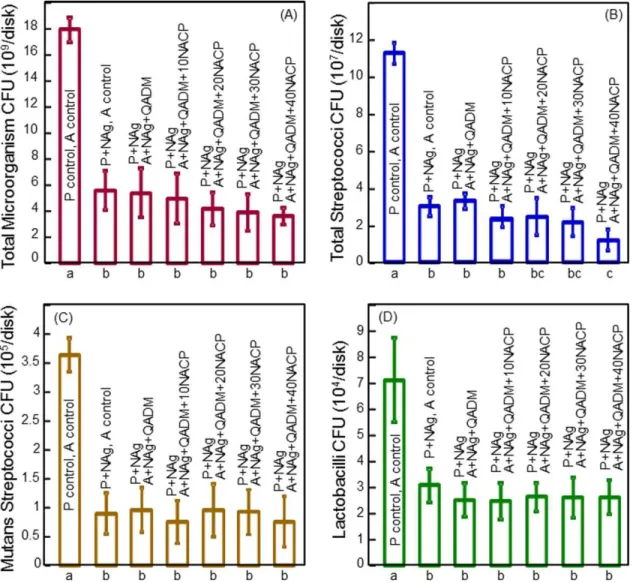
Biofilm CFU counts are plotted in Figure 6 for: (A) Total microorganisms, (B) total streptococci, (C) mutans strepto-cocci, and (D) lactobacilli (mean6 sd;n¼ 6). In each case, the control bonding agent had relatively high CFU, and the addition of NAg and QADM substantially reduced the CFU (p
< 0.01). In general, adding NACP from 10% to 40% into
the adhesive did not significantly affect the CFU counts, except for total streptococci where the 40% NACP group had a significantly lower CFU than that without NACP (p<
0.05). These results demonstrate that adding NAg and QADM into the experimental bonding agent was effective in reducing the microcosm biofilm viability and lactic acid production.
DISCUSSION
This study incorporated NAg into an experimental primer, and QADM and NACP into an experimental adhesive for the first time. A previous study incorporated NAg and QADM into a commercial bonding agent (SBMP, 3M),43 but it was unclear if the NAg and QADM could be similarly applied to other bonding systems to achieve potent antibacterial activ-ities without compromising the dentin bond strength. This study used a primer comprising of PMGDM and HEMA, and an adhesive comprising of BisGMA and TEGDMA, which are an experimental bonding system and not commercially available. The results showed that QADM and NAg were suc-cessfully incorporated into this experimental bonding sys-tem, exhibiting strong antibacterial effects, which suggest that QADM and NAg are promising for applications into other bonding agents. Although several previous studies used single-species bacteria models,19,26,42this study used a dental plaque microcosm model. Dental plaque is a compli-cated ecosystem with about 1000 bacterial species,11 hence microcosm models could maintain much of the complexity and heterogeneityin vivo.49The new bonding agent reduced the biofilm viability and CFU to about one-third of those of the control. CFU counts of total microorganisms, total strep-tococci, mutans strepstrep-tococci, and lactobacilli were all greatly reduced by the new bonding agents.
FIGURE 2.High magnification TEM examination of dentin-adhesive
interfaces. (A) Typical resin tag for the primer and adhesive group of PþNAg, AþNAgþQADMþ30NACP, shown as an example. Arrows indicate NACP that successfully flowed with the adhesive into denti-nal tubules. (B) Higher magnification TEM revealed NAg as well as NACP in the resin tags in the dentinal tubules. The NAg appeared as black dots in TEM images with sizes of less than 10 nm, similar to those observed in previous study.49[Color figure can be viewed in
The results showed that using the layered disks, anti-bacterial primer PþNAg had strong antibacterial effects.
Biofilms were inoculated on the primer layer, hence the adhesive underneath did not directly contact the biofilms. It should be noted that clinically, both antibacterial primers and adhesives are needed for caries inhibition. During a tooth cavity preparation, it is often difficult or impossible to remove all the infected and affected tissues, hence there are often residual bacteria left in the dentinal tubules of the prepared tooth cavity.15,22 This is especially true with the increased interest in less removal of tooth structure and the minimal intervention dentistry,54 which could leave
behind more carious tissues with active bacteria in the tooth cavity. These residual bacteria in the tooth cavity could contribute to caries and pulp damage. Therefore, an antibacterial primer could be beneficial because the primer directly contacts the dentin and infiltrates into dentinal tubules to help disinfect the prepared tooth cavity and erad-icate the residual bacteria.
In this study, the QADM was incorporated into the adhe-sive, while the primer contained only NAg but not QADM. This is because the addition of QADM into primer decreased the dentin bond strength in preliminary studies, while the addition of NAg into primer did not adversely affect the
FIGURE 3.Human saliva microcosm biofilm live/dead staining assay. (A) Schematic of biofilm on cured disk with three layers: Primer, adhesive,
and composite. (B–F) Representative live/dead images. Live bacteria were stained green, and dead bacteria were stained red. The live and dead bacteria in close proximity to each other produced orange/yellow colors. The biofilms were primarily alive on the control. In contrast, disks con-taining NAg and NAgþQADM had large amounts of dead bacteria. Incorporation of NACP into adhesive from 10% to 40% did not noticeably change the biofilm features. [Color figure can be viewed in the online issue, which is available at wileyonlinelibrary.com.]
dentin bond strength. QADM was incorporated into the adhesive. This is because an antibacterial adhesive is also beneficial clinically. Although a complete sealing of the tooth-restoration interface is highly desirable, it is difficult to achieve. Indeed, previous studies revealed microgaps between the adhesive and the primed dentin, or between the adhesive and the hybrid layer.55,56 This would suggest that a large portion of the marginal gap is surrounded by the adhesive resin, hence the invading bacteria would mostly come into contact with the adhesive surface.21
Therefore, both antibacterial primer and adhesive are needed to combat the residual bacteria, as well as the new invading bacteria along the tooth-restoration margins due to bacterial leakage, thereby to protect the pulp and inhibit recurrent caries.
The results of this study showed that incorporating 10% to 40% of NACP fillers into the BisGMA-TEGDMA adhesive did not adversely affect the dentin bond strength. The pur-pose of NACP incorporation was for the adhesive to obtain CaP ion release and remineralization capabilities. Previous studies showed that CaP-containing resins remineralized enamel and dentin lesions in vitro.29,30 Recent studies
showed that NACP-containing composites released high lev-els of Ca and P ions.32 Furthermore, NACP nanocomposite rapidly neutralized a cariogenic acid challenge and raised the pH from 4 to above 6.33Although this study focused on the effects of NACP-containing adhesive on dentin bond strength and NACP incorporation into dentinal tubules, fur-ther study should measure the mineral content of enamel and dentin around NACP adhesive under biofilms to investi-gate the caries-inhibition efficacy. In addition, the results of the present showed that the incorporation of NACP had little effect on the antibacterial effects. This is because CaP
activity was observed at 30% and 40% NACP. In Figure 5, there was a small reduction in lactic acid production at 40% NACP. These small reductions in bacteria activity may be due to the alkaline property of NACP leading to an increase in local pH.33 However, any antibacterial function from NACP appears to be minor, hence the incorporation of antibacterial agents such as QADM and NAg are needed in adhesives to achieve potent anti-biofilm capabilities.
Quaternary ammonium monomers (QAMs) have been incorporated into dental resins to obtain antibacterial func-tions.15–23,41,57The QAM is copolymerized with the resin by forming a covalent bonding with the polymer network and therefore is immobilized in the resin yielding a contact-inhi-bition effect against bacteria that attach to the surface.15,16,41
The antibacterial mechanism of QAMs was suggested to be that, when the negatively charged bacterial cell contacts the positively charged sites of QAM resin, the electric balance of cell membrane could be disturbed, and the bacterium could explode under its own osmotic pressure.23,58Extensive stud-ies have been performed on MDPB-containing dental compo-sites and bonding agents, which showed strong antibacterial effects.15,16,21,41MDPB was effective against various oral
bac-teria, including facultative and obligate anaerobe in coronal lesions, and actinomyces andCandida albicansisolated from root caries.59–61In other studies, a QAM chloride was incor-porated to develop an antibacterial bonding agent.22In
addi-tion, QAM bromides and chlorides were synthesized to de-velop antibacterial glass ionomer cements.57Recently, QADM was synthesized and incorporated into composites which hindered Streptococcus mutans growth.20 Furthermore, the
microcosm biofilm viability was greatly reduced when QADM was incorporated into a commercial primer.43
FIGURE 4.MTT metabolic activity of biofilms (mean 6sd;n ¼ 6).
The control disks had adherent biofilms with a relatively high meta-bolic activity. The modified bonding agents with QADM and NAg had much lower metabolic activity. The addition of NACP of 10% to 20% into the adhesive did not significantly change the metabolic activity, while 30% and 40% NACP significantly (p < 0.05) reduced the metabolic activity. Values with dissimilar letters at the bottom are significantly different from each other (p<0.05). [Color figure can be viewed in the online issue, which is available at wileyonlinelibrary.com.]
FIGURE 5.Microcosm biofilm lactic acid production (mean6sd;n¼
Besides QAMs, silver (Ag) is another effective antimicro-bial agent.62,63It was suggested that the Ag ions could inac-tivate the bacterial enzymes, causing the DNA to lose its replication ability, which leads to cell death.63 Ag has good
biocompatibility and low toxicity to human cells, has long-term antibacterial effects,62 and causes less bacterial resistance than antibiotics.64 Compared with traditional Ag particles of several micrometers in size, an advantage of NAg is their high surface area, so that a low filler level of NAg in resin is sufficient for the resin to be strongly anti-bacterial, without compromising the resin color or mechani-cal properties.20,26,43In this study, Ag salt was dissolved in TBAEMA which was then mixed with resin. Ag ions agglom-erated to form nanoparticles that became part of the resin on photo-polymerization.20,26,43 An advantage of this method was that it reduced the Ag salt to NAgin situin the resin, avoiding the mixing of preformed Ag nanoparticles which could form large agglomerates. Another advantage was that TBAEMA contained a reactive methacrylate
functionality, and could be chemically incorporated into resin on photo-polymerization. In this study, NAg was incor-porated into the experimental primer and adhesive, yielding potent antibacterial effects. TEM examination revealed suc-cessful incorporation of NAg and NACP into dentinal tubules. Further study should investigate the effect of NAg in the tubules on the killing of residual bacteria inside the tubules, and the effect of NACP in bonding agent on the remineralization of residual lesions in the tooth cavity.
SUMMARY
This study investigated an experimental primer comprising of PMGDM and HEMA, and an adhesive comprising of BisGMA-TEGDMA, with the incorporation of NAg, QADM and NACP for the first time. The purpose was to develop a new antibacterial primer using NAg, and a new antibacterial adhesive using NAg and QADM, with NACP for remineraliza-tion capability. The combinaremineraliza-tion of antibacterial agents (NAg and QADM) with remineralizing agent (NACP) did not
FIGURE 6.CFU counts for biofilms on the layered disks (mean6sd;n¼6). (A) Total microorganisms, (B) total streptococci, (C) mutans
strepto-cocci, and (D) lactobacilli. The CFU counts for biofilms adherent on the new bonding agents were reduced to about 20%–30% of the CFU of bio-films on the control. Hence, the new bonding agents had strong anti-biofilm effects. In each plot, values with dissimilar letters at the bottom are significantly different from each other (p<0.05). [Color figure can be viewed in the online issue, which is available at wileyonlinelibrary.com.]
dentinal tubules at the dentin-adhesive interface. Human saliva microcosm biofilm viability, metabolic activity, lactic acid, and CFU were greatly reduced by the new bonding agents. Therefore, the new formulations have the potential to kill residual bacteria in the tooth cavity and inhibit the invading bacteria along the tooth-restoration margins, with NACP for Ca and P ions to remineralize tooth lesions. The novel combination of antibacterial and remineralizing agents is promising for incorporation into a wide range of dental adhesives to inhibit caries.
ACKNOWLEDGMENTS
The authors thank Drs. Joseph M. Antonucci, Nancy J. Lin, and Sheng Lin-Gibson of the National Institute of Standards and Technology, and Dr. Gary E. Schumacher and William D. Schmuck of the Paffenbarger Research Center for discus-sions. They thank Esstech (Essington, PA) for donating the materials. This study was supported by NIH R01DE17974 and R01DE14190 (HX), a scholarship from the Coordination for Improvement of Higher Education/Fulbright Doctoral Program BEX0523/11-9 (MASM), National Natural Science Foundation of China grant-81100745 (LC), and the University of Maryland School of Dentistry.
REFERENCES
1. Drummond JL. Degradation, fatigue, and failure of resin dental composite materials. J Dent Res 2008;87:710–719.
2. Ferracane JL. Resin composite—State of the art. Dent Mater 2011; 27:29–38.
3. Demarco FF, Correa MB, Cenci MS, Moraes RR, Opdam NJM. Longevity of posterior composite restorations: Not only a matter of materials. Dent Mater 2012;28:87–101.
4. Bayne SC, Thompson JY, Swift EJ Jr, Stamatiades P, Wilkerson M. A characterization of first-generation flowable composites. J Am Dent Assoc 1998;129:567–577.
5. Lim BS, Ferracane JL, Sakaguchi RL, Condon JR. Reduction of polymerization contraction stress for dental composites by two-step light-activation. Dent Mater 2002;18:436–444.
6. Watts DC, Marouf AS, Al-Hindi AM. Photo-polymerization shrink-age-stress kinetics in resin-composites: Methods development. Dent Mater 2003;19:1–11.
7. Samuel SP, Li S, Mukherjee I, Guo Y, Patel AC, Baran GR, Wei Y. Mechanical properties of experimental dental composites contain-ing a combination of mesoporous and nonporous spherical silica as fillers. Dent Mater 2009;25:296–301.
8. Zalkind MM, Keisar O, Ever-Hadani P, Grinberg R, Sela MN. Accu-mulation ofStreptococcus mutanson light-cured composites and amalgam: An in vitro study. J Esthet Dent 1998;10:187–190. 9. Beyth N, Domb AJ, Weiss EI. An in vitro quantitative antibacterial
analysis of amalgam and composite resins. J Dent 2007;35: 201–206.
10. Featherstone JD. The continuum of dental caries—Evidence for a dynamic disease process. J Dent Res 2004;83:C39–C42.
11. ten Cate JM. Biofilms, a new approach to the microbiology of dental plaque. Odontology 2006;94:1–9.
12. Deligeorgi V, Mjor IA, Wilson NH. An overview of reasons for the placement and replacement of restorations. Prim Dent Care 2001; 8:5–11.
13. Jokstad A, Bayne S, Blunck U, Tyas M, Wilson N. Quality of den-tal restorations. FDI Commision Projects 2–95. Int Dent J 2001;51: 117–158.
14. NIDCR (National Institute of Dental and Craniofacial Research) announcement # 13-DE-102, Dental Resin Composites and Caries, Bethesda, MD, USA, March 5, 2009.
containing antibacterial monomer MDPB. Dent Mater 2003;19: 313–319.
16. Imazato S. Bioactive restorative materials with antibacterial effects: New dimension of innovation in restorative dentistry. Dent Mater J 2009;28:11–19.
17. Xu X, Wang Y, Liao S, Wen ZT, Fan Y. Synthesis and characteriza-tion of antibacterial dental monomers and composites. J Biomed Mater Res B 2012;100:1511–1162.
18. Weng Y, Howard L, Guo X, Chong VJ, Gregory RL, Xie D. A novel antibacterial resin composite for improved dental restoratives. J Mater Sci Mater Med 2012;23:1553–1561.
19. Antonucci JM, Zeiger DN, Tang K, Lin-Gibson S, Fowler BO, Lin NJ. Synthesis and characterization of dimethacrylates containing quaternary ammonium functionalities for dental applications. Dent Mater 2012;28:219–228.
20. Cheng L, Weir MD, Xu HHK, Antonucci JM, Kraigsley AM, Lin NJ, Lin-Gibson S, Zhou XD. Antibacterial amorphous calcium phosphate nanocomposite with quaternary ammonium salt and silver nanoparticles. Dent Mater 2012;28:561–572.
21. Imazato S, Tay FR, Kaneshiro AV, Takahashi Y, Ebisu S. An in vivo evaluation of bonding ability of comprehensive antibacterial adhesive system incorporating MDPB. Dent Mater 2007;23: 170–176.
22. Li F, Chen J, Chai Z, Zhang L, Xiao Y, Fang M, Ma S. Effects of a dental adhesive incorporating antibacterial monomer on the growth, adherence and membrane integrity of Streptococcus mutans. J Dent 2009;37:289–296.
23. Namba N, Yoshida Y, Nagaoka N, Takashima S, Matsuura-Yoshi-moto K, Maeda H, Van Meerbeek B, Suzuki K, Takashida S. Antibacterial effect of bactericide immobilized in resin matrix. Dent Mater 2009;25:424–430.
24. Morones JR, Elechiguerra JL, Camacho A, Holt K, Kouri JB, Ramirez JT, Yacaman MJ. The bactericidal effect of silver nano-particles. Nanotechnology 2005;16:2346–2353.
25. Fan C, Chu L, Rawls HR, Norling BK, Cardenas HL, Whang K. Development of an antimicrobial resin—A pilot study. Dent Mater 2011;27:322–328.
26. Cheng YJ, Zeiger DN, Howarter JA, Zhang X, Lin NJ, Antonucci JM, Lin-Gibson S. In situ formation of silver nanoparticles in photocrosslinking polymers. J Biomed Mater Res 2011;97B: 124–131.
27. Allaker RP. The use of nanoparticles to control oral biofilm forma-tion. J Dent Res 2010;89:1175–1186.
28. Skrtic D, Antonucci JM, Eanes ED, Eichmiller FC, Schumacher GE. Physiological evaluation of bioactive polymeric composites based on hybrid amorphous calcium phosphates. J Biomed Mater Res B 2000;53:381–391.
29. Dickens SH, Flaim GM, Takagi S. Mechanical properties and biochemical activity of remineralizing resin-based Ca-PO4
cements. Dent Mater 2003;19:558–566.
30. Langhorst SE, O’Donnell JNR, Skrtic D. In vitro remineralization of enamel by polymeric amorphous calcium phosphate compos-ite: Quantitative microradiographic study. Dent Mater 2009;25: 884–891.
31. Xu HHK, Weir MD, Sun L, Moreau JL, Takagi S, Chow LC, Anto-nucci JM. Strong nanocomposites with Ca, PO4and F release for
caries inhibition. J Dent Res 2010;89:19–28.
32. Xu HHK, Moreau JL, Sun L, Chow LC. Nanocomposite containing amorphous calcium phosphate nanoparticles for caries inhibition. Dent Mater 2011;27:762–769.
33. Moreau JL, Sun L, Chow LC, Xu HHK. Mechanical and acid neutralizing properties and inhibition of bacterial growth of amorphous calcium phosphate dental nanocomposite. J Biomed Mater Res 2011;98B:80–88.
34. Spencer P, Wang Y. Adhesive phase separation at the dentin interface under wet bonding conditions. J Biomed Mater Res 2002;62:447–456.
36. Ritter AV, Swift EJ Jr, Heymann HO, Sturdevant JR, Wilder AD Jr. An eight-year clinical evaluation of filled and unfilled one-bottle dental adhesives. J Am Dent Assoc 2009;140:28–37.
37. Park J, Eslick J, Ye Q, Misra A, Spencer P. The influence of chemical structure on the properties in methacrylate-based dentin adhesives. Dent Mater 2011;27:1086–1093.
38. Pashley DH, Tay FR, Breschi L, Tjaderhane L, Carvalho RM, Carrilho M, Tezvergil-Mutluay A. State of the art etch-and-rinse adhesives. Dent Mater 2011;27:1–16.
39. Spencer P, Ye Q, Park JG, Topp EM, Misra A, Marangos O, Wang Y, Bohaty BS, Singh V, Sene F, Eslick J, Camarda K, Katz JL. Ad-hesive/dentin interface: The weak link in the composite restora-tion. Ann Biomed Eng 2010;38:1989–2003.
40. Van Meerbeek B, Yoshihara K, Yoshida Y, Mine A, De Munck J. State of the art of self-etch adhesives. Dent Mater 2011;27:17–28. 41. Imazato S, Ehara A, Torii M, Ebisu S. Antibacterial activity of
dentine primer containing MDPB after curing. J Dent 1998;26: 267–271.
42. Hiraishi N, Yiu CK, King NM, Tay FR. Effect of chlorhexidine incor-poration into a self-etching primer on dentine bond strength of a luting cement. J Dent 2010;38:496–502.
43. Cheng L, Zhang K, Melo MAS, Weir MD, Zhou XD, Xu HHK. Anti-biofilm dentin primer with quaternary ammonium and silver nanoparticles. J Dent Res 2012;91:598–604.
44. Dickens SH, Cho BH. Interpretation of bond failure through conversion and residual solvent measurements and Weibull analyses of flexural and microtensile bond strengths of bonding agents. Dent Mater 2005;21:354–364.
45. Sadek FT, Mazzoni A, Breschi L, Tay FR, Braga RR. Six-month evaluation of adhesives interface created by a hydrophobic adhe-sive to acid-etched ethanol-wet bonded dentine with simplified dehydration protocols. J Dent 2010;38:276–283.
46. Chow LC, Sun L, Hockey B. Properties of nanostructured hydroxy-apatite prepared by a spray drying technique. J Res NIST 2004; 109:543–551.
47. Antonucci JM, O’Donnell JN, Schumacher GE, Skrtic D. Amor-phous calcium phosphate composites and their effect on compos-ite-adhesive-dentin bonding. J Adhes Sci Technol 2009;23: 1133–1147.
48. Van Meerbeek B, Yoshida Y, Lambrechts P, Vanherle G, Duke ES, Eick JD, Robinson SJ. A TEM study of two water-based adhesive systems bonded to dry and wet dentin. J Dent Res 1998;77:50–59. 49. McBain AJ. In vitro biofilm models: An overview. Adv Appl
Micro-biol 2009;69:99–132.
50. McBain AJ, Sissons C, Ledder RG, Sreenivasan PK, De Vizio W, Gilbert P. Development and characterization of a simple perfused oral microcosm. J Appl Microbiol 2005;98:624–634.
51. Lima JP, Sampaio de Melo MA, Borges FM, Teixeira AH, Steiner-Oliveira C, Nobre Dos Santos M, Rodrigues LK, Zanin IC. Evalua-tion of the antimicrobial effect of photodynamic antimicrobial therapy in an in situ model of dentine caries. Eur J Oral Sci 2009; 117:568–574.
52. Hildebrandt GH, Bretz WA. Comparison of culture media and chairside assays for enumerating mutans streptococci. J Appl Microbiol 2006;100:1339–1347.
53. Park JH, Tanabe Y, Tinanoff N, Turng BF, Lilli H, Minah GE. Evalu-ation of microbiological screening systems using dental plaque specimens from young children aged 6–36 months. Caries Res 2006;40:277–280.
54. Murdoch-Kinch CA, McLean ME. Minimally invasive dentistry. J Am Dent Assoc 2003;134:87–95.
55. Duarte SJ, Lolato AL, de Freitas CR, Dinelli W. SEM analysis of internal adaptation of adhesive restorations after contamination with saliva. J Adhes Dent 2005;7:51–56.
56. Loguercio AD, Reis A, Bortoli G, Patzlaft R, Kenshima S, Rodrigues Filho LE, Accorinte Mde L, van Dijken JW. Influence of adhesive systems on interfacial dentin gap formation in vitro. Oper Dent 2006;31:431–441.
57. Xie D, Weng Y, Guo X, Zhao J, Gregory RL, Zheng C. Preparation and evaluation of a novel glass-ionomer cement with antibacterial functions. Dent Mater 2011;27:487–496.
58. Beyth N, Yudovin-Farber I, Bahir R, Domb AJ, Weiss EI. Antibacte-rial activity of dental composites containing quaternary ammonium polyethylenimine nanoparticles againstStreptococcus mutans. Biomaterials 2006;27:3995–4002.
59. Imazato S, Ebi N, Tarumi H, Russell RRB, Kaneko T, Ebisu S. Bactericidal activity and cytotoxicity of antibacterial monomer MDPB. Biomaterials 1999;20:899–903.
60. Imazato S, Torii Y, Takatsuka T, Inoue K, Ebi N, Ebisu S. Bacteri-cidal effect of dentin primer containing antibacterial monomer methacryloyloxydodecylpyridinium bromide (MDPB) against bacteria in human carious dentin. J Oral Rehabil 2001;28:314–319. 61. Yoshikawa K, Clark DT, Brailsford SR, Beighton D, Watson TF,
Imazato S, Momoi Y. The effect of antibacterial monomer MDPB on the growth of organisms associated with root caries. Dent Mater J 2007;26:388–392.
62. Damm C, Munsted H, Rosch A. Long-term antimicrobial polyam-ide 6/silver nanocomposites. J Mater Sci 2007;42:6067–6073. 63. Rai M, Yada A, Gade A. Silver nanoparticles as a new generation
of antimicrobials. Biotechnol Adv 2009;27:76–83.
64. Percival SL, Bowler PG, Russell D. Bacterial resistance to silver in wound care. J Hosp Infect 2005;60:1–7.