Abstract
Objective: To compare the severity of single respiratory syncytial virus (RSV) infections with that of coinfections.
Methods: A historical cohort was studied, including hospitalized infants with acute RSV infection. Nasopharyngeal aspirate samples were collected from all patients to detect eight respiratory viruses using molecular biology techniques. The following outcomes were analyzed: duration of hospitalization and of oxygen therapy, intensive care unit admission and need of mechanical ventilation. Results were adjusted for confounding factors (prematurity, age and breastfeeding).
Results: A hundred and seventy six infants with bronchiolitis and/or pneumonia were included in the study. Their median age was 4.5 months. A hundred and twenty one had single RSV infection and 55 had coinfections (24 RSV + adenovirus, 16 RSV + human metapneumovirus and 15 other less frequent viral associations). The four severity outcomes under study were similar in the group with single RSV infection and in the coinfection groups, independently of what virus was associated with RSV.
Conclusion: Virus coinfections do not seem to affect the prognosis of hospitalized infants with acute RSV infection.
J Pediatr (Rio J). 2011;87(4):307-313: Coinfections, respiratory viruses, infants.
ORiginAl ARtiCle
Copyright © 2011 by Sociedade Brasileira de Pediatria307 introduction
Most infections of the lower respiratory tract in infants are caused by viruses. Respiratory syncytial virus (RSV) infections are the major cause of hospitalization among infants and are responsible for at least 3.4 million hospital admissions of children younger than 5 years all over the
world.1 Other viruses are also important etiological factors of
respiratory infections in infancy: human metapneumovirus
(hMPV); adenovirus (ADV); parainluenza (PIV) 1, 2 and 3; inluenza (Flu) A and B; rhinovirus; bocavirus; and
coronavirus.2-4 Viral coinfections gained greater attention
Severity of viral coinfection in hospitalized infants
with respiratory syncytial virus infection
Milena De Paulis,1 Alfredo elias gilio,2 Alexandre Archanjo Ferraro,3Angela esposito Ferronato,4 Patrícia Rossi do Sacramento,5 Viviane Fongaro Botosso,6 Danielle Bruna leal de Oliveira,7
Juliana Cristina Marinheiro,8 Charlotte Marianna Hársi,9 edison luiz Durigon,10 Sandra elisabete Vieira11
1. Mestre, Ciências. Assistente, Pronto Socorro de Pediatria, Hospital Universitário, Universidade de São Paulo (USP), São Paulo, SP, Brazil. 2. Diretor, Divisão de Clínica Pediátrica, Hospital Universitário, USP, São Paulo, SP, Brazil.
3. Professor, Departamento de Pediatria, Faculdade de Medicina, USP, São Paulo, SP, Brazil. 4. Assistente, Enfermaria de Pediatria, Hospital Universitário, USP, São Paulo, SP, Brazil.
5. Mestre, Biotecnologia, USP, São Paulo, SP. Instituto de Ciências Biomédicas, USP, São Paulo, SP, Brazil. 6. Doutora, Ciências, Microbiologia, USP, São Paulo, SP. Pesquisadora, Instituto Butantan, São Paulo, SP, Brazil. 7. Doutora, Ciências, Instituto de Ciências Biomédicas, USP, São Paulo, SP, Brazil.
8. Doutora, Ciências, Microbiologia, USP, São Paulo, SP, Brazil.
9. Doutora, Ciências, Microbiologia. Professora, USP, São Paulo, SP, Brazil.
10. Doutor, Ciências. Professor, Instituto de Ciências Biomédicas, USP, São Paulo, SP, Brazil. 11. Doutora, Ciências. Professora, Departamento de Pediatria, USP, São Paulo, SP, Brazil.
No conflicts of interest declared concerning the publication of this article.
Suggested citation: De Paulis M, Gilio AE, Ferraro AA, Ferronato AE, do Sacramento PR, Botosso VF, et al. Severity of viral coinfection in hospitalized infants with respiratory syncytial virus infection. J Pediatr (Rio J). 2011;87(4):307-13.
Manuscript submitted Dec 14 2010; accepted for publication Mar 30 2011.
after the introduction of molecular biology techniques, such as polymerase chain reaction (PCR), which can detect not only a larger number of viruses, but also more than one virus using the same respiratory secretion specimen. These techniques have been used to show the variable prevalence
of coinfections by respiratory viruses. In children hospitalized
due to severe bronchiolitis, coinfection may reach 70% according to some reports, although most studies show that prevalence rates range from 15 to 39%.2-5
Presently, the importance of detecting multiple viral
agents in respiratory secretions remains unclear. In clinical
practice, the presence of more than one viral agent generates uncertainties about the prognosis of infections. Some authors reported similar clinical progression for coinfections and infections by a single virus, whereas others suggested that, in infants with bronchiolitis, coinfection may increase the severity of the disease. This controversy became even greater after studies found lower severity rates in cases of coinfection.5-7
In this study, severity of RSV coinfections with other
viruses is compared with severity of infections caused by a single pathogen in infants hospitalized due to acute lower respiratory tract disease. Molecular biology techniques were used to detect 8 respiratory tract viruses.
Patients and methods
This study included a historical cohort of infants with acute lower respiratory tract infections admitted to the Pediatric Clinic Division of the University Hospital of Universidade de
São Paulo from February to November 2005. This hospital
provides secondary care to a population of about 400,000 people in the western region of the city of São Paulo,
Brazil. Inclusion criteria were: (a) age below 2 years;
(b) respiratory symptoms for up to 7 days at admission, characterized by tachypnea and adventitious lung sounds at physical examination; and (c) positive detection of RSV
in nasopharyngeal aspirate (NPA) collected in the irst 5
hospitalization days.
First, all infants that underwent NAP collection for viral
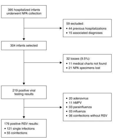
testing were selected. Patients were excluded if they had diagnoses of other associated morbidities or infection by bacteria, fungi or any other microorganism other than respiratory viruses. Patients were also excluded if they had
other hospitalizations in the previous 30 days (Figure 1).
Data were collected from patient charts by one of the authors according to standardized protocols. The variables analyzed were demographic characteristics (age and sex), signs and symptoms at admission, discharge diagnosis, prematurity (gestational age < 37 weeks), bronchopulmonary dysplasia, heart disease, immunosuppression and neuropathy. Severity was assessed according to the following outcomes: total hospitalization
time; oxygen therapy duration, admission to intensive care
unit (ICU) and mechanical ventilation. In the service where
this study was conducted, oxygen therapy is prescribed to keep oxygen saturation above 92% according to
pulse oximetry. Criteria for ICU admission were clinical and laboratory signs of respiratory insuficiency that
indicated the imminent need of mechanical ventilation and maintenance of oxygen saturation levels below 92% in patients receiving an inspired fraction of oxygen greater than or equal to 60%.
During the study, NPA of infants with respiratory problems
was routinely collected for viral testing. Because of the hours of the Virology Laboratory of the Institute of Biomedical
Sciences of Universidade de São Paulo, patients admitted
between Sunday and Friday (up to 5 pm) had NPA collected on the irst hospitalization day and sent to the virology
laboratory on the same day. The material from patients
hospitalized after 5 pm on Fridays, Saturdays and holidays was collected and sent to the laboratory on the irst working
day after hospitalization. The cases in which material was
collected on the irst 5 hospitalization days were analyzed.
Laboratory tests were conducted using PCR/RT-PCR for RSV,
hMPV, PIV 1, 2, and 3, Flu A and B, and ADV. Oligonucleotide
primers were used for each virus (Table 1). RT-PCR assays were performed using the High Capacity cDNA Archive kit
(Applied Biosystems, Carlsbad, USA). Ampliications were run separately (not multiplex). After RT-PCR, ampliication products (plate column) were puriied and transferred to sequencing tubes (Applied Biosystems, Carlsbad, USA). Ampliied fragments were analyzed using an ABI Prism 310 genetic analyzer (Applied Biosystems, Carlsbad, USA) and
the GeneScan 3.1.2 software.8
The study was approved by the Ethics in Research Committee of the University Hospital of Universidade de São Paulo.
Statistical analysis
Categorical variables were analyzed using a chi-square test, and continuous variables, the Mann-Whitney test. The association between explanatory variables and outcomes
was analyzed using irst univariate and then multivariate logistic regression. For that purpose, continuous outcome variables were treated as binary variables and classiied
according to their median value. The confounding variables age and breastfeeding duration were entered as continuous variables. These variables were also analyzed as categories.
The level of statistical signiicance was set at p > 0.05. The
Stata 10.0® software was used for statistical analyses.
Results
hMPV = human metapneumovirus; NPA = nasopharyngeal aspirate; RSV = respiratory syncytial virus.
Figure 1 - Flowchart of patient inclusion/exclusion in the study viral infections. RSV was the most frequent virus identiied
in this group (80.4%). The 176 infections by RSV were
analyzed in this study (Figure 1).
Eighty-ive per cent of the infants had a diagnosis of
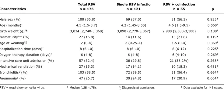
bronchiolitis or pneumonia. Other less frequent diagnoses were asthma and persistent or recurrent wheezing (14.8%). Chronic diseases were not frequent: in the group of single RSV infection, there were 2 patients with heart disease, 2 with neuromuscular diseases, and 1 with chronic pulmonary disease; in the group with RSV and coinfections, there was 1 patient with heart disease, and 2 with neuromuscular diseases. The characteristics of the 176 infants are summarized in Table 2.
RSV infections occurred as single infections in 121 infants (68.8%) and as coinfections in 55 infants (31.2%). The most frequent viruses associated with RSV were ADV (43.6%) and hMPV (29.1%). Other coinfections with RSV and different respiratory viruses were less frequent and could not be analyzed statistically because of the small
number of cases (Flu A = 4; PIV 3 = 3; PIV 2 = 1; PIV 3 + hMPV = 1; Flu A + ADV = 1; and ADV + hMPV = 5).
Infection severity was analyzed by comparing severity
outcomes between groups: single RSV infection (RSV); coinfection by RSV and any other virus (RSVCo); coinfection by RSV and adenovirus (RSV + ADV); and coinfection by RSV and human metapneumovirus (RSV + hMPV). Of the confounding variables, prematurity, age and breastfeeding were determinants of severity (Table 3). Prematurity increased absolute risk of coinfection by any virus, which was 28 and 48% for non-premature and premature patients
(absolute risk increase 20%; p = 0.04), and of coinfection
by RSV + hMPV, from 9.4% to 26.3% (absolute risk
increase = 16.9%; p = 0.04). The increase in absolute risk of coinfection by RSV + ADV was not statistically signiicant (absolute risk = 16 and 26%; absolute risk increase = 10%; p = 0.26).
Size
of ampliied
Virus/Primer gene Sequences (5’ > 3’) fragment (bp)
RSV
VSRAB-F1 F AACAGTTTAACATTACCAAGTGA 380
VSRAB-R1 TCATTGACTTGAGATATTGATGC
PIV 1
HPIV1-F1 HN CCGGTAATTTCTCATACCTATG 317
HPIV1-R1 CCTTGGAGCGGAGTTGTTAAG
PIV 2
HPIV2-F1 HN CCATTTACCYAAGTGATGGAAT 203
HPIV2-R1 GCCCTGTTGTATTTGGAAGAGA
PIV 3
HPIV3-F1 HN ACTCCCAAAGTTGATGAAAAGAT 102
HPIV3-R1 TAAATCTTGTTGTTGAGATTGA
Inluenza A
FLUA-F1 NS1 CTAAGGGCTTTCACCGAAGA 192
FLUA-R1 CCCATTCTCATTACTGCTTC
Inluenza B
FLUB-F1 NS1 ATGGCCATCGGATCCTCAAC 241
FLUB-R1 TGTCAGCTATTATGGAGCTG
Adenovirus
ADENO-F1 Hexon CCC(AC)TT(CT)AACCACCACCG 167
ADENO-R1 ACATCCTT(GCT)C(GT)GAAGTTCCA
hMPV
MPV-F1 F GAGCAAATTGAAAATCCCAGACA 347
MPV-R1 GAAAACTGCCGCACAACATTTAG
bp = base pairs; hMPV = human metapneumovirus; PIV = parainfluenza virus; RSV = respiratory syncytial virus. table 1 - Polymerase chain reaction assay panel for each virus
total RSV Single RSV infectio RSV + coinfection
Characteristics n = 176 n = 121 n = 55 p
Male sex (%) 100 (56.8) 69 (57.0) 31 (56.3) 0.935*
Age (months)† 4.5 (1.5-8.7) 4.2 (1.45-8.55) 4.6 (1.5-9.5) 0.560‡
Birth weight (g)†¶ 3,034 (2,740-3,360) 3,090 (2,778-3,367) 2,980 (2,580-3,300) 0.138‡
Prematurity** (%) 27 (16.8) 14 (11.6) 13 (23.6) 0.119*
Age at weaning†║ 2 (0-4) 2 (0.25-4) 1.5 (0-4) 0.369‡
Hospitalization time (days)† 8 (6-10) 8 (6-10) 8 (6-12) 0.225‡
Oxygen therapy duration (days)† 6 (4-8) 6 (4-8) 6 (4-10) 0.269‡
Intensive care unit admission (%) 57 (32.4) 36 (29.8) 21 (38.2%) 0.268*
Mechanical ventilation (%) 27 (15.3) 17 (14.1) 10 (18.2) 0.481*
Bronchiolitis§ (%) 103 (58.5) 72 (59.5) 31 (56.4) 0.664*
Pneumonia§ (%) 47 (26.7) 30 (24.8) 17 (30.9) 0.664*
table 2 - Demographic and medical characteristics of 176 infants included in the study according to absence or presence of coinfections
RSV = respiratory syncytial virus. * Chi-square.
† Median (p25 - p75). ‡ Mann-Whitney test.
§ Diagnosis at admission. ║Data available for 155 cases.
Analysis Hospitalization time * O2 duration* iCU admission Mechanical ventilation
OR 95%Ci p OR 95%Ci p OR 95%Ci p OR 95%Ci p
Univariate analysis
Male sex 0.78 0.42-1.42 0.41 0.90 0.49-1.65 0.74 0.94 0.49-1.77 0.84 1.51 0.66-3.44 0.33 Prematurity 0.45 0.23-0.88 0.02 0.43 0.22-0.83 0.01 0.44 0.22-0.88 0.02 0.32 0.14-0.75 0.01 Age† 0.94 0.89-0.99 0.04 0.91 0.86-0.98 0.01 0.98 0.92-1.04 0.49 0.89 0.81-0.99 0.04
Breastfeeding duration† 0.80 0.65-0.98 0.04 0.78 0.63-0.96 0.02 0.79 0.64-0.98 0.04 0.86 0.65-1.14 0.31
RSVCo 1.32 0.70-2.51 0.39 1.46 0.77-2.78 0.25 1.46 0.75-2.85 0.27 1.36 0.58-3.20 0.48 RSV + ADV 1.62 0.67-3.91 0.28 1.93 0.80-4.66 0.15 1.42 0.57-3.53 0.46 0.87 0.23-3.25 0.84 RSV + hMPV 0.82 0.28-2.41 0.72 1.27 0.44-3.64 0.66 1.07 0.35-3.31 0.90 0.87 0.18-4.19 0.87
Multivariate analysis‡
Model 1
RSV 1.00 1.00 1.00 1.00
RSVCo 0.58 0.21-1.55 0.27 0.88 0.33-2.33 0.80 0.97 0.36-2.60 0.96 1.05 0.29-3.85 0.94
Model 2
RSV 1.00 1.00 1.00 1.00
RSV + ADV 0.97 0.23-4.00 0.96 1.21 0.29-5.13 0.79 1.50 0.35-6.42 0.59 1.29 0.21-7.86 0.78
Model 3
RSV 1.00 1.00 1.00 1.00
RSV + hMPV 0.21 0.04-1.17 0.07 0.48 0.10-2.21 0.34 0.30 0.05-1.80 0.19 0.88 0.58-1.32 0.54
table 3 - Univariate and multivariate analyses of possible determinants of infection severity
95%CI = 95% confidence interval; ICU = Intensive care unit; O2 = oxygen therapy; OR = odds ratio; RSV = respiratory syncytial virus; (n = 121); RSV + ADV = RSV and coinfection by adenovirus (n = 24); RSVCo = RSV and coinfection by any other virus (n = 55); RSV + hMPV = RSV and coinfection by human metapneumovirus (n = 16).
* Dichotomized according to median value. † Treated as continuous variables.
‡ Outcomes adjusted for prematurity, age and breastfeeding duration.
Discussion
This cohort of hospitalized infants with RSV infection had a high rate of coinfections by other respiratory viruses (31%). Coinfections were not associated with disease severety, regardless of the virus in association with RSV and the presence of any counfounding factor, such as prematurity, age and breastfeeding duration.
Evidence in current literature indicates that the clinical
meaning of the simultaneous identiication of more than one
virus in respiratory secretions is controversial. Cilla et al.
also did not ind any differences in the prognosis of children
infected by one or more viruses according to hospitalization
time, ICU admission and oxygen therapy. Other reports found different and conlicting results. Some suggest that
severity is greater in viral coinfections, whereas others found greater severity in infections by a single pathogen. Semple et al. found that RSV + hMPV coinfection increased 10 times
the relative risk of ICU admission for mechanical ventilation (relative risk = 10.99, 95%CI = 5.0-24.12, p < 0.001). As
the studies were retrospective, characteristics not evaluated in the populations under study or even differences in viral subtypes and the interaction with environmental factors may explain the differences found.5,9
Interesting evidence, though weaker, has been produced
by case reports.10 Greensill et al. evaluated children with
bronchiolitis caused by RSV that received mechanical ventilation; 70% had coinfection by hMPV, which suggested
greater severity in these cases. In contrast, Canducci found
lower severity in cases of coinfection by RSV + hMPV than in infections by a single virus in hospitalized children.3,10
Conlicting results may be explained by several factors,
such as the fact that different pathogenic mechanisms may be triggered by different viruses that mutually potentialize or mitigate each other’s effects. Moreover, the actual pathogenic role of each virus may be unclear. The simultaneous detection of one or more pathogenic virus,
such as those investigated in this study, is usually classiied
as coinfection. However, the presence of viral genome detected using molecular biology techniques may indicate
viral persistence, with no signiicant pathogenic effect at
the time of detection.2,9,10 A recent study evaluated the
presence of ADV DNA in respiratory secretions of children that had recurrent infections and found both recurrent infections due to different ADV genotypes and persistence of viral DNA for a long time, which stresses the importance
References
1. Nair H, Nokes DJ, Gessner BD, Dherani M, Madhi SA, Singleton RJ, et al. Global burden of acute lower respiratory infections due to respiratory syncytial virus in young children: a systematic review and meta-analysis. Lancet. 2010;375:1545-55. 2. Stempel HE, Martin ET, Kuypers J, Englund JA, Zerr DM. Multiple
viral respiratory pathogens in children with bronchiolitis. Acta Paediatr. 2009;98:123-6.
3. Canducci F, Debiaggi M, Sampaolo M, Marinozzi MC, Berrè S, Terulla C, et al. Two-year prospective study of single infections and co-infections by respiratory syncytial virus and viruses identiied recently in infants with acute respiratory disease. J Med Virol. 2008;80:716-23.
4. Calvo C, García-García ML, Blanco C, Vázquez MC, Frías ME, Pérez-Breña P, et al. Multiple simultaneous viral infections in infants with acute respiratory tract infections in Spain. J Clin Virol. 2008;42:268-72.
5. Semple MG, Cowell A, Dove W, Greensill J, McNamara PS, Halfhide C, et al. Dual infection of infants by human metapneumovirus and human respiratory syncytial virus is strongly associated with severe bronchiolitis. J Infec Dis. 2005;191:382-6.
6. Wolf DG, Greenberg D, Kalkstein D, Shemer-Avni Y, Givon-Lavi N, Saleh N, et al. Comparison of human metapneumovirus, respiratory syncytial virus and inluenza A virus lower respiratory tract infections in hospitalized young children. Pediatr Infect Dis J. 2006;25:320-4.
7. Cuevas LE, Nasser AM, Dove W, Gurgel RQ, Greensill J, Hart CA.
Human metapneumovirus and respiratory syncytial virus, Brazil.
Emerg Infect Dis. 2003;9:1626-8.
8. Thomazelli LM, Vieira S, Leal AL, Sousa TS, Oliveira DB, Golono MA, et al. Surveillance of eight respiratory viruses in clinical samples of pediatric patients in southeast Brazil. J Pediatr (Rio J). 2007;83:422-8.
9. Cilla G, Oñate E, Perez-Yarza EG, Montes M, Vicente D, Prez-Trallero E. Viruses in community-acquired pneumonia in children aged less than 3 years old: high rate of viral coinfection. J Med Virol. 2008;80:1843-9.
10. Greensill J, McNamara PS, Dove W, Flanagan B, Smyth RL, Hart CA. Human metapneumovirus in severe respiratory syncytial virus bronchiolitis. Emerg Infect Dis. 2003;9:372-5.
11. Kalu SU, Loeffelholz M, Beck E, Patel JA, Revai K, Fan J, et al. Persistence of adenovirus nucleic acids in nasopharyngeal secretions: a diagnostic conundrum. Pediatr Infect Dis J. 2010;29:746-50.
signs and symptoms.11 RSV + ADV coinfection was the most
frequent in this study, as well as in studies conducted by other authors, who found coinfection rates of up to 43% when this pathogen was involved.2 Infections by ADV alone
have been frequent in the service where this study was
conducted. In a previous study, the authors found that ADV
was the second most frequent agent of single infections in hospitalized infants with acute respiratory disease, with prevalence rates ranging from 5.6 to 9.6%. The occurrence of less aggressive genotypes may explain the mild features of ADV infection, but viral genotyping was not performed in this study.12,13 The overlapping of RSV and hMPV infection
seasons, previously demonstrated in a 4-year surveillance study in the same service where this study was conducted, also explains the high RSV + hMPV coinfection rates. Viral coinfections are frequently caused by the pathogens that are predominant in single infections in the period under study.14 Interestingly, prematurity increased the risk to coinfection by hMPV (absolute risk increase = 16.9%; p = 0.04), but severity remains similar to that of single
infection by RSV.
The high prevalence of viral coinfection by ADV and hMPV may also be explained by the characteristics of the study population, composed of infants, most of them in
their irst year of life, with bronchiolitis and pneumonia,
and hospitalized, in particular, during the season of the respiratory viruses.9 The use of molecular biology methods
also had high diagnostic sensitivity, and positive results were
found in 72% of the samples. This inding is in agreement
with those in the literature, as studies found viral detection rates ranging from 45 to 70% under the same conditions and viral coinfection rates from 15 to 39%.2-4,15-17
This study included only hospitalized children, and its results cannot be extrapolated to children with less severe conditions. Tests to detect bocavirus and rhinovirus were not performed. The inclusion of these viruses in this study might have resulted in a greater prevalence of coinfections.9,18
However, these two pathogens are usually less frequent causes of bronchiolitis and pneumonia in infants than the viruses selected for this study. The role of the rhinovirus is more remarkable, as it is an important trigger of wheezing episodes in atopic children, but is also usually isolated in about 30% of asymptomatic individuals. Moreover, in the same way as the bocavirus, it often has an unclear pathogenic role.9,18 Had these pathogens been included, the analysis
of coinfection prognosis in the study population might not have been different.
This study had some limitations due to its retrospective design. The parameters used were selected because they are carefully evaluated and recorded in the service’s charts, and, therefore, data are adequate to assess clinical severity by means of medical chart reviews, not subject to any retrospective interpretation bias, as would be the case with clinical scores. Other factors, such as passive smoking,
daycare center attendance, and contact with school age children could not be assessed because these data are not as carefully collected and recorded in medical charts as the variables included in this study. The agreement between the different severity outcomes and the similar clinical progression of patients with infection by RSV as a single agent or coinfection by other virus further support to our results.
Conclusion
12. Vieira SE, Stewien KE, Queiroz DA, Durigon EL, Török TJ, Anderson LJ, et al. Clinical patterns and seasonal trends in respiratory syncytial virus hospitalizations in São Paulo, Brazil. Rev Inst Med Trop Sao Paulo. 2001;43:125-31.
13. Moura PO, Roberto AF, Hein N, Baldacci E, Vieira SE, Ejzenberg B, et al. Molecular epidemiology of human adenovirus isolated from children hospitalized with acute respiratory infection in São Paulo, Brazil. J Med Virol. 2007;79:174-81.
14. Oliveira DB, Durigon EL, Carvalho AC, Leal AL, Souza TS, Thomazelli LM, et al. Epidemiology and genetic variability of human metapneumovirus during a 4-year-long study in Southeastern Brazil. J Med Virol. 2009;81:915-21.
15. Jennings LC, Anderson TP, Werno AM, Beynon KA, Murdoch DR. Viral etiology of acute respiratory tract infections in children presenting to hospital: role of polymerase chain reaction and demonstration of multiple infections. Pediatr Infect Dis J. 2004;23:1003-7.
16. Straliotto SM, Siqueira MM, Machado V, Maia TM. Respiratory viruses in the pediatric intensive care unit: prevalence and clinical aspects. Mem Inst Oswaldo Cruz. 2004;99:883-7.
17. van der Zalm MM, van Ewik BE, Wilbrink B, Uiterwaal CS, Wolfs TF, van der Ent CK. Respiratory pathogens in children with and without respiratory symptoms. J Pediatr. 2009;154:396-400. 18. Lemanske RF Jr, Jackson DJ, Gangnon RE, Evans MD, Li Z, Shult
PA, et al. Rhinovirus illnesses during infancy predict subsequent childhood wheezing. J Allergy Clin Immunol. 2005;116:571-7.
Correspondence: Sandra Elisabete Vieira
Av. Dr. Eneas Carvalho de Aguiar, 647 CEP 05403-000 – São Paulo, SP - Brazil Tel.: +55 (11) 3069.8803

