PÓS-GRADUAÇÃO EM GENÉTICA E BIOQUÍMICA
IDENTIFICAÇÃO DE SOROTIPOS DE ADENOVÍRUS EM ASPIRADOS DE NASOFARINGE DE CRIANÇAS COM DOENÇA RESPIRATÓRIA AGUDA,
ATENDIDAS EM UBERLÂNDIA, MG
Aluno: Lysa Nepomuceno Luiz
Orientador: Dr. Júlio César Nepomuceno
Co-orientadora: Dra. Divina Aparecida Oliveira Queiróz
UNIVERSIDADE FEDERAL DE UBERLÂNDIA INSTITUTO DE GENÉTICA E BIOQUÍMICA PÓS-GRADUAÇÃO EM GENÉTICA E BIOQUÍMICA
IDENTIFICAÇÃO DE SOROTIPOS DE ADENOVÍRUS EM ASPIRADOS DE NASOFARINGE DE CRIANÇAS, COM DOENÇA RESPIRATÓRIA AGUDA,
ATENDIDAS EM UBERLÂNDIA, MG
LYSA NEPOMUCENO LUIZ
Orientador: Dr. Júlio César Nepomuceno
Co-orientadora: Dra. Divina Aparecida Oliveira Queiróz
Dissertação apresentada à Universidade Federal de Uberlândia como parte dos requisitos para obtenção do Título de Mestre em Genética e Bioquímica (Área Genética)
Dados Internacionais de Catalogação na Publicação (CIP)
L953i Luiz, Lysa Nepomuceno, 1980-
Identificação de sorotipos de adenovírus em aspirados de
nasofaringe de crianças, com doença respiratória aguda, atendidas em Uberlândia, MG / Lysa Nepomuceno Luiz. - 2008.
42 f.
Orientador: Júlio César Nepomuceno.
Co-orientadora: Divina Aparecida Oliveira Queiroz.
Dissertação (mestrado) - Universidade Federal de Uberlândia, Pro-grama de Pós-Graduação em Genética e Bioquímica.
Inclui bibliografia.
1. 1. Doença respiratória infantil - Teses. I. Nepomuceno, Júlio César. II. Queiroz, Divina Aparecida Oliveira. III. Universidade Fede-ral de Uberlândia. Programa de Pós-Graduação em Genética e
Bioquí-mica. IV. Título.
CDU: 616.2-053.2
ii
UNIVERSIDADE FEDERAL DE UBERLÂNDIA INSTITUTO DE GENÉTICA E BIOQUÍMICA PÓS-GRADUAÇÃO EM GENÉTICA E BIOQUÍMICA
IDENTIFICAÇÃO DE SOROTIPOS DE ADENOVÍRUS EM ASPIRADOS DE NASOFARINGE DE CRIANÇAS, COM DOENÇA RESPIRATÓRIA AGUDA,
ATENDIDAS EM UBERLÂNDIA, MG
LYSA NEPOMUCENO LUIZ
COMISSÃO EXAMINADORA
Presidente: Dr. Júlio César Nepomuceno
Examinadores: Dr. José Paulo Gagliardi Leite
Dr. Foued Salmen Espindola
Data da defesa:
iv
AGRADECIMENTOS
A Deus, pela vida e pela fé que suavizou minhas lutas e permitiu muitas vitórias
Aos meus pais, pela dedicação, compreensão, carinho e principalmente, pelo exemplo. A
vocês, minha eterna gratidão!
Aos meus irmãos Marcos e Bruno pelo companheirismo, compreensão e carinho de
sempre.
Ao Miro, Lorena Landerdahl e Carlos Roberto por fazerem parte de minha vida de uma
maneira tão especial. Obrigada por tudo!
À Profa Dra. Divina Aparecida Oliveira Queiróz pelos ensinamentos, carinho e confiança.
Ao Prof. Dr. Júlio César Nepomuceno por acreditar em meus esforços e me aceitar, formalmente, como orientanda.
Ao Dr. Jonny Yokosawa pela atenção, carinho, ensinamentos e sugestões.
Ao Lourenço Costa, Bruno Carneiro, Thelma Mattos de Oliveira, Guilherme Freitas,
Nayhanne Tizzo, Lucas Zimon, Paulo, Tatiany Calegari e Gabriela Dyonísio pela amizade,
ensinamentos, inúmeras sugestões e auxílio em diferentes etapas laboratoriais como coleta,
À Juliana Ribeiro pela ajuda na análise estatística
Ao Dr. José Paulo Gagliardi Leite, Edson Pereira Filho e demais membros do Laboratório
de Virologia Comparada/ Fiocruz-RJ, por viabilizarem grande parte dos experimentos,
pelo carinho e pelos ensinamentos.
Aos médicos do Hospital de Clínicas de Uberlândia, Dr. Orlando C. Mantese, Dr. Hélio L.
Silveira, Dr. Francisco C. Diniz e todos os demais médicos e residentes, pela seleção dos
pacientes.
Aos responsáveis pelos laboratórios de Imunologia, Parasitologia, Genética Molecular,
Biologia Molecular, Fisiologia, pelos equipamentos e espaço físico cedidos, sem os quais
não seria possível a realização deste trabalho.
Às crianças que participaram deste trabalho e aos responsáveis que consentiram com a
coleta dos espécimes clínicos.
Às amigas Mariana Moreira, Luciana Karen, Renata Brito, Sinara, Marine, Marília e
Silmara Parreira, pelo apoio nos momentos difíceis e pelos momentos inesquecíveis da
vi
ÍNDICE
LISTA DE ABREVIATURAS, SIGLAS E SÍMBOLOS ... vii
LISTA DE TABELAS... viii
LISTA DE FIGURAS... ix
INTRODUÇÃO GERAL... 1
REFERÊNCIAS BIBLIOGRÁFICAS ... 5
CAPÍTULO ÚNICO ... 10
Resumo ... 11
Abstract ... 12
Introduction... 13
Material and Methods ... 14
Results... 18
Discussion ... 20
Acknowlegments ... 23
LISTA DE ABREVIATURAS, SIGLAS E SÍMBOLOS
L: microlitro
Células A-549: linhagem de células de carcinoma de pulmão humano
Células HEp-2: linhagem de células de carcinoma epidermóide de laringe humana
CPE: “cytopathic effect” (efeito citopático)
DNA: ácido desoxirribonucléico
DRA/ARD: doença respiratória aguda/“acute respiratory disease”
HAdV: “human adenovirus” (adenovírus humano)
IFA: “immunofluorescence assay” (ensaio de imunofluorescência)
IRA: infecção respiratória aguda
MAbs: “monoclonal antibodies” (anticorpos monoclonais)
NFA: “nasopharyngeal aspirate” (aspirado de nasofaringe)
PCR: “polymerase chain reaction” (reação em cadeia pela polimerase)
RT-PCR: “reverse transcription-polymerase chain reaction” (transcrição reversa
da reação em cadeia pela polimerase)
viii
LISTA DE TABELAS
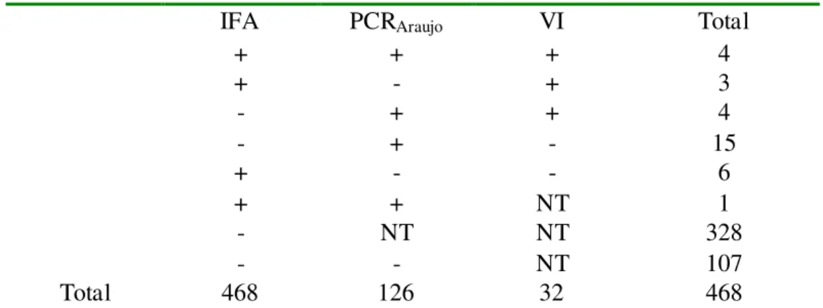
Table 1:Results obtained by IFA, VI and PCR for adenoviruses detection in 468 specimens
of NFA collected from children less than 5 years old, with acute respiratory disease ... 26
Table 2: Comparison of two PCR assays with 33 specimens that were previously positive
specimens for presence of adenovirus ... 27
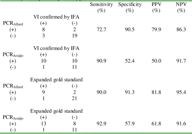
Table 3: Comparison of the results obtained, from DNA extracted from in natura samples,
by the two PCR assays in 33 specimens previously positive for adenovirus ... 28
Table 4: Distribution of the 12 serotyped cases according to the involvement of symptoms
LISTA DE FIGURAS
INTRODUÇÃO
Fig. 1: Partícula do adenovírus ... 2
CAPÍTULO ÚNICO
Fig. 1: Seasonal distribution of adenovirus-positive cases compared with the total number of collected samples during the study period. ... 30
INTRODUÇÃO GERAL
Os vírus constituem importante causa de doenças respiratórias agudas (DRAs) em
crianças menores de cinco anos de idade, respondendo por elevadas taxas de morbidade e
mortalidade em todo o mundo (WILLIAMS et al., 2002), sendo que aproximadamente 5%
desses casos estão associados aos adenovírus (BRANDT et al., 1969).
Na década de 50 estes vírus foram isolados e caracterizados por dois grupos de
pesquisadores que estudavam a etiologia de infecções respiratórias agudas (ROWE et al.,
1953; HILLEMAN & WERNER, 1954). Diferentes denominações foram adotadas antes da
atual: agentes da degeneração da adenóide, da doença da adenóide-faringe-conjuntiva ou
da doença respiratória aguda. Somente em 1956 passaram a ser denominados adenovírus,
tendo em vista que o isolamento original foi realizado a partir de tecidos de adenóides
(ENDERS et al, 1956).
Os adenovírus humanos pertencem ao gênero Mastadenovirus, da família
Adenoviridade. São conhecidos 51 sorotipos, classificados em 6 espécies (A a F) (de
JONG et al., 1999), baseado na sua capacidade de aglutinar vários tipos de eritrócitos.
Outras propriedades virais tais como relações antigênicas, tamanho da fibra, grau de
homologia do DNA, percentual das bases nitrogenadas guanina e citosina, número de
fragmentos de clivagem após digestão com a endonuclease SmaI e massa molecular de
certas proteínas internas tendem a coincidir com esta classificação (SWENSON et al.,
2003).
São vírus DNA de fita dupla, de simetria icosaédrica, de 70 a 90 nm de diâmetro,
formado por 252 capsômeros, sendo 240 hexons e 12 pentons. Cada unidade do penton é
constituída por uma base da qual se projeta a fibra (HIERHOLZER et al., 1989) (Fig. 1).
Fig. 1: Partícula do adenovírus
Http://iavireport.org/Issues/Issue 11-1/Winner.asp
Além de acometerem o trato respiratório, os adenovírus causam uma variedade de
outras síndromes, incluindo doenças oculares, gastroenterites e infecções urinárias
(SHENK, 2001). Embora a maioria seja autolimitante, esses vírus podem estar associados
a doenças graves ou letais tanto em indivíduos imunocomprometidos quanto em indivíduos
saudáveis (MUNOZ et al., 1998).
Nas décadas de 50 e 60, esses vírus foram reconhecidos como um dos mais
importantes responsáveis por doenças respiratórias entre recrutas militares nos Estados
Unidos (TOP, 1975). O impacto dos adenovírus nesta população estimulou o
desenvolvimento de vacinas contra os sorotipos 4 e 7. Entretanto, a produção das duas
vacinas foi descontinuada em 1996, acarretando no ressurgimento de epidemias em vários
centros de treinamento militares por esses vírus (GRAY et al., 1999).
O diagnóstico laboratorial pode ser feito por isolamento viral em cultura de células,
3
crescente o emprego da reação em cadeia pela polimerase (PCR) tendo em vista a rapidez e
a elevada sensibilidade deste método (OSIOWY, 1998).
Trabalhos realizados em diferentes países têm mostrado um percentual de detecção
desses vírus de aproximadamente 7,0% em crianças com doença respiratória aguda (DRA)
(ROCHOLL et al., 2004; VIEGAS et al., 2003). No Brasil, resultados similares têm sido
reportados. Em Porto Alegre, Stralliotto et al. (2002), encontraram um percentual de 6% de
positividade em crianças com DRA. Em Salvador, o percentual encontrado por Moura et
al. (2003) foi de 7,1% em crianças atendidas em emergência e em enfermarias de um
centro pediátrico da cidade, e em São Paulo, outro grupo de pesquisadores encontrou um
percentual de 8,2% em crianças internadas, sendo a maioria com IRA e menores de 5 anos
de idade (MOURA et al. 2007).
Diferentes fatores devem ser considerados ao se analisar a prevalência destes vírus,
incluindo o método de diagnóstico empregado, a gravidade da doença, a idade dos
pacientes e o período da coleta dos espécimes clínicos (ECHAVARRIA et al., 2006).
Referente à DRA, além da detecção viral, a determinação dos sorotipos constitui
outro dado importante, considerando-se que certos sorotipos como o 7 e 3 são mais
freqüentemente associados a surtos de infecções graves do trato respiratório (HONG et al.,
2001; KIM et al., 2003), embora outros sorotipos da espécie B (AdV 14, 16, 21, 34, 35), da
espécie C (Ad1, 2, 5, 6 ) e da espécie E (Ad4) também sejam responsáveis por DRAs em
crianças e militares (WADELL, 1984; HORWITZ, 2001).
Um estudo realizado a partir dos casos positivos para adenovírus coletados de
crianças hospitalizadas por doença respiratória aguda inferior nas cidades de Buenos Aires,
Santiago e Montevidéu, demonstrou que 71,0% dos isolados pertenciam à espécie B,
28,5% à espécie C e somente 0,6% à espécie E, sendo o sorotipo 7, mais especificamente o
estudo (KAJON et al, 1996). Entretanto, além do sorotipo, outros fatores podem
influenciar na gravidade da doença, como: idade, condição socioeconômica e fatores
ambientais (HONG et al, 2001).
A transmissão nosocomial constitui outro problema freqüentemente associado aos
adenovírus. O fato de responderem por cerca de 10,0% dos casos de pneumonia, que
freqüentemente necessitam de suporte de terapia intensiva, associado à excreção destes
vírus pelas fezes por longos períodos e a transmissão tanto por fômites quanto por
aerossóis ajudam a explicar tal transmissão (HIERHOLZER, 1989). Além disso, tem sido
observado que muitas crianças com DRA por esses vírus desenvolvem alguma seqüela
pulmonar (LANG et al.,1969; SIMILA et al, 1981).
Neste trabalho, pesquisou-se adenovírus em aspirados de nasofaringe de crianças
menores de 5 anos de idade com DRA, atendidas em Uberlândia, MG, no período de
REFERÊNCIAS BIBLIOGRÁFICAS
BRANDT, C. D.; KIM, H. W.; VARGOSKO, A. J.; JEFFRIES, B. C.; ARROBIO, J. O.,
RINDGE, B. PARROTT, R. H.; CHANOCK, R. M. Infections in 18,000 infants and
children in a controlled study of respiratory tract disease. I. Adenovirus pathogenicity in
relation to serologic type and illness syndrome. Am J Epidemiol, v.90, n.6, p.484-500,
Dec. 1969.
DE JONG, J. C.; WERMENBOL, A. G.; VERWEIJ-UIJTERWAAL, M. W.; SLATERUS,
K. W.; WERTHEIM-VAN DILLEN, P.; VAN DOORNUM, G. J.; KHOO, S. H.;
HIERHOLZER, J. C. Adenoviruses from human immunodeficiency virus-infected
individuals, including two strains that represent new candidate serotypes Ad50 and Ad51
of species B1 and D, respectively. J Clin Microbiol, v.37, n.12, p.3940-3945, Dec. 1999.
ECHAVARRIA, M.; MALDONADO, D.; ELBERT, G.; VIDELA, C.; RAPPAPORT, R.;
CARBALLAL, G. Use of PCR to demonstrate presence of adenovirus apecies B, C or F as
well as coinfection with two adenovirus species in children with flu-like symptoms. J.
Clin. Microbiol, v.44, n.2, p.625-627, Feb. 2006.
ENDERS, J. F.; BELL, J. A.; DINGLE, J. H.; FRANCIS, T. JR.; HILLEMAN, M. R.;
HUEBNER, R. J.; PAYNE, A. M. Adenoviruses: group name proposed for new
GRAY, G. C.; GOSWAMI, P. R.; MALASIG, M. D; HAWKSWORTH, A. H.; TRUMP,
D. H.; RYAN, M. A.; SCHNURR, D. P. Adult adenovirus infections: loss of orphaned
vaccines precipitates military respiratory disease epidemics. Clin. Infect. Dis, v.31,
p.663-670. 2000.
HILLEMAN, M. R.; WERNER, J. H. Recovery of new agent from patients with acute
respiratory illness. Proc Soc Exp Biol Med, v.85, n.1, p.183-188, Jan. 1954.
HIERHOLZER, J. C. Adenoviruses. In: Achmidt, N. J., Emmons, R. W. (Ed.). Diagnostic
procedures for viral, rickettsial and chlamydial infections. Washington, D.C.:
American Public Health Association, 1989. p.219-264.
HONG, J. Y.; LEE, H. J.; PIEDRA, P. A.; CHOI, E. H.; PARK, K. H.; KOH, Y. Y.; KIM,
W. S. Lower respiratory tract infections due to adenovirus in hospitalized Korean children:
epidemiology, clinical features, and prognosis. Clin Infect Dis, v.32, n.10, p.1423-1429,
May. 2001.
HORWITZ, M. S. ADENOVIRUSES. IN: KNIPE, D. M.; HOWLEY, P. M. (Ed.). Fields
Virology. Philadelphia, Pa: Lippincott/The Williams & Wilkins Co., 2001. p.2301-2326
KAJON, A. E.; MISTCHENKO, A. S.; VIDELA, C.; HORTAL, M.; WADELL, G.;
AVENDANO, L. F. Molecular epidemiology of adenovirus acute lower respiratory
infections of children in the south cone of South America (1991-1994). J Med Virol, v.48,
7
KIM, Y. J.; HONG, J. Y.; LEE, H. J.; SHIN, S. H.; KIM, Y. K.; INADA, T.; HASHIDO,
M.; PIEDRA, P. A. Genome type analysis of adenovirus types 3 and 7 isolated during
successive outbreaks of lower respiratory tract infections in children. J Clin Microbiol,
v.41, n.10, p.4594-4599, Oct. 2003.
LANG, W. R.; HOWDEN, C. W.; LAWS, J.; BURTON, J. F. Bronchopneumonia with
serious sequelae in children with evidence of adenovirus type 21 infection. Br Med J, v.1,
n.5636, p.73-79, Jan. 1969.
MOURA, F. E. A.; BORGES, L. C.; SOUZA, L. S. F.; RIBEIRO, D. H.; SIQUEIRA, M.
M.; RAMOS, E. A. G. Estudo de infecções respiratórias agudas virais em crianças
atendidas em um centro pediátrico em Salvador (BA). Jornal Brasileiro de Patologia e
Medicina Laboratorial, v.39, n.4, p.275, 2003.
MOURA, P. O.; ROBERTO, A. F.; HEIN, N.; BALDACCI, E.; VIEIRA, S. E.;
EJZENBERG, B.; PERRINI, P.; STEWIEN, K. E.; DURIGON, E. L.; MEHNERT, D. U.;
HARSI, C. M. Molecular epidemiology of human adenovirus isolated from children
hospitalized with acute respiratory infection in Sao Paulo, Brazil. J Med Virol, v.79, n.2,
p.174-181, Feb. 2007.
MUNOZ, F. M.; PIEDRA, P. A.; DEMOLER, G. J. Disseminated adenovirus disease in
immunocompromised and immunocompetent children. Clin Infect Dis, v.27, n.5,
OSIOWY, C. Direct detection of respiratory syncytial virus, parainfluenza virus, and
adenovirus in clinical respiratory specimens by a multiplex reverse transcription-PCR
assay. J Clin Microbiol, v.36, n.11, p.3149-3154, Nov. 1998.
ROCHOLL, C.; GERBER, K.; DALY, J.; PAVIA, A. T.; BYINGTON, C. L. Adenoviral
infections: the impactc of rapid diagnosis. Pediatrics, v.113, n.1, p. 51-56, Jan. 2004.
ROWE, W. P.; HUEBNER, R. J.; GILMORE, L. K. Isolation of a cytopathogenic agent
from human adenoids undergoing spontaneous degeneration in tissue culture. Proc Soc
Exp Biol Med, v.84, p.570-573, 1953.
SHENK, T. E. Adenoviridae: the viruses and their replication. In: KNIPE, D. M.;
HOWLEY, P. M. E. (Ed.). Fields Virology. Philadelphia, Pa: Lippincott/The Williams &
Wilkins Co., 2001. p.2265-2300
SIMILA, S.; LINNA, O.; LANNING, P.; HEIKKINEN, E.; ALA-HOUHALA, M. Chronic
lung damage caused by adenovirus type 7: a ten-year follow-up study. Chest, v.80, n.2,
p.127-31, Aug. 1981.
STRALLIOTTO, S. M.; SIQUEIRA, M. M.; MULLER, R. L.; FISCHER, G. B.; CUNHA,
M. L.; NESTOR, S. M. Viral etiology of acute respiratory infections among children in
Porto Alegre, RS, Brazil. Revista da Sociedade Brasileira de Medicina Tropical, v.35,
9
SWENSON, P. D.; WADELL, G.; ALLARD, A.; HIERHOLZER, J. C. Adenoviruses. In:
P. R. MURRAY, E. J. BARON, et al (Ed.). Manual of Clinical Virology. Washington,
D.C.: ASM Press, 2003. p.1404-1417.
TOP, F. H., Jr. Control of adenovirus acute respiratory disease in U.S. Army trainees. Yale
J Biol Med, v.48, n.3, p.185-195, Jul. 1975.
VIEGAS, M.; BARRERO, P. R.; MAFFEY, A. F.; MISTCHENKO, A. S. Respiratory
viruses seasonality in children under five years of age in Buenos Aires, Argentina. A
five-year analysis. Journal of Infection, v. 49, p.222–228, 2004.
WADELL, G. Molecular epidemiology of human adenoviruses. Curr Top Microbiol
Immunol, v.110, p.191-220, 1984.
WILLIAMS, B. G.; GOUWS, E.; BOSCHI-PINTO, C.; BRYCE, J.; DYE, C. Estimates of
world-wide distribution of child deaths from acute respiratory infections. Lancet Infect
Capítulo Único
ADENOVIRUS SEROTYPES IN NASOPHARINGEAL ASPIRATES OF CHILDREN PRESENTING ACUTE RESPIRATORY DISEASE, ATTENDED IN
UBERLÂNDIA, MG
Manuscrito a ser submetido ao Journal of Clinical Virology
11
Resumo
Introdução: Adenovírus (AdVs) constituem importante causa de doença respiratória
aguda (DRA), gastroenterites, conjuntivites e infecções urinárias em humanos.
Objetivos: Detecção de AdVs por imunofluorescência indireta (IFI) e por reação em
cadeia pela polimerase (PCR) em aspirados de nasofaringe de crianças menores de 5 anos de idade, com DRA, atendidas em Uberlândia, MG, assim como, comparação de dois testes de PCR e identificação de sorotipos de AdVs circulantes na região.
Material e Métodos: Um total de 468 espécimes clínicos obtidos de novembro de 2000 a
abril de 2007 foi testado pela IFI para detecção de AdVs e outros vírus respiratórios. Em seguida, o DNA das 126 amostras in natura que obtiveram resultado negativo
/inconclusivo pela IFI e que também foram negativas para rinovírus pela RT-PCR, foi extraído por Trizol® e testado pela PCRAraújo (Araújo et al., 2001) para os AdVs. Os espécimes positivos para AdVs foram inoculados em células HEp-2 e A549. Além disso, o DNA das amostras positivas, tanto in natura quanto do raspado de cultura de células, foi
extraído utilizando-se o kit “QIAmp DNA Mini Kit” (QIAGENTM Valencia, CA) e testado novamente pela PCRAraujo e pela PCRAllard (Allard et al., 2001), afim de comparar a sensibilidade/especificidade dos dois testes. Além disso, os sorotipos foram identificados a partir do seqüenciamento dos nucleotídeos de produtos de PCR positivos para AdVs.
Resultados: Das 468 amostras, 33 (7,1%) foram positivas para AdVs, sendo 14 pela IFI e
19 pela PCRAraujo. De 32 espécimes inoculados em cultura de células, foi possível isolar os AdVs de 16. A comparação dos resultados obtidos a partir do DNA das 33 amostras in
natura, extraídas por coluna, mostrou que a sensibilidade da PCRAraujo foi ligeiramente
superior à da PCRAllard (92,9% and 90,0%, respectivamente). Entretanto, a primeira PCR apresentou uma especificidade consideravelmente menor que a segunda PCR (57,9% and 91,3% respectivamente). O sorotipo AdV2 foi detectado em quase 60,0% (7/12) dos identificados.
Conclusões: Os AdVs foram detectados em 7,1% das amostras clínicas de crianças com
DRA mediante a combinação de dois métodos, IFI e PCR. A análise da sensibilidade e da especificidade dos dois métodos de PCR empregados, mostrou que a PCRAllard, apresentou sensibilidade ligeiramente inferior à PCRAraujo e especificidade consideravelmente superior. O sorotipo AdV2 foi identificado em 7 das 12 amostras de AdVs seqüenciadas.
Abstract
Background: Adenoviruses (AdVs) are important cause of acute respiratory disease
(ARD), gastroenteritis, conjunctivitis and urinary infections in humans.
Objectives: Detection of AdVs by immunofluorescence assay (IFA) and by polymerase
chain reaction (PCR) in nasopharyngeal aspirates of children less than 5 years old presenting ARD in Uberlândia, MG, as well as, comparison of two PCR assays and identification of serotypes that circulated in this region.
Study Design: A total of 468 clinical specimens was collected from November 2000 to
April 2007 and tested by IFA for adenovirus detection and other viruses. After that, the DNA of the 126 in natura negative/inconclusive samples by IFA which were also negative
for rhinovirus by RT-PCR, were extracted by Trizol® and tested by PCR
Araújo (Araújo et al, 2001) for adenovirus detection. The positive specimens for adenovirus were inoculated into HEp-2 and A-549 continuous cell lineages. In addition, the DNA of either, in natura
samples and cell culture-scrapped samples, were extracted by using the QIAmp DNA Mini Kit (QIAGENT M Valencia, CA) and tested again by PCR
Araujo and by PCRAllard (Allard et al., 2001) in order to compare the sensitivity/specificity of both tests. In addition, the serotypes were identified from the nucleotide sequencing of PCR products positive for adenovirus.
Results: From the 468 samples, 33 (7.1%) were positive for AdVs, 14 by IFA and 19 by
PCRAraujo. From the 32 specimens inoculated in cell culture, it was possible to isolate AdVs in 16. The comparison of the results obtained from the DNA of the 33 in natura samples
extracted by the QIAmp DNA Mini Kit (QIAGENTM Valencia, CA) showed that the sensibility of PCRAraujo was a little higher than PCRAllard (92.9% and 90.0%, respectively). However, the first PCR presented a lower specificity than the second one (57.9% and 91.3%, respectively). The serotype AdV2 was detected in almost 60.0% (7/12) of those identified.
Conclusions: AdVs were detected in 7.1% of the clinical samples in children with ARD
throught the combination of two methods, IFA and PCR. The analysis of the sensibility and specificity of the two PCR assays, showed that PCRAllard presented a little lower sensibility than PCRAraujo and higher specificity. The serotype AdV2 was identified in 7 of the 12 AdVs sequenced samples.
13
Introduction
Acute respiratory diseases (ARDs) are the main cause of morbidity and mortality
among children younger than 5 years old (Garbino et al., 2004), and about 5% of these
cases are associated with adenovirus (Brandt et al., 1969). This virus belongs to the
Adenoviridae family, which comprises 51 human serotypes, classified into six species
(A-F) (De Jong et al., 1999).
Conventional methods of direct diagnosis for adenovirus detection include the
isolation in cell culture and immunofluorescence assay. Isolation in cell culture presents a
good sensitivity; however, it demands time (O'Neill et al 1996). On the other hand,
although the immunofluorescence constitutes a rapid diagnosis method, it is relatively
insensitive (Swenson et al., 2003). Considering this, the use of techniques such as
polymerase chain reaction (PCR) for adenovirus detection is a good alternative since it
offers both, fast diagnostic result and high sensitivity (Osiowy, 1998).
In addition to adenovirus detection, determination of serotypes is of great clinical
interest, since specific serotypes are frequently associated to the manifestation and severity
of the disease (Hong et al., 2001). Indeed, among all identified serotypes, adenovirus
serotype 7 (HAdV7) is the most frequently associated with serious or fatal respiratory
disease (Carballal et al., 2002).
The purpose of this study was to detect adenovirus by immunofluorescence assay
and by polymerase chain reaction in Uberlândia, MG, Brazil, as well as, to compare two
Material and Methods
Patients and specimens - From April 2000 to April 2007, a total of 468 nasopharyngeal
aspirate (NFA) samples were collected from children less than five years old presenting
acute respiratory disease (ARD), in order to investigate respiratory viruses.
NFA were collected and processed according to Ribeiro et al., 2007 at Hospital de
Clínicas de Uberlândia (HCU) and Laboratório de Virologia/Universidade Federal de
Uberlândia (UFU), respectively.
The project was submitted and approved by the Ethics and Research Consil of UFU
(annex A, B, C and D). A written consent was obtained from each child’s parent or foster
parent (annex E).
Detection of adenovirus by indirect immunofluorescence assay (IFA) – An initial
screening was carried out by IFA using the Respiratory Panel I Viral Screening and
Identification kit (Chemicon International, Inc. Temecula, CA. USA), following
manufacturer’s instructions. The definition of results (positive, negative and inconclusive)
followed the criteria established by Queiroz et al. (2002). Samples that were negative or
inconclusive by IFA were tested by reverse transcription-polymerase chain reaction
(RT-PCR) to detect rhinovirus and the negative ones were tested by PCR for adenovirus
detection.
Detection of adenovirus by polymerase chain reaction (PCR) - DNA of the 126
negative or inconclusive samples by IFA and negative for rhinovirus by RT-PCR was
extracted by Trizol® (Invitrogen Corp., Carlsbad, CA) method, following the
15
PCR was carried out using the primers described by Araújo et al, (2001), with one
modification: an addition of one nucleotide in the reverse primer. These primers were
based on the Hierholzer et al. (1993) ones. The sequences of the inner and outer pair of
primers are: 5' - TGA CTT TTG AGG TGG ATC CCA TGG –3’, 5' - GGT CTC GAT
GAC GCC GCG GTG C - 3' and 5' - GCC GAG AAG GGC GTG CGC AGG TA- 3', 5' -
TAC GCC AAC TCC GCC CAC GCG CT - 3', respectively. After PCR, 5 l of the
product generated by the amplification reaction was submitted to electrop horesis on 1.5%
agarose gel stained with ethidium bromide (0.5 g/ml) and bands were visualized under
UV light.
Isolation of adenovirus in cell culture – Adenovirus-positive samples by IFA and/or PCR
according to Araújo et al, 2001, were inoculated into HEp-2 and A-549 continuous cell
lineages. Briefly, 100 µl of the NFA was inoculated into each well of a 12-well polystyrene
microplate containing a semi-confluent cellular monolayer of each cell line. The plates
were incubated under a 5% CO2 atmosphere and at 37°C for viral adsorption. After 30
minutes, 900 µL of Dulbecco’s minimum essential medium (Gibco-BRL Life
Technologies) supplemented with 2% fetal calf serum was added. Cultures were observed
daily until observation of cytopathic effect (CPE) and then cells were collected for IFA and
PCR; When CPE was not observed cultures were maintained up to seven days and a
sub-culture was done to confirm the negativity – by IFA and PCR.
Characterization of the adenoviruses: Considering that the Trizol-extracted DNA was
insufficient for a molecular characterization, a new DNA extraction of the positive samples
was carried out, using the QIAmp DNA Mini Kit (QIAGENT M Valencia, CA), according to
according to (Allard et al., 2001), in order to characterize the adenovirus specimens by
nucleotide sequencing. In addition, to compare the sensitivity/specificity of the two PCR
assays, the first one was repeated using the same DNA aliquot extracted by the kit above
mentioned. DNA of cell culture-scrapped samples was also extracted and tested by the two
PCR assays.
After PCR, 10 l of the amplified product were subjected to electrophoresis on 2%
agarose gel stained with SYBR Safe™ DNA Gel Stain (Invitrogen Corp.,
Carlsbad, CA). Bands were visualized under UV light.
Purification of PCR products: Purification of PCR products was carried out using the
QIAquickT M PCR purification kit (QIAGEN, Inc. Valencia, CA. USA). Then purified
DNA was submitted to agarose gel electrophoresis and DNA concentration was
determined by comparison to Low DNA Mass Ladder (Invitrogen Corp. Carlsbad, CA).
Sequencing and phylogenetic analyses: Nucleotide sequencing of both strands of PCR
products positive for adenovirus was carried out by using ABI Prism Big Dye Terminator
Cycle Sequencing Ready Reaction Kit (PE Applied Bio Systems. Foster City, CA. USA).
Nucleotide sequencing edition and phylogenetic analysis were carried out using the
programs BioEdit (Hall, 1999) and MEGA 3.1 (Kumar et al., 2004), respectively.
Statistical analysis: To analyze the sensitivity/specificity of the two PCR assays we
compare the results obtained by these assays with the results obtained by cell culture and
with an expanded gold standard. The last one defined a true positive sample as one that
17
analyzed PCR. Just the results obtained by PCR assays from in natura samples extracted
Results
From the total of 468 children nasopharyngeal aspirates tested by IFA,
approximately 3% (14/468) were positive for adenovirus. Using PCR in the
Trizol-extracted DNA of the 126 in natura negative/inconclusive samples by IFA which were also
negative for rhinovirus by RT-PCR, 19 additional adenoviruses were detected, resulting in
a total of 33 adenoviruses samples (approximately 7.1% for these viruses). Of the 32
inoculated in cell culture, adenovirus was confirmed by IFA in 34.4% (11/32) (Table 1).
Besides that, adenovirus was confirmed by PCR in 5 additional samples, totalizing 16
(50%) isolates.
Seasonal distribution of the 33 adenovirus positive cases showed that almost 50%
(16/33) were detected during autumn, although this virus circulated in all year seasons.
None case was detected in the studied months of 2007 (Fig. 1). The median age was 13
months old year. No significant difference between the genders was observed.
When submitting the DNA of the 33 in natura samples extracted by column
method to the two PCR assays, we noticed that using primers described by Allard et al.
(2001), 32.3% (11/33) was positive. On the other hand, when using primers described by
Araújo et al. (2001), the detection rate increased to 63.6% (21/33). Furthermore, when
DNA from scrapped cells of inoculated cultures of negative samples was tested, five
additional cases were detected in both reactions (Table 2). Also, 3 in natura samples that
were positive by IFA were not positive by none of the PCR assays. In addition, results of 4
in natura samples that were positive by PCR from Trizol-extracted DNA were not
reproduced by none of PCR assays that were carried out from column-extracted DNA
19
The PCR assay described by Araújo et al. (2001) showed higher sensitivity thanthe
one described by Allard et al. (2001), however its specificity and positive predictive value
were lower, according to Table 3.
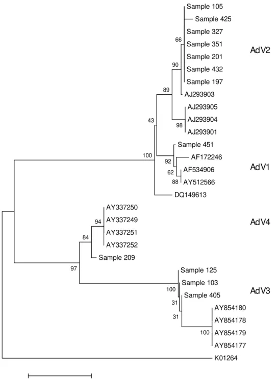
Of the 16 adenovirus positive samples by PCR according to Allard et al. (2001), we
were able to obtain nucleotide sequence of 12 specimens. Analyses by BLAST and by
comparison to sequences obtained from GenBank revealed that the specimens of our study
showed high identity to sequences of species B, C and E. Also, a phylogenetic tree with the
nucleotide sequences obtained in our study and the sequences from GenBank that showed
high identity with ours was constructed (Fig. 2). These analyses showed that the
characterized adenovirus specimens that circulated in this region are distributed according
to the following: eight specimens of species C (seven AdV2 and one AdV1), three of
species B (AdV3) and one of species E (AdV4). Upper respiratory tract involvement was
Discussion
The percentage of approximately 7.1% (33/468) observed for adenovirus is similar
to earlier studies that have documented that these viruses are responsible for about 7% to
8% of the acute respiratory disease in children (Brandt et al., 1969; Foy, 1997), which
reinforces its importance.
Although IFA constitutes a not expensive and rapid diagnostic method, the use of
PCR was of great importance in the detection of these viruses, considering its higher
sensitivity rate, which was increased from 3% to 7%. In addition, 5 samples that were
inoculated in cell culture and had a negative result by IFA, had its isolation confirmed by
PCR.
Although other reports demonstrated a specificity of approximately 99% of IFA
(Mitchell et al., 2003; Larrañaga, 2007), 21.4% (3/14) of the positive samples for
adenovirus by IFA was negative by all other tested methods (virus inoculation in cell
culture and PCR assay), which suggest that these results might be false-positives. This fact
can be attributed to mistakes in the interpretation of IFA, considering the subjectivity of
the method (Ascher & Wilber 1990; Malan, et al. 2003) and the alternation of the
personnel during the period of the study. In addition, 4 positive cases by PCR from
Trizol-extracted DNA were not positive by the 2 PCR assays tested by using column-Trizol-extracted
DNA, even when using the same conditions of the first assay. These results indicated that
difference in DNA extraction method may affect the sensitivity of the detection of the
virus.
The calculation of the sensitivity of the two PCR assays show that the reaction
using the conditions described by Araújo et al. (2001) was more sensitive than using those
described for Allard et al. (2001) (90.9% and 72.7%, respectively, using the virus isolation
21
expanded gold standard). The fact that 4 DNA samples extracted from in natura specimens
were positive by Araújo et al. (2001), and by Allard et al.(2001) were positive only after
testing the DNA extracted from cell culture, emphasize these data. However, the number of
positive cases here detected according to Allard et al. (2001) may have been
underestimated since the Nested-PCR described by these authors, was not carried out due
to primers shortage.
On the other side, the specificity of PCR according to Allard et al. (2001) was much
higher (90.5% and 52.4%, using the virus isolation as a gold standard and 91.3% and
57.9%, using the previously defined expanded gold standard).
It is important to mention that detection of adenovirus in samples does not mean
that it is the etiologic agent of the disease in all cases, considering that species C
adenoviruses can be excreted for long periods after the infection (Fox et al, 1977) and it
was not our objective to investigate cases of coinfections.
The detection of adenovirus circulation throughout the year is similar to what was
reported by other authors (Viegas et al., 2004; Cabello et al., 2006). However,
approximately one half of the cases was detected in autumn. It may have happened due to
the higher number of specimens collected in this season.
The serotypes identified in this study are in accordance with the literature, in which
the more frequently involved in respiratory tract infections are AdV3, 7, 14, 16, 21 and 35
(of species B), AdV1, 2, 5 and 6 (of species C) and AdV4 (of species E) (Hierholzer, 1989;
Swenson et al., 2003). However, AdV4 is not very common in children (Schmitz et al.,
1983). This serotype is most frequently associated with acute respiratory disease among
military recruits (Gaydos & Gaydos, 1995, Dingle & Langmuir, 1968). In our study, the
serotype 4 isolated from basic training recruits in United States in the end of 90’s (Choi et
al, 2006).
In addition, we observed 100% identity between the nucleotide sequence of AdV1
and a feline adenovirus reported by Lakatos et al., (1999) and Pring-Akerblom & Ongradi
(only available on GenBank) (Acession numbers: AF172246 and AY512566,
respectively). In Japan one adenovirus strain bearing feline adenovirus gene was also
detected in a fecal specimen collected from a 1-year old child with gastroenteritis,
providing evidence of adenovirus type 1 transmission between humans and animals (Phan
et al., 2006).
From the total of 4 lower respiratory tract infections cases, 3 were associated with
specie C serotypes, although some reports have found a higher association between lower
respiratory tract infections and specie B adenoviruses (Kajon et al., 1996, Carballal et al.,
2002, Hong et al., 2001).
In conclusion, through the combination of IFA and PCR, it was possible to detect
adenoviruses in 7.1% of the clinical samples in children presenting ARD. The analysis of
the sensibility and specificity of the two PCR assays, showed that PCR using the
conditions described by Araújo et al. (2001) presented a sensibility higher than PCR using
the conditions described by Allard et al. (2001), however its specificity was considerable
lower. The serotype AdV2 was identified in 7 of the 12 PCR products positive for
adenovirus sequenced.
It is important to continue detecting and characterizing adenovirus in children with
acute respiratory disease in order to better understand the association between adenovirus
23
Acknowlegments
We thank Dr Dean D Erdman, Centers for Disease Control and Prevention (CDC), Atlanta,
GA and Dr. Marilda Siqueira, Laboratório de Vírus Respiratórios e Sarampo, FIOCRUZ,
Rio de Janeiro, for providing the Respiratory Panel I Viral Screening and Identification
kit; the laboratories of Immunology, Parasitology, Molecular Biology, Physiology, and
Genetics, UFU, MG, for their equipments and the health care professionals of the Hospital
References
Allard A, Alb insson B, Wadell G. Rapid typing of human adenoviruses by a general PCR co mbined with restriction endonuclease analysis. J Clin Microbiol 2001; 39:498-505.
Araújo AA, Yokosawa, J., Durigon, E. L., Ventura, A. M. Polymerase chain reaction detection of adenovirus DNA sequences in human ly mphocytes. Brazilian Journal of Microbiology 2001; 32: 1886-1891. Ascher MS, W ilber JC. Immunofluorescence for serodiagnosis of retrovirus infect ion. Arch Pathol Lab Med.
1990;114:246-248.
Brandt CD, Kim HW, Vargosko AJ, Jeffries BC, Arrobio JO, Rindge B, Parrott RH, Chanock RM. Infections in 18,000 infants and children in a controlled study of respiratory tract disease. I. Adenovirus pathogenicity in relation to serologic type and illness syndrome. A m J Ep idemio l 1969; 90:484-500. Cabello C, Manjarrez ME, Olvera R, Villalba J, Valle L, Paramo I. Frequency of viruses associated with acute respiratory infections in children younger than five years of age at a locality of Mexico City. Mem Inst Oswaldo Cru z 2006; 101:21-24.
Carballal G, Videla C, Misirlian A, Requeijo PV, Aguilar Mdel C. Adenovirus type 7 associated with severe and fatal acute lower respiratory infections in Argentine children. BMC Pediatr 2002; 2:6.
Choi EH, Kim HS, Park KH, Lee HJ. Genetic heterogeneity of the hexon gene of adenovirus type 3 over a 9-year period in Korea. J Med Virol 2006; 78:379-383.
De Jong JC, Wermenbol A G, Verweij-Uijterwaal MW, Slaterus KW, Wertheim-Van Dillen P, Van Doornum GJ, Khoo SH, Hierho lzer JC. Adenoviruses from human immunodeficiency virus -infected individuals, including two strains that represent new candidate serotypes Ad50 and Ad51 of species B1 and D, respectively. J Clin Microbiol 1999; 37:3940-3945.
Dingle JH, Lang muir A D. Ep idemiology of acute, respiratory disease in military recruits. A m Rev Respir Dis 1968; 97(6):Suppl:1-65.
Foy HM. Adenoviruses. In: Evans AS, Kaslow RA, editors. Viral infections of humans: epidemiology and control. 4th ed. Plenu m Publishing; 1997; 119-138.
Fo x JP, Hall CE, Cooney MK. The Seattle Virus Watch. VII. Observations of adenovirus infections. Am J Ep idemio l, 1977; 105: 362-86.
Garbino J, Gerbase MW, Wunderli W, Ko larova L, Nicod LP, Rochat T, Kaiser L. Respiratory viruses and severe lower respiratory tract comp lications in hospitalized patients. Chest 2004; 125:1033-1039. Gaydos CA, Gaydos JC. Adenovirus vaccines in the U.S. military. Mil Med 1995; 160:300-304.
Hall TA. 1999. Bio Edit: a user-friendly b iological sequence alignment edit and analysis program for Windows 95/98/NT. Nucleic Acids Symp Ser 41, 95-98.
Hierholzer JC. Adenoviruses. In: Achmidt NJ, Emmons RW, editors. Diagnostic procedures for viral, rickettsial and chlamydial infections. 6th ed. A merican Public Health Association; 1989. p 219-264. Hierholzer JC, Halonen PE, Dahlen PO, Bingham PG, McDonough MM. Detection of adenovirus in clinical
specimens by polymerase chain reaction and liquid-phase hybridization quantitated by time-resolved fluoro metry. J Clin Microbio l 1993; 31:1886-1891.
Hong JY, Lee HJ, Piedra PA, Choi EH, Park KH, Koh YY, Kim WS. Lo wer respiratory tract infections due to adenovirus in hospitalized Korean children : epidemiology, clin ical features, and prognosis. Clin Infect Dis 2001; 32:1423-1429.
Kajon, AE, Mistchenko, AS, Videla C, Hortal M, Wadell G, Avendano LF. Molecular epidemiology of adenovirus acute lower respiratory infections of children in the south cone of South America (19 91-1994). J Med Virol 1996;.48:151-156.
Ku mar S, Tamura K, Nei M. 2004. MEGA3: Integrated software for Molecu lar Evo lutionary Genetics Analysis and sequence alignment. Briefings in Bio informatics 5:150-163.
Lakatos B, Farkas J, Egberin k HF, Vennema H, Horzinek MC, Ben ko M. Detection of adenovirus hexon sequence in a cat by poly merase chain reaction (short communication). Acta Vet Hung 1999; 47:493-497.
Larrañaga C, Jorge Martínez H., Angélica Palo mino M., Mónica Peña C., Flavio Carrión A., Lu ís Fidel Avendaño. Molecular characterizan of hospital-acquired adenovirus infantile respiratory infection in Chile using species-specific PCR assays. Journal of Clinica Viro logy 2007; 39:175-181.
Malan, AK, Martins TB, Jaskowski TD, Hill HR, Litwin CM. Co mparison of two commercial enzy me -linked immunosorbent assays with an immunofluorescence assay for detection of Legionella pneumophila types 1 to 6. J Clin Microbio l 2003;41:3060-3063.
25
O’Neill HJ, Russell JD, Wyatt DE, McCaughey C, Coyle PV. Isolation of viruses fro m clin ical specimens in microtitre plates with cells inoculated in suspension. J Virol Methods 1996, 62:169 -78.
Osiowy C. Direct detection of respiratory syncytial virus, parainfluen za virus, and adenovirus in clinical respiratory specimens by a multiplex reverse transcription-PCR assay. J Clin Microb iol 1998; 36:3149-3154.
Phan TG, Shimizu H, Nishimura S, Okitsu S, Maneekarn N, Ushijima H. Hu man adenovirus type 1 related to feline adenovirus: evidence of interspecies transmission. Clin Lab 2006; 52:515-518.
Queiró z DA O, Du rigon, EL., Botosso, VF., Ejzemberg B., Vieira, SE., Mineo, JR., Yamashita, C., Hein, N., Lopes, CL., Cacharro, A L., Stewien, KE. Immune response to respiratory syncytial virus in young Brazilian ch ildren. Brazilian Journal o f Medical and Biolog ical Research 2002; 35:1183 - 1193. Ribeiro LZG, Tripp RA, Rossi, LMG, Palma PVB, Yokosawa J, Mantese OC, Oliveira TFM, Nepomuceno
LL, Queiro z, DAO.Seru m mannose-binding lectin levels are lin ked with respiratory syncytial vírus (RSV) d isease. J Clin Immunol, 2008; 20: p.(in press).
Schmitz H, Wigand R, Heinrich W. Worldwide epidemio logy of human adenovirus infections. Am J Ep idemio l 1983; 117:455-466.
Swenson PD, Wadell G, A llard A, Hierholzer JC. Adenoviruses. In: Murray PR, Baron EJ, Baron EJ, Jorgensen JH, Pffaler MA, Yolken RH, editors. Manual of Clinical Virology. 8th ed. ASM Press; 2003. p 1404-1417.
Table 1: Results obtai ned by IFA, VI and PCR for adenoviruses detection in 468 specimens of NFA
collected from chil dren less than 5 years ol d, wi th acute res piratory disease
IFA PCRAraujo VI Total
+ + + 4
+ - + 3
- + + 4
- + - 15
+ - - 6
+ + NT 1
- NT NT 328
- - NT 107
Total 468 126 32 468
27
Table 2: Comparison of two PCR assays with 33 speci mens that were previ ously positi ve s peci mens for
presence of adenovirus
Sample no. PCRAllard PCRAraújo
7 - +
41 - +
47 - +
53 - -
56 +* +
103 + +
105 + +
121 - -
124 - -
125 + +
128 +* +*
137 - +*
147 - -
153 - +*
157 + +
179 - -
197 + +
201 + +
209 +* +
210 - +
245 - +*
247 - -
251 +* +
313 - +
327 + +
351 + +
405 + +
410 - +*
412 - -
415 - +
425 +* +
432 + +
451 + +
PCRAllard: Poly merase chain reaction using the conditions described by Allard et al, 2001; PCRAraújo:
Poly merase chain reaction using the conditions described by Araújo et al 2001. Both PCRs were perfo rmed at column-extracted DNA.
+: Positive by testing DNA extracted fro m in natura samples
Table 3: Comparison of the results obtained, from DNA extracted from in natura samples, by the two PCR assays in 33 speci mens pre viously positive for adenovirus
Sensitivity
(%) Specificity (%) PPV (%) NPV (%) VI confirmed by IFA
PCRAllard (+) (-)
(+) 8 2 72.7 90.5 79.9 86.3
(-) 3 19
VI confirmed by IFA
PCRAraújo (+) (-)
(+) 10 10 90.9 52.4 50.0 91.7
(-) 1 11
Expanded gold standard
PCRAllard (+) (-)
(+) 9 2 90.0 91.3 81.8 95.4
(-) 1 21
Expanded gold standard
PCRAraújo (+) (-)
(+) 13 8 92.9 57.9 61.8 91.6
(-) 1 11
29
Table 4: Distributi on of the 12 serotyped cases according to the invol vement of symptoms
Species and serotypes URTI LRTI URTI and LRTI
B AdV3 2 - 1
C AdV1 AdV2 1 4 3 - - -
E AdV4 1 - -
Total 8 3 1
Fig. 1: Seasonal distributi on of adenovirus -positi ve cases compared wi th the total number of collected samples during the study peri od.
-1 1 3 5 7 9 11 13 15
2000 2001 2002 2003 2004 2005 2006 2007
N o. of A D V -po si ti ve s am pl e -15 5 25 45 65 85 105 125 145 165 N o. of s am pl es t es te d
31
Fig. 2: Phyl ogenetic tree referring to parti al nucleoti de sequences of the adenovirus hexon gene built wi th the program MEGA 3.1 by neighbor-joining method. B ootstrap val ues (from 1000 replicates) are indicated as a percentage in each node. The following published sequences were used: AdV1 (Accession numbers: AF 534906, AY 512566, AF 172246, DQ 149613), AdV2 (Acession numbers: AJ 293903, AJ293901, AJ 293905, AJ 293904), AdV3 (Acession numbers: AY 854180, AY 854179, AY 854178, AY 854177), AdV 4 (Acession numbers: AY 337252, AY 337251, AY 337250, AY 337249) and for the outgroup (Acession number: KD 1264)
33
35
Annex E
Termo de consentimento livre e esclarecido
Pesquisa de vírus respiratórios e aspectos da resposta i mune em es pécimes clínicos obti dos de crianças
de 0-5 anos de i dade de regiões do Triângul o Mi neiro, MG
Hospital de Clínicas da Universidade Federal de Uberlândia
Sr. Pais/Responsáveis,
Os vírus respiratórios são os principais agentes causadores de doença respiratória aguda em crianças menores de cinco anos de idade, razão pela qual a equipe de Virolog ia da UFU vem desenvolvendo pes quisa nesse assunto.
A detecção do agente viral que está causando a infecção em seu filho, além de au xiliar o médico a tratá-lo, irá fornecer in formações para trabalhos sobre mecanismos de infecção e sobre a circulação dos principais vírus respiratórios em nossa região.
É importante lemb rar que a participação neste estudo é voluntária, que o nome de seu filho não será divulgado e que as amostras clínicas não serão utilizadas para nenhum outro estudo.
Se for do seu consentimento a coleta de secreção de nasofaringe (aspirado) e de 2-3mL de sangue de seu filho, favor assinar este documento.
Uberlândia: ___/___/___
No me do pai/mãe ou responsável: _____________________________
Assinatura: _____________________________
Médicos responsáveis: _______________________________ Profs. Drs. Orlando César Mantese e Hélio Lopes da Silveira
Coordenadora do Projeto:
______________________________ Profa. Dra. Divina A. O. Queiróz (34) 3218-2664