Received on 02/22/2011. Approved on 11/02/2011. Authors declare no confl ict of interests. Financial Support: Fundação de Amparo à Pesquisa (FAPERJ). Ethics Committee: 870-CEP/HUPE.
Faculdade de Odontologia da Universidade do Estado do Rio de Janeiro – UERJ. 1. Master in Periodontology, Universidade Veiga de Almeida – UVA
2. PhD in Periodontology, Universidade do Estado do Rio de Janeiro – UERJ
3. PhD candidate in Internal Medicine, Universidade Federal do Rio de Janeiro – UFRJ; Responsible for the Department of Rheumatology of the NESA, UERJ; As-sisting Professor, Pediatrics Department, UFRJ and Universidade do Grande Rio – Unigranrio; Physician of the Department of Pediatric Rheumatology, IPPMG-UFRJ 4. PhD in Periodontology, UERJ; Adjunct Professor of Odontology, Universidade Federal Fluminense – UFF
5. PhD in Periodontology, UERJ; Assistant Professor of Periodontology, UVA
6. PhD in Periodontology, Lund University, Sweden; Full Professor of Periodontology, UERJ
7. Post-doctor in Periodontology, Karolinska Institute, Sweden; Assistant Professor of Periodontology, UERJ
Correspondence to: Flávia Silva Farah Ferreira Braga. Rua Almirante Sadock de Sá, 26/201 – Ipanema. CEP: 22411-040. Rio de Janeiro, RJ, Brasil. E-mail: fl aviasffb@yahoo.com.br
Reduction in alveolar bone density of
patients with juvenile idiopathic arthritis
Tânia Lúcia de Oliveira Silva1, Flávia Silva Farah Ferreira Braga2, Flavio Roberto Sztajnbok3,
Alessandra Areas e Souza4, Fernanda de Brito Silva5, Ricardo Guimarães Fischer6, Carlos Marcelo da Silva Figueredo7
ABSTRACT
Objectives: To evaluate the alveolar bone density (ABD) in the upper fi rst molars of patients with juvenile idiopathic arthritis (JIA) and to compare their ABD with that of healthy controls. Secondary objectives included the evaluation of the infl uence of medication and rheumatic disease activity on ABD, in addition to the correlation between ABD and periodontal and rheumatologic clinical parameters. Patients and methods: This study assessed 16 patients with JIA (six boys and 10 girls; mean age, 16.2 ± 2 years) and 11 controls (six boys and fi ve girls; mean age, 16.4 ± 2.1 years). Probing depth (PD), visible plaque index, gingival bleeding on probing (GBP), and the clinical insertion level (CIL) were recorded. Bite-wing radiographs were obtained and ABD was measured in the upper molars by use of the Kodak RVG 6100 Digital Radiography System. Results: ABD, the percentage of sites with PD ≥ 4 mm, and GBP were signifi cantly lower in patients with JIA than in controls (P = 0.001; P = 0.019; P = 0.011, respectively). ABD was infl uenced by neither medication nor JIA activity, and showed no correlation with periodontal and rheumatologic clinical parameters. Conclusion: ABD was lower in patients with JIA and seemed to be infl uenced by neither medication nor rheumatic disease activity. In addition, no correlation was observed between ABD and periodontal and rheumatologic clinical parameters.
Keywords: juvenile idiopathic arthritis, alveolar bone loss, bone density, periodontitis.
© 2012 Elsevier Editora Ltda. All rights reserved.
INTRODUCTION
Periodontitis, a form of periodontal disease, is a destructive chronic infl ammation leading to tooth supporting tissue loss and, eventually, tooth loss and edentulism. The periodontal ligament and bone tissue are destroyed by an immune-infl ammatory response to the presence of bacteria, especially gram-negative ones, in the gingival sulcus.1,2 This destruction
is probably mediated by an altered host response, making him/ her susceptible to the bacterial challenge.1–3
It has not been clarifi ed why, in some individuals, peri-odontal infl ammation evolves to periodontitis, while in others
it is restricted to gingivitis. Studies have demonstrated that rheumatologic conditions, such as rheumatoid arthritis and juvenile idiopathic arthritis (JIA), can affect periodontal health and disease,4 increasing the susceptibility to destructive
periodontal disease in both adults5 and younger individuals.6
It has been suggested that an increase in cytokine circulation can play a key role in this process.7
The term JIA refers to a group of diseases that have in common chronic arthritis of unknown etiology and several systemic manifestations, beginning before 16 years of age.8
sites from the primary arthritis site, causing osteopenia in other skeletal bones, is a common complicating aspect of JIA.9–12 This change can affect up to 40% of the patients with
JIA and relates to factors, such as rheumatic disease activ-ity,10 nutritional aspects, and chronic corticosteroid use.9 The
systemic reduction in bone density in JIA can lead adolescents to osteopenia, reduced skeletal growth, and greater risk of systemic osteoporosis,12 which can accelerate periodontal
bone loss,13 functioning as a link between arthritis and
peri-odontitis.14 These changes can lead to functional loss of both
involved joints and dental elements, with direct impact on the patients’ quality of life.
Although osteopenia in patients with JIA has been mainly investigated in long bones and spine, to our knowledge, noth-ing is known about any change in alveolar bone (the bone that supports the teeth) and its role in periodontal disease.
The primary hypothesis of the present study was that patients with JIA would have lower alveolar bone density (ABD) when compared with healthy individuals of the same age, similarly to what is seen in long bones and axial skeleton. The secondary hypothesis was that ABD could be related to clinical periodontal infl ammation and rheumatologic param-eters. Thus, the objective of the present study was to radio-graphically evaluate ABD of the upper fi rst molars in patients with JIA and to compare it with that of healthy controls. The secondary objectives included the evaluation of the infl uence of the rheumatic disease activity and medication on ABD, and of the correlation of ABD with periodontal and rheumatologic clinical parameters.
MATERIAL AND METHODS
Subjects
This study evaluated 16 patients with JIA (10 girls and six boys), mean age of 16.2 ± 2 years, and 11 healthy controls (six boys and fi ve girls), mean age of 16.4 ± 2.1 years. All subjects were consecutively cared for at the rheumatology outpatient clinic of the Adolescent Health Study Center of the Universidade do Estado do Rio de Janeiro (UERJ). This study was approved by the Ethics Committee of the Hospital Pedro Ernesto, UERJ, Rio de Janeiro, Brazil. All subjects, as well as their legal guardians, were informed about the objectives and methods of the study, and they all gave their written consent. Adolescents volunteered to participate in the study and met the inclusion criteria. The JIA diagnosis was performed by a single pediatric rheumatologist, according to the International League of Association for Rheumatology (ILAR) classifi ca-tion criteria.8
None of the subjects were smokers, had taken antibiotics in the last three months, and had any other systemic condition besides JIA. All girls had their menarche at least two years before the evaluation. In the control group, none of the subjects had taken anti-infl ammatories in the three months preceding the exams. No patient in the study had eating disorders, such as bulimia or anorexia.
Questionnaires
Subjects answered a questionnaire about their personal data and diet. In addition, patients with JIA answered the Brazilian version of the Childhood Health Assessment Questionnaire (CHAQ),15 which evaluates the functional capacity of JIA
patients with a score ranging from 0 to 3.
Rheumatologic evaluation
Rheumatologic evaluation, consisting of the number of tender joints (TJ), and presence of edema and limitation of movements (LOM), was performed by a single pediatrician. Physician’s Global Assessment (MDGA) and Patient’s Global Assessment (PGA) were recorded in a Visual Analogue Scale (VAS), rang-ing from 0 to 10. Patients were divided into two groups (medi-cated and non-medi(medi-cated) and classifi ed as active and inactive. For this classifi cation and following the same criteria adopted by Miranda et al.,7 parameters such as MDGA, PGA, CHAQ,
number of joints involved, and erythrocyte sedimentation rate (ESR)16 were considered. Active patients had ESR > 20 mm/h
and at least one tender joint with pain and/or edema. ESR was determined by use of the Westergren method on the fi rst hour.
Clinical periodontal exam
Clinical periodontal exam was performed by a single cali-brated examiner [probing depth (PD) kappa = 0.92; clinical insertion level (CIL) kappa = 0.89], consisting of records of PD, gingival bleeding on probing (GBP),17 visible plaque
index (VPI),17 and CIL in six sites of each tooth, except for
third molars, by using a 0.2-N pressure-controlled Williams’ periodontal probe (DB76R, Aesculap AG & Co., Tuttlingen, Germany). Individuals showing proximal CIL ≥ 2 mm in one or more sites were considered to have loss of periodontal clinical insertion.18 In addition, the number of lost teeth in each group
was recorded.
Alveolar bone density evaluation
Kodak fi lm, and the digital Elitys Dental X-ray device (Trophy Trex, Beaubourg, France), with 8 mA, 70 KV, and 0.3-second exposure time. An AT 2000 XR (Air Techniques Hicksville, New York) automatic processor was used to develop the X-ray fi lms. Then, the radiographs were digitalized with a high-resolution scanner (HP Scanjet 5590, Hewlett-Packard Company, USA) and stored in the jpeg format for the radio-graphic analysis of ABD. ABD was measured with the Kodak RVG 6100 Digital Radiography System (Rochester, New York, USA), which quantifi es the gray scale of the pixels that form the digitized image, ranging from 0 (darker) to 256 (lighter). The region of interest (ROI) for assessing the radiographic density was established 1 mm below the alveolar bone crest on the mesial surface (closer to the midline) of the upper fi rst molars, which are the teeth with the greatest prevalence of alveolar bone crest height loss in adolescents.16 Radiographic evaluation
of ABD was performed by a single calibrated examiner. Mean densities of the teeth 16 (right upper fi rst molar) and 26 (left upper fi rst molar) were calculated for each patient. To calibrate the measurement of alveolar bone crest density, the examiner performed 52 measurements on the mesial surface of the right and left upper molars in the JIA and control groups. Those measurements were repeated 24 hours later, and, considering a variation of ± 5 in the pixel gray scale, which ranges from 0 to 256, agreement was observed in 100% of the measurements.
Statistical analysis
The individual was considered as the unit of analysis, and the level of signifi cance was established at 5%. Data were expressed as means and standard deviations or as medians and interquartile range. The Mann-Whitney test was used for as-sessing the differences between JIA and control groups, and for comparing active versus inactive, and medicated versus non-medicated subjects, for all variables analyzed. The Spearman’s correlation coeffi cient was calculated to assess the degree of association between ABD and periodontal and rheumatologic clinical parameters. Non-parametric tests were used because the variables did not show normal distribution (Gaussian) due to data dispersion, lack of symmetry in distribution, and/or the small sample size of some subgroups. Statistical analysis was performed with the SAS 6.11 software (SAS Institute, Inc., Cary, North Carolina).
RESULTS
Females represented 62.5% in the JIA group and 45.4% in the control group. Considering the ethnic distribution of the sample, Caucasians represented 46.5% of the JIA group
and 55% of the control group. Of the 16 patients with JIA, eight had persistent oligoarthritis, two had enthesitis-related arthritis, three had systemic arthritis, three had RF-negative polyarthritis, and 10 were on medication. The drugs used in-cluded methotrexate, naproxen, prednisone, cyclosporine, and infl iximab. The mean duration of medication use was 4.2 ± 3.6 years. The methotrexate dosage ranged from 10 to 15 mg/m2/
week. The prednisone dosage ranged from 1 to 2 mg/kg/day. Medians and interquartile range for age of JIA onset (years), duration of JIA (years), ESR (mm/h), CHAQ (score), PGA (score), pain (score), MDGA (score), number of TJ, LOM, and edema were 10 (7), 6 (7), 17.5 (19.5), 0.5 (0.64), 1.1 (4.07), 2.1 (4.9), 1.95 (5.75), 1 (2), 2 (4), and 1 (1.75), respectively.
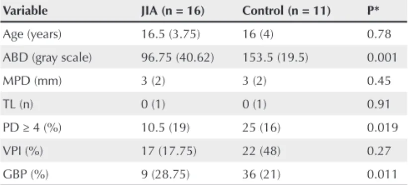
Table 1 shows periodontal clinical parameters and medians of ABD in both groups. Median ABD (P = 0.001), percentage of sites with GBP (P = 0.011), and PD ≥ 4 mm (P = 0.019) were lower in the JIA group. Of all subjects studied, only three in the JIA group had a loss of proximal clinical insertion (PCI) ≥ 2 mm. No signifi cant differences were observed between the groups in the remaining variables.
No statistically significant differences were observed between active and inactive patients with JIA, regarding the periodontal clinical and radiographic variables analyzed. However, inactive patients showed a tendency (P = 0.06) to-wards a higher percentage of sites with PD ≥ 4 mm (Table 2). Regarding the rheumatologic data, the active subgroup had MDGA (P = 0.001), TJ (P = 0.014), edema (P = 0.001), and pain (P = 0.038) signifi cantly higher than those of the inactive subgroup (Table 2). No statistically signifi cant differences in periodontal and rheumatologic clinical variables and ABD were observed between the medicated and non-medicated JIA subgroups (Table 2).
Table 1
Median and interquartile range for age, alveolar bone density (ABD), and periodontal clinical variables in the JIA and control groups
Variable JIA (n = 16) Control (n = 11) P*
Age (years) 16.5 (3.75) 16 (4) 0.78
ABD (gray scale) 96.75 (40.62) 153.5 (19.5) 0.001
MPD (mm) 3 (2) 3 (2) 0.45
TL (n) 0 (1) 0 (1) 0.91
PD ≥ 4 (%) 10.5 (19) 25 (16) 0.019
VPI (%) 17 (17.75) 22 (48) 0.27
GBP (%) 9 (28.75) 36 (21) 0.011
MPD = mean probing depth on mesial surface of upper fi rst molars; TL = teeth lost; PD = probing depth (percentage per individual of sites with PD ≥ 4); VPI = visible plaque index; GBP = gingival bleeding on probing.
Signifi cant correlations between ABD and age were ob-served in the control group (rs = 0.63; P = 0.03), indicating that the higher the age, the greater the ABD in this group. When the infl uence of gender in ABD was evaluated, no signifi cant difference between girls and boys was observed (P = 0.47).
DISCUSSION
In the present study, a signifi cantly lower ABD in the upper fi rst molars was observed in patients with JIA when compared with healthy controls. Some studies have demonstrated a re-duced bone density in sites distant from the primary arthritis site.9–11 Recently, Hämäläinen et al.11 have reported that male
patients with JIA have reduced mineral bone content in the femoral neck. In addition, Stagi et al.,12 using dual-energy
X-ray absorptiometry (DXA), have reported reduced mineral bone density in the spine of patients with JIA. However, to our knowledge, the present study is the fi rst to assess bone density of the alveolar crest in patients with JIA by use of intraoral radiograph. The systemic reduction in bone density in JIA
might lead those adolescents to develop osteopenia, reduced skeletal growth, and greater risk of systemic osteoporosis,12
which might accelerate periodontal bone loss,13,19 acting as a
link between rheumatoid arthritis and periodontitis.14
The true cause of the reduction in systemic bone density in patients with JIA is not known. The generalized bone loss in arthritis has been suggested to be related to increased osteoclas-tic activity or a reduction in the process of bone formation.20,21
Reduced physical activity and inadequate calcium and vitamin D intake can infl uence this reduction in bone density.22,23 In
ad-dition, C-reactive protein levels have been correlated to mineral bone density loss during active arthritis.20 This result suggests
that proinfl ammatory cytokines released in infl amed joints, which induce systemic acute response, can also contribute to generalized bone resorption in rheumatoid arthritis.14 However,
in the present study, the rheumatic disease activity showed no infl uence on ABD. Drugs used to control JIA in this study, including corticosteroids, also did not infl uence ABD. These results are in accordance with previous studies that demon-strated reduced bone mass in patients with JIA, regardless of Table 2
Median and interquartile range for age, alveolar bone density (ABD), and periodontal and rheumatologic clinical parameters in active, inactive, medicated, and non-medicated JIA groups
Variable Active JIA
n = 10
Inactive JIA
n = 6 P*
Medicated n = 10
Non-medicated
n = 6 P**
Age (years) 17 (4.5) 16 (2.75) 0.70 17 (3.25) 15.5 (5.25) 0.58
ABD (gray scale) 100.75 (46.62) 95.75 (54.12) 0.91 96.75 (29) 107.75 (58.25) 0.91
MPD (mm) 2.5 (2) 3 (2.25) 0.43 2.5 (2.25) 3 (1.5) 0.61
TL (n) 0 (0.25) 1 (1) 0.071 0 (1) 0 (1) 0.80
PD ≥ 4 (%) 5 (12.7) 21 (28) 0.064 10.5 (29) 8.5 (21) 0.79
VPI (%) 16 (16.5) 19.5 (36) 0.45 22.5 (22) 13 (16) 0.55
GBP (%) 6.5 (21) 10.5 (43) 0.62 8 (35) 9 (23) 0.99
JIA duration (years) 4 (7.25) 8.5 (6.25) 0.28 5.5 (7) 7.5 (10.68) 0.74
Onset (years) 13 (6.7) 7.5 (5.25) 0.11 11.5 (6.75) 8 (7.5) 0.27
ESR (mm/h) 25 (44.25) 11 (12.5) 0.19 23 (24.5) 10 (24) 0.33
CHAQ (score) 0.5 (0.44) 0.43 (1.5) 0.91 0.5 (0.76) 0.12 (0.68) 0.17
GPA (score) 1.55 (4.32) 1.1 (3.87) 0.83 3.05 (4.62) 0.5 (1.92) 0.18
Pain (score) 5 (4.97) 1 (1.6) 0.038 2.75 (5.75) 1.6 (5.05) 0.44
MDGA (score) 4.75 (4.27) 0 (0) 0.001 3.25 (5.35) 0 (3.37) 0.13
TJ (n) 1.5 (2.5) 0 (0.25) 0.014 1 (2.5) 0 (1.5) 0.19
LOM (n) 2 (3.25) 3.5 (13.5) 0.44 3 (4.25) 1.5 (3.75) 0.38
Edema (n) 1 (1.75) 0 (0) 0.001 1 (2.75) 0 (1) 0.08
MPD = mean probing depth on the mesial surface of the upper fi rst molars; TL = teeth lost; PD = probing depth; VPI = visible plaque index; GPB = gingival bleeding on probing; JIA = juvenile idiopathic arthritis; ESR = erythrocyte sedimentation rate; CHAQ = childhood health assessment questionnaire; MDGA = physician global assessment; PGA = patient global assessment; TJ = tender joint; LOM = limitation of movement.
corticosteroid therapy.10,24 It is worth emphasizing that, due to
the reduced number of participants, the results of the present study should be assessed with caution. The fact that we could only enroll girls two years or more after menarche, when hormonal levels are stabilized, to minimize the infl uence of sexual hormones on bone density, represents one of the limiting factors of the present study. Because JIA is more common in girls,25 the selection of patients was hampered by that factor.
In addition, the percentages of sites with GBP and of PD ≥ 4 mm were signifi cantly lower in the JIA group (P = 0.01; P = 0.01, respectively). A possible explanation for those differences can be associated with the anti-rheumatic drug regimens used to control JIA. Similarly to the fi ndings of this study, studies carried out by our group have suggested a pos-sible protective action of anti-rheumatic drugs on periodontal tissue.26,27 A previous study has shown that, after two years,
the rheumatologic and periodontal conditions of patients with JIA were similar to those of the control group.26 In another
study comparing patients with juvenile systemic lupus ery-thematosus (SLE) and healthy control subjects, the percentage of sites with greater PD was signifi cantly higher in healthy individuals. Comparing infl ammatory markers in the gingival fl uid of SLE and control groups, patients with SLE showed signifi cantly lower levels of IL-1β and IL-18.27 However, the
specifi c effect of anti-rheumatic drugs on the periodontal tissue is not fully known.
Methotrexate was the major anti-rheumatic drug used by patients with JIA in the present study. The mechanisms of ac-tion of that drug include reducac-tions in the producac-tion of TNF-α by T cells, and in the levels of IL-1β and IL-18. However, the
production of IFN-γ and IL-4 does not seem to be affected by methotrexate, which shows a selective effect on cytokines, be-ing able to modulate the immunologic response.28–30 Therefore,
those drugs might have improved periodontal clinical infl am-mation parameters in patients with JIA.
ABD showed a positive correlation with age only in the control group (r = 0.63, P = 0.03). Because patients with JIA might have several changes in bone maturation and growth,10
the same correlation is accepted to not exist in adolescents with JIA. Another important fi nding of the present study is that ABD showed no correlation with periodontal clinical parameters in the sites where bone density was measured. This suggests that the change in ABD might be associated with the presence of JIA and not with the local periodontal infl ammation.
The long-term consequence of the reduction in ABD in patients with JIA has not been determined. Whether arthritis has any infl uence on periodontitis and vice-versa or whether the individual is susceptible to chronic osteolytic disorders in general, manifesting both arthritis and periodontitis or osteoporosis, is yet to be clarifi ed. Our group is conducting a prospective study to assess the possible outcomes of this reduction in ABD in patients with JIA, regarding both the speed of alveolar bone crest resorption and the early diagnosis of changes in systemic bone density.
REFERENCES REFERÊNCIAS
2. Bartold PM, Cantley MD, Haynes DR. Mechanisms and control of pathologic bone loss in periodontitis. Periodontol 2000 2010; 53:55–69.
3. Figueredo CM, Fischer RG, Gustafsson A. Aberrant neutrophil reactions in periodontitis. J Periodontol 2005; 76(6):951–5. 4. Braga FSFF, Miranda LA, Miceli VC, Áreas A, Figueredo CMS,
Fischer RG et al. Artrite crônica e periodontite. Rev Bras Reumatol 2007; 47(4):276–80.
5. Mercado FB, Marshall RI, Klestov AC, Bartold PM. Relationship between rheumatoid arthritis and periodontitis. J Periodontol 2001; 72(6):779–87.
6. Miranda LA, Fischer RG, Sztajnbok FR, Figueredo CM, Gustafsson A. Periodontal conditions in patients with juvenile idiopathic arthritis. J Clin Periodontol 2003; 30(11):969–74.
7. Miranda LA, Fischer RG, Sztajnbok FR, Johansson A, Figueredo CM, Gustafsson A. Increased interleukin-18 in patients with juvenile idiopathic arthritis and early attachment loss. J Periodontol 2005; 76(1):75–82.
8. Petty RE, Southwood TR, Manners P, Baum J, Glass DN, Goldenberg J et al. International League of Associations for Rheumatology classiication of juvenile idiopathic arthritis: second revision, Edmonton, 2001. J Rheumatol 2004; 31(2):390– 2. 9. Celiker R, Bal S, Bakkaloğlu A, Ozaydin E, Coskun T, Cetin A et al.
Factors playing a role in the development of decreased bone mineral density in juvenile chronic arthritis. Rheumatol Int 2003; 23(3):127–9.
10. Lien G, Flato B, Haugen M, Vinje O, Sorskaar D, Dale K et al. Frequency of osteopenia in adolescents with early-onset juvenile idiopathic arthritis: a long-term outcome study of one hundred ive patients. Arthritis Rheum 2000; 48(8):2214–23.
11. Hämäläinen H, Arkela-Kautiainen M, Kautiainen H, Haapasaari J, Leirisalo-Repo M. Bone mineral content in young adults with active or inactive juvenile idiopathic arthritis and in controls. Scand J Rheumatol 2010; 39(3):219–22.
12. Stagi S, Masi L, Capannini S, Cimaz R, Tonini G, Matucci-Cerinic M et al. Cross-sectional and longitudinal evaluation of bone mass in children and young adults with juvenile idiopathic arthritis: the role of bone mass determinants in a large cohort of patients. J Rheumatol 2010; 37(9):1935–43.
13. Payne JB, Reinhardt RA, Nummikoski PV, Patil KD. Longitudinal alveolar bone loss in postmenopausal osteoporotic/osteopenic women. Osteoporos Int 1999; 10(1):34–40.
14. Golub LM, Payne JB, Reinhardt RA, Nieman G. Can systemic diseases co-induce (not just exacerbate) periodontitis? A hypothetical “two-hit” model. J Dent Res 2006; 85(2):102–5.
15. Machado CS, Ruperto N, Silva CH, Ferriani VP, Roscoe I, Campos LM et al. The Brazilian version of the Childhood Health Assessment Questionnaire (CHAQ) and the Child Health Questionnaire (CHQ). Clin Exp Rheumatol 2001; 19(4):25–9.
16. Ravelli A,Viola S, Ruperto N, Corsi B, Ballardini G, Martini A. Correlation between conventional disease activity measures in juvenile chronic arthritis. Ann Rheum Dis 1997; 56(3):197–200. 17. Ainamo J, Bay I. Problems and proposals for recording gingivitis
and plaque. Int Dent J 1975; 25(4):229–35.
18. Jenkins WM, Papapanou PN. Epidemiology of periodontal disease in children and adolescents. Periodontol 2000. 2001; 26:16–32. 19. Al Habashneh R, Alchalabi H, Khader YS, Hazzaa AM, Odat Z,
Johnson GK. Association between periodontal disease and osteoporosis in postmenopausal women in Jordan. J Periodontol 2010; 81(11):1613–21.
20. Gough A, Sambrook P, Devlin J, Huissoon A, Njeh C, Robbins S et al. Osteoclastic activation is the principal mechanism leading to secondary osteoporosis in rheumatoid arthritis. J Rheumatol 1998; 25(7):1282–9.
21. Compston JE, Vedi S, Croucher PI, Garrahan NJ, O'Sullivan MM. Bone turnover in non-steroid treated rheumatoid arthritis. Ann Rheum Dis 1994; 53(3):163–6.
22. Hopp R, Degan J, Gallagher JC, Cassidy JT. Estimation of bone mineral density in children with juvenile rheumatoid arthritis. J Rheumatol 1991; 18(8):1235–9.
23. Cassidy JT, Langman CB, Allen SH, Hillman LS. Bone mineral metabolism in children with juvenile rheumatoid arthritis. Pediatr Clin North Am 1995; 42(5):1017–33.
24. Henderson CJ, Specker BL, Sierra RI, Campaigne BN, Lovell DJ. Total-body bone mineral content in non-corticosteroid- treated postpubertal females with juvenile rheumatoid arthritis. Arthritis Rheum 2000; 43(3):531–40.
25. Macaubas C, Nguyen K, Milojevic D, Park JL, Mellins ED. Oligoarticular and polyarticular JIA: epidemiology and pathogenesis. Nat Rev Rheumatol 2009; 5(11):616–26.
26. Miranda LA, Braga F, Fischer RG, Sztajnbok FR, Figueredo CM, Gustafsson A. Changes in periodontal and rheumatological conditions after 2 years in patients with juvenile idiopathic arthritis. J Periodontol 2006; 77(10):1695–700.
27. Figueredo CM, Areas A, Sztajnbok FR, Miceli V, Miranda LA, Fischer RG et al. Higher elastase activity associated with lower IL-18 in GCF from juvenile systemic lupus patients. Oral Health Prev Dent 2008; 6(1):75–81.
28. Neurath MF, Hildner K, Becker C, Schlaak JF, Barbulescu K, Germann T et al. Methotrexate speciically modulates cytokine production by T cells and macrophages in murine collagen-induced arthritis (CIA): a mechanism for methotrexate-mediated immunosuppression. Clin Exp Immunol 1999; 115(1):42–55. 29. Thomas R, Carroll G J. Reduction of leukocyte and interleukin-1 beta
concentrations in the synovial luid of rheumatoid arthritis patients treated with methotrexate. Arthritis Rheum 1993; 36(9):1244–52. 30. Leung BP, Culshaw S, Gracie JA, Hunter D, Canetti CA, Campbell C