Cyclosporine-loaded delivery system for the
treatment of keratoconjunctivitis sicca: a pilot study
Gustavo de Oliveira Fulgêncio
1, Juliana Barbosa Saliba
1, Sílvia Ligório Fialho
2, Armando da Silva Cunha Júnior
11Ph.D., Faculty of Pharmacy, Federal University of Minas Gerais, Belo Horizonte/MG, Brazil.
2Ph.D., Division of Biotechnology and Pharmacotechnical Development, Ezequiel Dias Foundation, Belo Horizonte/MG, Brazil.
Funding sources: National Council for Scientific and Technological Development (CNPq) and Research Support Foundation of the State of Minas Gerais (Fapemig).
The authors declare no conflict of interest.
Received for publication: 15/9/2012 - Accepted for publication: 28/2/2013
R
ESUMOObjetivo: Este trabalho objetivou o desenvolvimento de um sistema mucoadesivo de liberação de ciclosporina A (CsA) para o tratamento de ceratoconjuntivite seca (CCS). Métodos: O sistema mucoadesivo foi preparado na forma de filme utilizando o polímero quitosana e CsA (25%p/v). Foram administrados no saco conjuntival do olho direito de coelhos normais (n=6) e a aferição da produção de lágrimas foi realizada diariamente antes e após a aplicação, de forma bilateral, durante sete dias, por meio do teste lacrimal de Schirmer. Avaliação oftalmológica foi realizada diariamente durante todo o estudo e seguido da análise histológica. Resultados: Os valores médios de produção de lágrimas foram alterados de 9,88 ± 0,37 mm/min para 16,02 ± 0,38 mm/min antes e após a administração do sistema respectivamente, significando um aumento de aproximadamente 60%. Todos os coelhos apresen-taram hiperemia da conjuntiva palpebral e lacrimejamento. A hiperemia permaneceu durante 48 h após administração dos sistemas com resolução espontânea e o lacrimejamento foi diagnosticado até o final do experimento. Não foram observados outros sinais de reações indesejáveis. Nenhuma alteração histológica foi observada na mucosa conjuntival bulbar e palpebral à histopatologia. Conclusão: Os sistemas desenvolvidos são aparentemente seguros e eficientes criando expectativa para o tratamento da CCS. Novos estudos são necessários para avaliar a concentração de CsA liberada, assim como aceitabilidade e toxicidade dos sistemas em tratamentos mais prolongados.
Descritores: Ceratoconjuntivite seca/terapia; Ciclosporina/uso terapêutico; Sistemas de liberação de medicamentos
A
BSTRACTPurpose: The present work aimed to present the development of a conjunctival mucosa system for the controlled delivery of cyclosporine A (CsA) in the treatment of keratoconjunctivitis sicca (KCS). Methods: The conjunctival mucosa system was prepared in the form of films containing chitosan as the polymer and CsA as the drug (25%w/v). The films were applied to the conjunctival sac of one eye from normal rabbits (n=6), and the evaluation of lachrymal production was performed daily, before and after application, for seven days. Clinical examination was executed daily on the eyes of each animal during the entire period of study. Histological analyses were carried out at the end of the study. Results: The average amount of lachrymal production changed from 9.88 ± 0.37 mm/min to 16.02 ± 0.38 mm/ min, respectively, before and after applying the systems, which indicates an increase of approximately 60%. All rabbits presented hyperemia in the palpebral conjunctiva and tearing. Hyperemia continued for 48h after the application of the systems with spontaneous resolution, and tearing was diagnosed throughout the entire study. No other sign of undesirable reactions could be observed. Moreover, no histological changes could be identified in the bulbar and palpebral conjunctival mucosa. Conclusion: The developed systems proved to be safe and efficient in this pilot study and present a promising future for the treatment of KCS. Other studies are warranted to evaluate the released concentration of CsA as well as the feasibility and toxicity of these systems in a more prolonged treatment.
Keywords: Keratoconjunctivitis sicca/therapy; Cyclosporine/therapeutic use; Drug delivery system
I
NTRODUCTIONK
eratoconjunctivitis sicca ( KCS) or dry eye syndrome is a multifactorial disease related to greater evaporation or decreased production of tears, with consequent tear hyperosmolarity(1). Its main symptoms include eye discomfort,photophobia, tear film instability, blurred vision, and visual impairment(2). KCS is an important public health problem that
can affect up to 15% of the population in specific groups such as the elderly(3). Its aetiology is varied and is associated with
immunological, genetic, therapeutic, hormonal, and environmental factors(4). The treatment of KCS depends on its
severity; mild KCS is treated with artificial tears, while therapy with immunosuppressive drugs such as topical cyclosporin A (CsA) is recommended in moderate and severe forms(5,6).
CsA is a potent immunosuppressive agent isolated from the fungus Tolypocladium inflatum gams, which acts by inhibiting T-cell activity and suppressing inflammatory cytokines in the conjunctiva and lacrimal gland(7,8). Furthermore, it promotes
increased goblet cell density and reduced apoptosis of epithelial cells in the conjunctiva(9). Thus, by suppressing inflammation in
the eye and lacrimal gland, CsA has a lacrimogenic effect, i.e. it increases tear production(4,6).
The work of Coster et al. (1979) pioneered the use of CsA in ophthalmology, administering the drug to rabbits that had received corneal transplants(10). This study was followed by
research in humans, and the drug was subsequently found to be effective in the treatment of various inflammatory processes on the ocular surface(11). Since 1995 it is commercially available as a
CsA-containing ointment (Optimmune™, Schering-Plough, Brazil) for veterinary use in the treatment of KCS. Another topical preparation in the form of an emulsion was approved in 2003 by the Food and Drug Administration (FDA) for the treatment of dry eye syndrome in humans (Restasis™ eye drops, Allergan, Brazil). CsA has since been widely prescribed topically for various eye diseases and systemically for autoimmune conditions with ocular involvement(5,6).
Due to its low water solubility, CsA is prepared using oily vehicles or as an emulsion. Such eye drops are not well absorbed by the corneal epithelium, with more than 95% of the drug reaching the systemic circulation through transnasal or conjunctival absorption(12). Thus, frequent administration is
required to control KCS, which leads to adverse effects including redness, itching, keratitis, blurred vision, and toxic effects on the cornea, limiting its use and reducing adherence to treatment(1,6).
Extended release systems provide increased bioavailability of drugs in eye tissues, reducing systemic absorption and therefore adverse effects. Such systems provide more sustained drug concentrations in ocular tissues, reducing the need for frequent administration and the discomfort associated with topical application(13).
Colloidal systems such as nanoparticles are able to encapsulate and protect the drug, increasing tolerance, penetration, efficiency, and absorption by the cornea(1,11). In a
study by Aksungur et al. cyclosporin nanoparticles were developed for the treatment of ocular surface inflammation. Such nanoparticles released 75-90% of the drug over 24 hours(1).
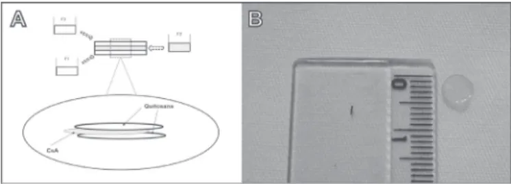
Non-colloidal systems can also be used for topical ocular administration. Such systems generally consist of rods, disks, or membranes and are produced through moulding, extrusion, and film preparation. Film preparation can be carried out by melting and pressing a powder mixture or by adding a solution. The latter is the most commonly-used method: the components are
dissolved in an appropriate solvent to form a solution which is then cast on a smooth, non-adhesive surface. The solvent evaporates, and the film is then removed from the surface(14).
Among the various polymers used in drug delivery systems, specifically in the form of films, chitosan is particularly suitable for topical ocular application. As the cornea and conjunctiva are negatively charged, the use of mucoadhesive polymers that can interact with these structures could extend the drug’s permanence in the pre-corneal cavity or cul-de-sac, thus increasing its absorption and therapeutic effect. Chitosan is a non-toxic biodegradable and biocompatible cationic polymer obtained by deacetylation of chitin, which is abundant in the exoskeleton of crustaceans and other marine animals(15,16).
Mucoadhesion and good ocular tolerance are important characteristics of this polymer, making it a promising substance as a polymer matrix for mucoadhesive ocular films(15). In
preliminary studies, chitosan showed promising results for ocu-lar drug transport by increasing the permanence time of antibiotics in the cornea and the intraocular penetration of nanocapsules.
Some studies in the literature have assessed delivery systems based on CsA-containing chitosan. Campos and colleagues developed chitosan nanoparticles containing CsA which achieved therapeutic concentrations of the drug in the cornea and conjunctiva for 48 hours after topical administration, while the drug was not detected in internal ocular structures or plasma (16). In another study, solid lipid nanoparticles containing
CsA showed good biocompatibility and increased drug penetration in vitro and ex vivo(17). To date, there are no studies
describing the use of mucoadhesive films with chitosan and CsA for the treatment of conditions affecting the ocular surface or KCS.
Thus, this study aimed to develop a drug delivery system in the form of a film made of chitosan and CsA and to assess the system’s ability to increase the production of tears in study subjects, followed by clinical and histological examination.
M
ETHODS MaterialsCyclosporin A [MW=1202.61; water solubility, 27.67 ìg/mL at 25°C(12), Sigma-Aldrich, Brazil], average molecular weight
chitosan (viscosity, 200-800 cP in 1% acetic acid at 25ºC, degree of deacetylation=85%, Sigma-Aldrich, Brazil), ultrafiltered water (Milli Q plus, Millipore, USA). The other reagents were of analytical grade.
Preparation of chitosan films containing CsA
In vivo study Animals
The study used six male New Zealand white rabbits aged approximately three months and weighing 1.9-2.3 kg, obtained from the Professor Hélio Barbosa Experimental Farm (Igarapé, Brazil). The animals were kept in individual cages throughout the adjustment and study period, in an environment with a mean temperature of 25 °C, constant ventilation, and brightness varying with sunlight. There was no food or water restriction during the experiment, and the animals received feed appropriate for the species.
The study was approved by the Ethics Committee on Ani-mal Experimentation of the Federal University of Minas Gerais (CETEA, Belo Horizonte, Brazil, Protocol 130/08). The entire experiment was conducted in accordance with the standards of the Association for Research in Vision and Ophthalmology (ARVO).
Film application
Prior to application, the films containing chitosan and CsA were immersed for 10 seconds in saline to facilitate administration and prevent conjunctival drying. Then, after administration of anaesthetic eye drops (proparacaine hydrochloride 0.5%, Anestalcon™, Allergan, São Paulo, Brazil) in the right eye (RE), the films were applied to the conjunctival sac of normal rabbits by tractioning the lower eyelid. The left eye (LE) received no treatment and was used as a control group.
Assessing the effect on tear production
Before applying the film, measurements of tear production were made for three days using the Schirmer tear test (STT) (Schirmer Tear Test™, Schering-Plough Animal Health, New Jersey, USA) in both eyes of each animal, daily at 2 pm prior to ophthalmic examination. After the third day, following application of the CsA and chitosan films, tear production was measured continually for seven days.
Clinical examination
The rabbits were submitted to a daily general and ophthalmic examination throughout the experiment. The ophthalmic examination used an appropriate light source (Maglite™ flashlight, Mag Instruments, Ontario, USA), an applanation tonometer (TonoPen™ XL, Reichert Technologies, Buffalo, USA), a direct ophthalmoscope (Heine™ K180, Heine Optotecnik, Herrsching, Germany), a slit lamp with biomicroscope (Kowa™ SL-15 Slit-lamp Biomicroscope, Tokyo, Japan), and an indirect ophthalmoscope (Binocular Indirect Ophthalmoscope, Opto Electronics Ltda, São Paulo, Brazil). All eyeballs and annexes underwent photographic monitoring using a digital camera (DSC HX-1™, Sony Company, Tokyo, Japan).
The examination assessed the main ophthalmic parameters, including inspection of the eyelids, the lacrimal system, and the eyeball. Superficial ocular conditions such as oedema, corneal ulceration or neovascularisation, and conjunctival hyperaemia were investigated. Nasolacrimal drainage was assessed with fluorescein dye (Fluoresceína™ - Allergan, São Paulo, Brazil). The lens and posterior segment (vitreous, retina, and choroid) were examined to identify cataract, vitreous opacities, and retinal detachment.
Histological examination
After seven days measuring tear production, the animals were sacrificed using a lethal dose of pentobarbital (100 mg/kg) (Hypnol™, Cristália Produtos Químicos Farmacêuticos Ltda., Itapira, Brazil) for removal of the eyeball and subsequent histological examination of the site of film application. Eyeballs were removed by incision in the conjunctival fornix. Samples of the lower eyelids were also collected. Sections of the eyeball were made in the antero-posterior plane, forming a meridian from the optic nerve to the cornea. The sections of the eyeball and eyelid were then fixed in a buffered 10% formaldehyde solution (pH 7.2), dehydrated, diaphanised, embedded in paraffin, and finally cut into 4-5 mM sections using a microtome, with subsequent mounting on slides. The slides were stained with haematoxylin-eosin and examined by light microscopy (optical light microscope, Carl Zeiss, Herrsching, Germany).
R
ESULTS In vivo studyAssessing the effect on tear production
Average tear production was 9.88 ± 0.37 mm/min and 10.29 ± 0.31 mm/min in the RE and LE, respectively, during the three days prior to film administration. There was no significant difference in tear production between the eyes during this period (p=0.1372) (Figure 2A).
After film application, there was an increase of approximately 60% in tear production in the RE, with mean values rising from 9.88 ± 0.37 to 16.02 ± 0.38 mm/min (Figures 2A and 2B) — a statistically-significant increase (p<0.0001). The LE, used as a control, showed no significant change (p=0.7332) in tear production (mean, 10.00 ± 0.39 mm/min). Previous studies had shown that a similar type of system containing no drug caused no increase in tear production when applied to rabbit eyes(18).
The results were analysed using GraphPad Prism 4.00 (GraphPad Software Inc., USA) and were expressed as mean tear production. The statistical significance of differences was assessed using by Student’s unpaired t test. p-values <0.05 were considered statistically significant.
Ophthalmic examination
On ophthalmic examination, all animals showed hyperaemia of the palpebral conjunctiva and tearing (Figure 3A). The hyperaemia persisted for 48h after film administration and resolved spontaneously, while epiphora was still present by the end of the experiment (Figure 3B). There were no signs of ocu-lar pain or discomfort such as itching, photophobia, and blepharospasm; also, we found no corneal oedema or neovascularisation. The film had the appearance of a gel a few hours after administration, and remained adhered to the conjunctival mucosa in all animals. The fluorescein dye showed no ulcerative keratitis or nasolacrimal duct obstruction.
Assessment of intraocular media showed no signs of cataract, vitreous opacities, or retinal detachment.
Histological examination
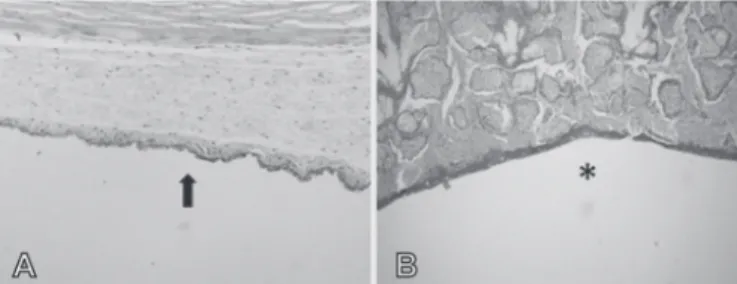
No histological changes were observed in the palpebral and bulbar conjunctival mucosa, i.e., the structures in direct contact with the film. The epithelium and adjacent cells were normal, presenting the typical features of each structure with no signs of cell toxicity or vascular changes such as acanthosis, hyperkeratosis, inflammatory infiltrate, vascular congestion, or haemorrhage (Figures 4A and 4B).
D
ISCUSSIONIn recent decades, many studies have assessed drug delivery systems for ophthalmic administration. Specifically with regard to topical administration, the aim is to increase drug penetration into the cornea to improve treatment efficacy against different eye conditions(16,19).
CsA is one of the main drugs prescribed for moderate and severe KCS. It is administered twice daily and may be associated with other therapies(4-8,20). Thus, extended release systems can
increase the interval between CsA doses(6,21).
This study assessed the potential use of chitosan film with mucoadhesive properties for sustained release of CsA. Increased tear production was observed in the RE of healthy rabbits that received the mucoadhesive film compared to the contralateral, control eye. These results support the findings of Toshida and colleagues(22), who found increased tear production in the eyes
of normal rabbits treated with CsA. However, in that study no increase in tear production was seen in animals with denervated eyes, suggesting that the drug stimulates the tear reflex. In another study, CsA not only increased tear production but also reduced the intensity of keratoconjunctivitis in animals with induced autoimmune dacryoadenitis(23).
In a study by De Campos et al.(16), CsA was detected in the
cornea and conjunctiva of rabbits 48 hours after administration of chitosan nanoparticles, in significantly higher concentrations compared to animals that received a CsA-containing solution and suspension. There were no signs of the drug in the intraocular structures or plasma. These promising results created expectations regarding the development of implants for local drug release minimising systemic absorption and, therefore, adverse effects. In this work, the release of cyclosporin was established from its pharmacodynamic effect, with a direct correlation being observed between the release of CsA on the ocular surface and increased tear secretion.
The process of film adhesion to the ocular mucosa is important not only to ensure sustained drug release but also to maintain corneal integrity. The lack of proper adhesion can cau-se ulcerative keratitis and compromicau-se therapy. The system developed here and applied into the conjunctival sac of normal rabbits after topical anaesthesia remained adhered to the mucosa throughout the study period. This system has the advantage of simple application by cranial traction of the lower eyelid, exposing the conjunctival sac without the need for physical or pharmacological restraint, which facilitates its clinical use. After application of the film, the rabbits remained calm and did not require any protection against pruritus or self-mutilation.
No ocular abnormalities on ophthalmic examination were observed during the period when the chitosan films remained in contact with ocular tissues. This was confirmed by histological examination, which found no significant alterations. In a recent
study, the same type of chitosan-based system containing timolol remained in contact with the conjunctival mucosa for a period of 10 weeks, with no observed increase in tear production on STT nor any other significant changes(18). It is known that the presence
of a foreign body on the ocular surface can cause reflex tearing due to irritation. However, it is suggested that the tearing observed in our study animals is directly related to the increased tear production caused by the CsA released from chitosan films, which exceeded the drainage capacity of the lacrimal system, as the nasolacrimal duct showed no obstructions. No ocular abnormalities were found on ophthalmic and histopathological examination study that would justify the tearing due to any side effects of the drug or delivery system.
Figure 2: A. Average tear production (mm/min) using the Schirmer tear test in eyes that received the chitosan film containing CsA compared with control eyes (RE, right eye; LE, left eye). The arrow indicates the moment when the film was applied. Values are presented as mean ± standard deviation. The statistical significance of differences was assessed using Student’s unpaired t test. * p<0.05 compared to the control group after the 3rd day. B. Rabbit eye after the Schirmer
tear test showing an increase in tear production.
Figure 4 A. Rabbit bulbar conjunctiva. Note the absence of significant changes in the epithelium and adjacent structures (arrow). HE, 25x.
B. Rabbit eyelid. Note the absence of significant changes in the epithelium and adjacent structures (star). HE, 25x
Correspondence adrress:
Avenida Antônio Carlos, nº 6627 – Pampulha CEP 31270-901– Belo Horizonte (MG), Brasil Tel: 55 (31) 3409-6949 – Fax: 55 (31) 3409-6753 e-mail: armando@ufmg.br
For these systems to be applied in clinical ophthalmology, further studies are needed to confirm the current results and asses the drug’s bioavailability in ocular tissues, its pharmacokinetics of distribution and elimination, and ocular toxicity.
C
ONCLUSIONChitosan films containing CsA appear to increase tear production without observable harmful effects on the ocular mucosa, as assessed by macroscopic and histological examination. Such systems may reduce the frequency of drug administration and may represent a more effective treatment for KCS. More studies are needed to confirm their potential use and clinical application in the treatment of various eye conditions.
Acknowledgements
The authors would like to thank the National Council for Scientific and Technological Development (CNPq) and the Research Support Foundation of the State of Minas Gerais (Fapemig) for their financial support.
R
EFERENCES1. Aksungur P, Demirbilek M, Denkbas EB, Vandervoort J, Ludwig A, Unlü N. Development and characterization of Cyclosporine A loaded nanoparticles for ocular drug delivery: Cellular toxic-ity, uptake, and kinetic studies. J Control Release. 2011;151(3):286-94.
2. Goto E, Yagi Y, Matsumoto Y, Tsubota K. Impaired functional visual acuity of dry eye patients. Am J Ophthalmol. 2002;133(2): 181-6.
3. Schein OD, Muñoz B, Tielsch JM, Bandeen-Roche K, West S. Preva-lence of dry eye among the elderly. Am J Ophthalmol. 1997; 124(6):723-8.
4. Behrens A, Doyle JJ, Stern L, Chuck RS, McDonnell PJ, Azar DT, Dua HS, Hom M, Karpecki PM, Laibson PR, Lemp MA, Meisler DM, Del Castillo JM, O’Brien TP, Pflugfelder SC, Rolando M, Schein OD, Seitz B, Tseng SC, van Setten G, Wilson SE, Yiu SC; Dysfunctional tear syndrome study group. Dysfunctional tear syndrome: a Delphi approach to treatment recommendations. Cornea. 2006;25(8):900-7. Comment in Cornea. 2007;26(7):901.
5. Paiva CS, Pflugfelder SC. Rationale for anti-inflammatory therapy in dry eye syndrome. Arq Bras Oftalmol. 2008;71(6 Suppl):89-95. Review.
6. Kymionis GD, Bouzoukis DI, Diakonis VF, Siganos C. Treatment of chronic dry eye: focus on cyclosporine. Clin Ophthalmol. 2008;2(4):829-36.
7. Kunert KS, Tisdale AS, Stern ME, Smith JA, Gipson IK. Analysis of topical cyclosporine treatment of patients with dry eye syndrome: effect on conjunctival lymphocytes. Arch Ophthalmol. 2000; 118(11):1489-96.
8. Turner K, Pflugfelder SC, Ji Z, Feuer WJ, Stern M, Reis BL. Interleukin-6 levels in the conjunctival epithelium of patients with dry eye disease treated with cyclosporine ophthalmic emul-sion. Cornea. 2000;19(4):492-6.
9. Pflugfelder SC, De Paiva CS, Villarreal AL, Stern ME. Effects of sequential artificial tear and cyclosporine emulsion therapy on conjunctival goblet cell density and transforming growth factor-beta2 production. Cornea. 2008;27(1):64-9.
10. Coster DJ, Shepherd WF, Fook TC, Rice NS, Jones BR. Prolonged survival of corneal allografts in rabbits treated with cyclosporin A. Lancet. 1979;2(8144):688-9.
11. Utine CA, Stern M, Akpek EK. Clinical review: topical ophthalmic use of cyclosporin A. Ocul Immunol Inflamm. 2010;18(5):352-61. 12. Peng CC, Chauhan A. Extended cyclosporine delivery by sili-cone–hydrogel contact lenses. J Control Release. 2011;154(3): 267-74.
13. Alonso MJ, Sánchez A. The potential of chitosan in ocular drug delivery. J Pharm Pharmacol. 2003;55(11):1451-63.
14. Kimura H, Ogura Y. Biodegradable polymers for ocular drug delivery. Ophthalmologica. 2001;215(3):143-55.
15. Alpar HO, Groves MJ. Vaccines: ancient medicines to modern therapeutics. In: Groves MJ, editor. Pharmaceutical biotechnol-ogy. 2nd ed. Boca Raton: CRC Press; 2006. p. 307-32.
16. De Campos AM, Sánchez A, Alonso MJ. Chitosan nanoparticles: a new vehicle for the improvement of the delivery of drugs to the ocular surface. Application to cyclosporine A. Int J Pharm. 2001;224(1-2):159-68.
17. Sandri G, Bonferoni MC, Gökçe EH, Ferrari F, Rossi S, Patrini M, et al. Chitosan-associated SLN: in vitro and ex vivo characteriza-tion of cyclosporine A loaded ophthalmic systems. J Microencapsul. 2010;27(8):735-46.
18. Fulgêncio GO, Viana FA, Ribeiro RR, Yoshida MI, Faraco AG, Cunha-Júnior Ada S. New mucoadhesive chitosan film for oph-thalmic drug delivery of timolol maleate: in vivo evaluation. J Ocul Pharmacol Ther. 2012;28(4):350-8.
19. Fialho SL, Cunha-Júnior AS. New vehicle based on a microemulsion for topical ocular administration of dexamethasone. Clin Experi-ment Ophthalmol. 2004;32(6):626-32.
20. Jackson MA, Burrell K, Gaddie IB, Richardson SD. Efficacy of a new prescription-only medical food supplement in alleviating signs and symptoms of dry eye, with or without concomitant cyclosporine A. Clin Ophthalmol. 2011;5:1201-6.
21. Dastjerdi MH, Hamrah P, Dana R. High-frequency topical cyclosporine 0.05% in the treatment of severe dry eye refrac-tory to twice-daily regimen. Cornea. 2009;28(10):1091-6. 22. Toshida H, Nguyen DH, Beuerman RW, Murakami A. Neurologic
evaluation of acute lacrimomimetic effect of cyclosporine in an experimental rabbit dry eye model. Invest Ophthalmol Vis Sci. 2009;50(6):2736-41.