Comparação dos resultados do OPD-Scan e performance visual das lentes
intraoculares monofocal e multifocal
Trabalho realizado no Departamento de Oftalmologia da Faculdade de Medicina da Universidade de São Paulo -USP - São Paulo (SP) - Brazil.
1Pesquisador do Setor de Catarata da Faculdade de Medi-cina da Universidade de São Paulo - USP - São Paulo (SP) - Brazil; Assistente do Setor de Catarata da Santa Casa de Misericórdia de São Paulo - São Paulo (SP) - Brazil. 2Pesquisador do Setor de Catarata da USP - São Paulo
(SP) - Brazil.
3 Livre-docente, Assistente do Setor de Catarata do Departamento de Oftalmologia da USP São Paulo (SP) -Brazil.
4Estagiário do Setor de Catarata da Faculdade de Medi-cina da USP - São Paulo (SP) - Brazil.
5Estagiário do Setor de Catarata da USP - São Paulo (SP ) - Brazil.
6Estagiário do Setor de Catarata da USP - São Paulo (SP) - Brazil.
7Pesquisador do Setor de Catarata da USP - São Paulo (SP) - Brazil.
Address for correspondence: Wilson Takashi Hida. Rua Afonso de Freitas, 488 - Apto. 61 - São Paulo (SP) CEP 04006-052
E-mail: [email protected] Recebido para publicação em 15.07.2007 Última versão recebida em 11.05.2009 Aprovação em 20.05.2009
Nota Editorial: Depois de concluída a análise do artigo sob sigilo editorial e com a anuência da Dra. Adriana dos Santos Forsetto sobre a divulgação de seu nome como revisora, agradecemos sua participação neste processo. Wilson Takashi Hida1
Antonio Francisco Pimenta Motta2
Newton Kara-José Junior3
Emerson Alves4
Marcel Tadeu5
Lívio Neiva Cordeiro6
Celso Takashi Nakano7
outcomes of monofocal and multifocal
intraocular lenses
Keywords: Intraocular lenses; Lens implantation, intraocular; Cataract; Phacoemulsification;
Visual acuity
Purpose: To compare the visual outcome, contrast sensitivity and
wave-front analysis of patients that underwent cataract surgery and implantation of AcrySof SN60D3 multifocal intraocular lens with those who received the AcrySof SN60AT monofocal IOL. Methods: This was a prospective clinical trial of forty eyes that received the multifocal IOL and thirty-two eyes that received the monofocal IOL after phacoemulsification. Results: Values for total and spherical aberrations in the multifocal group were statistically lower than in the monofocal group. In the monofocal group, 75% achieved uncorrected intermediate visual acuities between Jaeger 1 and 6. In the multifocal group, 75% of the eyes achieved more than Jaeger 6. At least 87.5% of the multifocal group and 6.3% of the monofocal group achieved monocular uncorrected near acuity of 20/30 (J2, N5) or better. And 90.0% of the eyes in the multifocal group and 37.5% in the monofocal group achieved an uncorrected near acuity of 20/40 (J3, N6) or better. The mean spherical error was 0.11 D in the multifocal group and -0.18 D in the monofocal group (p=0.0379). The SN60D3 group compared to SN60AT group had low contrast sensitivity (log units) with statistically significant differences in 6.0 cpd in photopic conditions (p=0.014) and the SN60D3 group compared to SN60AT group had higher contrast sensitivity (log units) under mesopic conditions (p=0.044). Conclusion: The multifocal IOLs induced less spherical aberration than monofocal IOLs and predictably good uncorrected distance and uncorrected near acuities. However, contrast sensitivity was lower in the multifocal group.
ABSTRACT
INTRODUCTION
Cataract surgery has evolved over the past few years with new surgical techniques, instrumentals, devices and intraocular lenses (IOLs) designs(1).
allow for distance and near viewing according to pupill size and object position. AcrySof Restor multifocal IOL is an apo-dized diffractive multifocal IOL. For these IOLs, the incoming light is divided between the apodized powers corresponding to both distance and near vision. When viewing an object, the portion of the AcrySof multifocal IOL used for focusing it depends on the distance of the object from the viewer. For example, a distant image only stays in focus through the por-tion of the IOL devoted for distance viewing, whereas it re-mains defocused through the portion of the IOL designed for near viewing(2-6). This approach conserves efficiency for
me-sopic activities when the pupil is larger, such as night driving, but reduces near vision under mesopic conditions(7).
For phakic eyes, the worsening optical aberrations and contrast sensitivity associated with aging is primarily attribu-ted to various changes of the lens(7). With the rapid advances in
microsurgical technology, most patients contemplating ca-taract surgery have ever-higher expectations for the visual outcome following phacoemulsification and IOL placement(8).
Aspheric, multifocal, and accommodative IOLs are all excellent options that may provide improved quality of vision for these patients who may engage in a wide range of daily visual tasks(9).
The development of new devices such as the Hartmann-Shack aberrometer and the Optical Path Difference Scan (OPD-Scan) provide new opportunities to study the quality of vision associated with various types of IOLs. For the OPD-Scan, optical aberrations are measured via the distance that light travels through different paths as it traverses the eye(10-11). The
integration of wavefront technology and lens-based surgery constitutes a step forward for achieving improved functional vision and ultimately, the quality of life of cataract patients(12-13).
Improvement in ocular biometry and microsurgical techniques for cataract surgery has resulted in less refractive errors, qui-cker visual recovery, lower intraoperative complications, and better quality of vision(5,14-17).
AcrySof SN60D3 multifocal and AcrySof SN60AT mo-nofocal IOLs do not present specific design to correct high order aberrations (HOAs). However, some reports state that apodization may reduce spherical aberrations(10-13). Therefore,
HOAs should be assessed and compared when studying IOLs. The purpose of this study is to compare the visual outcome, contrast sensitivity and aberrometry using the OPD-Scan in patients with AcrySof SN60D3 multifocal IOL with those who received the AcrySof SN60AT monofocal IOL.
METHODS
This was a non-randomized comparative prospective stu-dy comprised of 72 eyes of 36 patients who underwent pha-coemulsification and PCIOL insertion from March 1st to
De-cember 28th, 2006. This study was conducted according to
established ethical standards for clinical research and appro-ved by ethical committees of the institutional review board of “Faculdade de Medicina da Universidade de São Paulo”.
Criteria for inclusion in the study were: 1) Ages between 45 and 75, 2) Less than 1.0 diopter of corneal astigmatism, 3) Lack of substantial ocular pathology other than cataract, 4) Absence of prior ocular surgery, 5) No history of topical hypotensive medications, and 6) Pupillary size of 3.5 mm or greater under mesopic and photopic conditions as measured by the Colvard pupillometer (Oasis Corporation, Glendora, CA, USA). Exclu-sion criteria included: 1) Any systemic or ocular condition (e.g. diabetes mellitus and age-related macular degeneration) that may affect visual acuity and contrast sensitivity, 2) Any intraoperative or postoperative complication, such as the lack of definite “secured in-the-bag” IOL fixation, or IOL decentra-tion of more than 0.5 mm.
The patients were divided into one of 2 groups of IOL implantation as follows: AcrySof® Natural® lens (SN60AT,
Alcon Laboratories, Fort Worth, TX, USA) for the monofocal group (32 eyes, 16 patients), AcrySof® Restor® lens (SN60D3,
Alcon Laboratories, Fort Worth, TX, USA) for the multifocal group (40 eyes, 20 patients).
All IOL calculations were performed with the immersion ultrasonic technique using the Ocuscan RXP biometer (Alcon Laboratories, Fort Worth, TX, USA) and IOL-Master Optical biometer (Carl Zeiss Meditec, Jena, Germany) by the author (AFPM) who has considerable experience with this technique. Depending on the axial length readings, IOL powers were selected according to Hoffer-Q, Holladay I or SRK/T formula(18).
Target refraction was plano (0 D), or the first positive value for the multifocal group and target refraction was plano (0 D), or the first negative value for the monofocal group.
All surgeries were performed by the same senior surgeon (CTN) with the same technique, described as follow: under topical anesthesia, a 2.75 mm self-sealing clear-cornea inci-sion on the steepest meridian axis was created. After injection of cohesive and dispersive viscoelastic material with soft-shell technique, a continuous curvilinear capsulorhexis was created and hydrodisection was achieved with a solution of 1% non-preserved lidocaine in balanced salt solution(19). Cataracts were
extracted with Akahoshi pre-chop technique and by conven-tional phacoemulsification with Infiniti Vision System (Alcon Laboratories, Fort Worth, TX, USA). After cortical aspiration, the IOL was placed in the bag with careful centration using Royale® (Asico, Chicago, CA, USA) delivery system. Starting
on the day of surgery, all operated eyes received a topical 4th
generation quinolone (0.3% gatifloxacin) 4 times a day for 7 days, and topical corticosteroid (0,1% dexamethasone) 4 times a day with a tapering dosage for 30 days.
measurements, indirect ophthalmoscopy and assessment of pa-tient’s with spectacle correction.
Pupillary diameters were measured under the same condi-tions for both groups, including identical background lumi-nance provided by the Ginsburg box photometer (85 cd/m2 and
6 cd/m2) by means of a Colvard pupillometer (Oasis
Corpora-tion, Glendora, CA, USA). Analysis of wavefront aberrations with the OPD-Scan (Nidek Co. Ltd., Gamagori, Japan) included one measurement of each eye at 3 months after surgery. The reporting of optical aberrations was made according to stan-dard and well-accepted methodology after appropriate calcu-lations were performed with specific software for pupils with a dilatation of at least 6 mm(20).
The RMS of total HOAs and other aberrations were calcu-lated at each control for each patient examination as the mean value of 3 consecutive measurements at 6 mm. Measurement by the OPD-Scan wavefront aberrometer from each eye was evaluated at 3 months. The parameters analyzed included: total aberration (TT), high root-mean-square (RMS) of HOA from the third to forth orders; RMS of the total spherical aberration (TSA); RMS of total coma (TC); RMS of total trefoil (3FOIL) and tetrafoil (4FOIL).
Contrast sensitivity was measured by the VCTS® 6000
(Vis-tech Consultants Incorporation, Dayton, OH, USA) under photopic and mesopic conditions. The chart used displays sine-wave gratings at 5 standard spatial frequencies, from 1.5 to 18 cycles/degree (cpd). Log calculations of the obtained values were then taken to obtain the contrast sensitivity values that were entered in the database for statistical analysis.
Data was analyzed using the Statistical Program for Social Sciences (SPSS, Chicago, USA) version 10.0. The following statistical analyses were performed: Kruskall Wallis test, Fisher exact test and the Student T-test. Mean and standard deviations were recorded. A P-value of less than 0.05 was considered to be significant.
RESULTS
There was no significant difference for the 2 groups in preo-perative outcome of wavefront analysis (p=0.817) and visual acuity (p=0.729). There was no statistical difference between the 2 groups for age (p=0.87), gender (p=0.92), and right or left eye (p=0.37). The mean age for the monofocal group was 65.13 ± 7.34 years, and the mean age for the mulfocal group was 62.65 ± 8.11 years. The postoperative course was une-ventful for both groups.
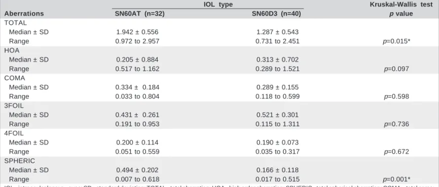
Postoperative wavefront analysis is shown in table 1. The-re weThe-re no significant diffeThe-rences in mean root-mean-squaThe-re values of higher-order aberration, total coma, trefoil and tetra-foil between both groups. The mean values for total aberra-tions (TOTAL; p=0.015) and total spherical aberraaberra-tions (TSA; p=0.001) in the SN60D3 multifocal group were statistically lower when compared to those for the SN60AT monofocal group.
All patients had distant monocular UCBVA (uncorrected best visual acuity) of 20/30 or better. Uncorrected distance acuity of 20/20 was achieved in 95% of the eyes in the multi-focal group and in 100% of the patients of the monomulti-focal group (p>0.017). Distance BCVA of 20/20 was achieved in all multifocal and monofocal eyes (Table 2).
In the monofocal group, 75% of the patients achieved an uncorrected intermediate visual acuity between Jaeger 1 and 6, but 25% of the patients achieved an intermediate visual acuity between Jaeger 1 and 6, with corrected distance vision. In the multifocal group, 75% of the patients were not able to see Jaeger 6 with and without optical correction for distance vision. A significantly higher proportion of eyes in the multi-focal group (85%) compared to the monomulti-focal group (6.3%) achieved an monocular uncorrected near acuity of 20/30 (J2, N5) or better, and all eyes in the multifocal group and 37.5% in the monofocal group achieved an uncorrected near acuity of 20/40 (J3, N6) or better. In the multifocal group, all patients achieved a binocular uncorrected near acuity of 20/30 or better (Table 3).
The mean spherical error was 0.11 D in the multifocal group and -0.18 D in the monofocal group (p=0.0377). There were no statistically significant differences in cylindrical errors between the multifocal and monofocal groups (0.29 D versus 0.38 D) (Table 4).
The results of contrast sensitivity under photopic condi-tions testing are shown in figure 1. The SN60D3 group presen-ted contrast sensitivity values (log units) lower in the 6.0 cpd spatial frequency compared to the SN60AT monofocal group. No statistical differences were detected at 1.5 cpd (KW=7.271; p=0.029), 3.0 cpd (KW=3.699; P=0.16), 12 cpd (KW=2.99; p=0.22) and 18 cpd (KW=4.85; p=0.089).
The results of contrast sensitivity under mesopic condi-tions testing are shown in figure 2. The SN60D3 group presen-ted contrast sensitivity values (log units) higher in the 6 cpd spatial frequency compared to the SN60AT group (p=0.01) with statistically significant differences (Figure 2). No statis-tical difference was detected in 1.5 cpd (KW= 0.59; p=0.721), 3.0 cpd (KW=2.63; p=0.31), 12 cpd (KW=3.43; p=0.21) and 18 cpd (KW=1.61; p=0.49).
All patients in both groups did well. As expected, the majo-rity of monofocal group (96.5%) required glasses for near vision. The multifocal group was satisfied and did not need glasses for distance, intermediate, or near vision.
DISCUSSION
Table 1. Comparison of mean root-mean-square values aberrations (µm) between SN60AT and SN60D3 IOLs
IOL type Kruskal-Wallis test
Aberrations SN60AT (n=32) SN60D3 (n=40) p value
TOTAL
Median ± SD 1.942 ± 0.556 1.287 ± 0.543
Range 0.972 to 2.957 0.731 to 2.451 p=0.015*
HOA
Median ± SD 0.205 ± 0.884 0.313 ± 0.702
Range 0.517 to 1.162 0.289 to 1.521 p=0.097
COMA
Median ± SD 0.334 ± 0.184 0.289 ± 0.155
Range 0.033 to 0.804 0.118 to 0.599 p=0.598
3FOIL
Median ± SD 0.431 ± 0.261 0.521 ± 0.301
Range 0.191 to 0.953 0.115 to 1.311 p=0.736
4FOIL
Median ± SD 0.200 ± 0.114 0.190 ± 0.073
Range 0.051 to 0.559 0.035 to 0.317 p=0.672
SPHERIC
Median ± SD 0.494 ± 0.202 0.166 ± 0.118
Range 0.007 to 0.618 0.017 to 0.515 p=0.001*
IOL= intraocular lens; n= eyes; SD= standard deviation; TOTAL= total aberration; HOA= high order aberration; SPHERIC= total spherical aberration; COMA= total coma; 3FOIL= trefoil; 4FOIL= tetrafoil
with spherical IOL’s in comparison to eyes with aspherical IOL’s(14). Rocha and associates reported the same results when
comparing multifocal aspheric IOL’s with monofocal spherical IOL’s using the Ladarvision aberrometer(21). It is possible to
hypothesize that the apodization, the gradual tapering of the diffractive steps from the center to the periphery of the Restor lens reduces spherical aberration in a way similar to that of an aspheric IOL. Regarding studies investigating ocular aberra-tions, a highly inverse relationship was found in the correla-tion between wavefront technology and visual performance only for those eyes with data sets of a high range of aberra-tions and acuities(12-13,21-25).
The effects of aberrations on visual function are complex and not completely understood(12-14,26). More attention should
be devoted to the relation between wavefront analysis and visual performance, and reassessment of their clinical signifi-cance is needed.
The difference in mean spherical errors between the groups could be explained by the slightly different target
postope-rative refraction. The postopepostope-rative refraction was intended to be slightly hyperopic or emmetropic for the SN60D3 group, whereas the postoperative refraction was targeted to be sli-ghtly myopic for the SA60AT group. In the monofocal group, 75% of the patients achieved an uncorrected intermediate visual acuity, but 25% of the patients achieved an interme-diate visual acuity, with corrected distance vision. In the multifocal group, 25% of the patients were able to interme-diate vision with and without optical correction for distance vision. This study found no significant differences in both uncorrected and corrected distance visual acuities. The mono-focal group was statistically superior for uncorrected mediate visual acuity, but no difference was found for inter-mediate visual acuity between both groups when corrected for distance vision. Previous studies comparing other types of diffractive multifocal IOLs and monofocal IOLs showed similar results(27-30).
Studies of different diffractive multifocal IOLs found that 86.8% to 91.3% of eyes had an uncorrected near visual acuity Table 2. Comparison of uncorrected and best corrected distance visual acuities (logMAR) between SN60AT and SN60D3 IOLs
IOL type t Student test
Distance visual acuities SN60AT (n=32) SN60D3 (n=40) p value
Uncorrected
Average ± SD (logMAR) 0.009 ± 0.027 0.028 ± 0.062 p=0.277
Range (logMAR) 0 to 0.100 0 to 0.222
Best corrected
Average ± SD (logMAR) 0.006 ± 0.160 0.015 ± 0.052 p=0.513
Range (logMAR) 0 to 0.046 0 to 0.180
Figure 2 - Comparison of contrast sensitvity test in mesopic conditions between Multifocal SN60D3 and Monofocal SN60AT intraocular lens Figure 1 - Comparison of contrast sensitvity in photopic conditions
between Multifocal SN60D3 and Monofocal SN60AT intraocular lens
Table 3. Comparison of uncorrected intermediate and near visual acuities (Jaeger chart) between SN60AT and SN60D3 IOLs
IOL type Fisher test
Visual acuities SN60AT (n=32) SN60D3 (n=40) p value
Intermediate % (n) % (n)
J1 - J2 6.25% ( 2) 0 p=0.4340
J3 - J4 25% ( 8) 5% ( 2) p=0.1390
J5 - J6 43.75% (14) 20% ( 8) p=0.0810
> J6 25% ( 8) 75% (30) P=0.0480*
Near % (n) % (n)
J1 - J2 6.25% ( 2) 85% (34)* p=0.0001*
J3 - J4 31.25% (10) 15% ( 6) p=0.2120
J5 - J6 12.5% ( 4) 0 p=0.1890
> J6 49.55 (16) 0 p=0.0003*
IOL= intraocular lens; n= eyes
of 20/40 (J3, N6) or better(3-4,30). In the present study, all
pa-tients in SN60D3 multifocal group achieved 20/40 (J3, N6) or better. These results were achieved by strict patient selection. Patients who did not want to use optical aids, particularly for the intermediate range of vision, were counseled appropria-tely regarding the multifocal IOL. With these points in mind, the AcrySof ReSTOR SN60D3 IOL can provide a higher de-gree of spectacle independence without intolerable visual symptoms. Published reports have consistently showed that multifocal IOL’s provide good near visual acuity(28-30).
According with this study and previous published re-views, the monocular contrast sensitivity at photopic and mesopic conditions with the SN60AT IOLs was higher than the SA60D3(17,30). Under mesopic conditions contrast
sensiti-vity showed lower values when compared to photopic condi-tions. Other study showed lower contrast sensitivity under mesopic conditions in multifocal IOLs with refractive tech-nology(28). The difference between both IOLs was higher in
photopic conditions, probably because the mesopic condition shows lower values than the photopic condition.
Table 4. Comparison of refractive results (diopters) between SN60AT and SN60D3 IOLs
IOL type t Student test
Refractive errors SN60AT (n=32) SN60D3 (n=40) p value
Spherical
Average ± SD -0.180 ± 0.377 0.110 ± 0.282 p=0.0377*
Range -1.25 to +0.25 -0.25 to +0.50
Cylindrical
Average ± SD 0.375 ± 0.242 0.288 ± 0.147 p=0.1790
Range 0 to +0.75 0 to +0.75
In conclusion, this study showed that the multifocal PCIOL (AcrySof SN60D3) provided as good distance visual acuity under high contrast and other conditions as the monofocal PCIOL (AcrySof SN60AT). However, contrast sensitivity was lower in the multifocal group, except in mesopic condition in low spatial frequency. There were also less spherical aberra-tions with the multifocal group in comparison to the monofo-cal group. This has yet to be correlated with visual performan-ce in further studies. Near-distanperforman-ce visual acuity was predic-tably achieved and higher in the multifocal group. Question-naires should be applied in further studies to assess the im-pact of spectacle independence in relation to differences in contrast sensitivity and spherical aberrations.
ACKNOWLEDGMENT
The authors acknowledge Drs. Alexandre Reis, Reinaldo Fujita, Maurício Pamplona, Danilo Fante, Amaryllis Avakian and Ricardo Holzchuh for the assistance in all phases of this study.
RESUMO
Objetivo: Comparar a performance visual, sensibilidade ao
contraste e de wavefront com OPD-Scan em pacientes subme-tidos a cirurgia de facoemulsificação com implante de lente intraocular AcrySof SN60D3 multifocal e AcrySof SA60AT monofocal. Métodos: Quarenta olhos com a lente intraocular multifocal e trinta e dois olhos com a lente intraocular monofo-cal. A avaliação oftalmológica contou com medida da acui-dade visual para longe, intermediária e curta distância, sem correção e com a melhor correção óptica, teste de sensibilida-de ao contraste e análise sensibilida-de frente sensibilida-de onda por meio do aberrô-metro OPD-Scan. Resultados: As aberração total e aberração esférica no grupo multifocal foi estatisticamente inferior com-parada com o grupo monofocal. No grupo monofocal 75% apresentaram acuidade visual monocular intermediária sem correção entre Jaeger 1 e 6, no grupo multifocal 75% apresen-taram mais que Jaeger 6. Aproximadamente 87,5% do grupo multifocal e 6,3% do grupo monofocal apresentaram acuidade visual monocular sem correção para perto de 20/30 (J2, N5), ou melhor, e 90,0% dos olhos do grupo multifocal e 37,5% do grupo monofocal apresentaram acuidade visual monocular sem correção para perto de 20/40 (J3, N6) ou melhor. A média de erro esférico foi de 0,11 D no grupo multifocal e -0,18 D no grupo monofocal (p=0,0379). O grupo monofocal apresentou superioridade estatística na sensibilidade ao contraste em condições fotópicas (p=0,014) e mesópicas (p=0,0044) a 6 cpg quando comparada ao grupo multifocal. Conclusão: A lente intraocular multifocal apresentou menos aberração esférica comparada à lente intraocular monofocal, da prevista multi-focalidade sem correção para longe e perto. Entretanto, o grupo multifocal apresentou baixa sensibilidade ao contraste.
Descritores: Lentes intraoculares; Implante de lente
intraocu-lar; Catarata; Facoemulsificação; Acuidade visual
REFERENCES
1. Obuchowska I, Mariak Z. [Sir Harold Ridley-the creator of modern cataract surgery]. Klin Oczna. 2005;107(4-6):382-4. Polish.
2. Davison JA, Simpson MJ. History and development of the apodized diffractive intraocular lens. J Cataract Refract Surg. 2006;32(5):849-58.
3. Sallet G. Refractive outcome after bilateral implantation of an apodized diffrac-tive intraocular lens. Bull Soc Belge Ophtalmol. 2006;(299):67-73. 4. Kohnen T, Allen D, Boureau C, Dublineau P, Hartmann C, Mehdorn E, et al.
European multicenter study of the AcrySof ReSTOR apodized diffractive intrao-cular lens. Ophthalmology. 2006;113(4):584.e1.
5. Bellucci R, Scialdone A, Buratto L, Morselli S, Chierego C, Criscuoli A, et al. Visual acuity and contrast sensitivity comparison between Tecnis and AcrySof SA60AT intraocular lenses: A multicenter randomized study. J Cataract Refract Surg. 2005;31(4):712-7. Erratum in: J Cataract Refract Surg. 2005;31(10):1857. 6. Rozema JJ, Van Dyck DE, Tassignon MJ. Clinical comparison of 6 aberrometers.
Part 1: Technical specifications. J Cataract Refract Surg. 2005;31(6):1114-27. 7. Alió JL, Schimchak P, Negri HP, Montés-Micó R. Crystalline lens optical
dysfunction through aging. Ophthalmology. 2005;112(11):2022-9. Comment in: Ophthalmology. 2007;114(3):618; author reply 619.
8. Rawer R, Stork W, Spraul CW, Lingenfelder C. Imaging quality of intraocular lenses. J Cataract Refract Surg. 2005;31(8):1618-31. Comment in: J Cataract Refract Surg. 2006;32(4):545-6; author reply 546.
9. Nio YK, Jansonius NM, Geraghty E, Norrby S, Kooijman AC. Effect of intrao-cular lens implantation on visual acuity, contrast sensitivity, and depth of focus. J Cataract Refract Surg. 2003;29(11):2073-81.
10. Dietze HH, Cox MJ. Limitations of correcting spherical aberration with aspheric intraocular lenses. J Refract Surg. 2005;21(5):S541-6.
11. Guirao A, Redondo M, Geraghty E, Piers P, Norrby S, Artal P. Corneal optical aberrations and retinal image quality in patients in whom monofocal intraocular lenses were implanted. Arch Ophthalmol. 2002;120(9):1143-51.
12. Packer M, Fine IH, Hoffman RS. Wavefront technology in cataract surgery. Curr Opin Ophthalmol. 2004;15(1):56-60.
13. Packer M, Fine IH, Hoffman RS, Piers P. Aberrations after intraocular lens implantation. J Cataract Refract Surg. 2006;32(2):184-5; author reply 185-6. 14. Marcos S, Barbero S, Jiménez-Alfaro I. Optical quality and depth-of-field of eyes implanted with spherical and aspheric intraocular lenses. J Refract Surg. 2005; 21(3):223-35.
15. Friström B, Lundh BL. Colour contrast sensitivity with different intraocular lens materials in the right and left eyes in same day surgery. Acta Ophthalmol Scand. 2005;83(4):443-7.
16. Rodríguez-Galietero A, Montés-Micó R, Muñoz G, Albarrán-Diego C. Compa-rison of contrast sensitivity and color discrimination after clear and yellow intraocular lens implantation. J Cataract Refract Surg. 2005;31(9):1736-40. 17. Oliveira F, Muccioli C, Silva LMP, Soriano ES, Souza CEB, Belfort Júnior R.
Avaliação da sensibilidade ao contraste e da estereopsia em pacientes com lente intraocular multifocal. Arq Bras Oftalmol. 2005;68(4):439-43.
18. Olsen T, Nielsen PJ. Immersion versus contact technique in the measurement of axial length by ultrasound. Acta Ophthalmol (Copenh). 1989;67(1):101-2. 19. Auffarth GU, Holzer MP, Visessook N, Apple DJ, Völcker HE. Removal times
for a dispersive and a cohesive ophthalmic viscosurgical device correlated with intraocular lens material. J Cataract Refract Surg. 2004;30(11):2410-4. 20. Jankov MR 2nd, Iseli HP, Bueeler M, Schor P, Seiler T, Mrochen M. The effect
of phenylephrine and cyclopentolate on objective wavefront measurements. J Refract Surg. 2006;22(5):472-81.
21. Rocha KM, Chalita MR, Souza CE, Soriano ES, Freitas LL, Muccioli C, Belfort R Jr. Postoperative wavefront analysis and contrast sensitivity of a multi-focal apodized diffractive IOL (ReSTOR) and three monomulti-focal IOLs. J Refract Surg. 2005;21(6):S808-12.
22. Alfonso JF, Fernández-Vega L, Baamonde MB, Montés-Micó R. Correlation of pupil size with visual acuity and contrast sensitivity after implantation of an apodized diffractive intraocular lens. J Cataract Refract Surg. 2007; 33(3):430-8. 23.Applegate RA, Hilmantel G, Howland HC, Tu EY, Starck T, Zayac EJ. Corneal first surface optical aberrations and visual performance. J Refract Surg. 2000; 16(5):507-14.
25. Bellucci R, Morselli S. Optimizing higher-order aberrations with intraocular lens technology. Curr Opin Ophthalmol. 2007;18(1):67-73.
26. Mayer S, Wirbelauer C, Pham DT. [Functional results after intraocular lens implantation with or without blue light filter: an intraindividual comparison]. Klin Monatsbl Augenheilkd. 2006;223(2):142-6. German.
27. Rossetti L, Carraro F, Rovati M, Orzalesi N. Performance of diffractive multi-focal intraocular lenses in extracapsular cataract surgery. J Cataract Refract Surg. 1994;20(2):124-8.
28. Souza CE, Muccioli C, Soriano ES, Chalita MR, Oliveira F, Freitas LL, et al.
Visual performance of AcrySof ReSTOR apodized diffractive IOL: a prospective comparative trial. Am J Ophthalmol. 2006;141(5):827-32.
29. Javitt J, Brauweiler HP, Jacobi KW, Klemen U, Kohnen S, Quentin CD, et al. Cataract extraction with multifocal intraocular lens implantation: clinical, func-tional, and quality-of-life outcomes. Multicenter clinical trial in Germany and Austria. J Cataract Refract Surg. 2000;26(9):1356-66.