ORIGINAL ARTICLE
Fluvoxamine alters the activity of energy metabolism
enzymes in the brain
Gabriela K. Ferreira,
1,2Mariane R. Cardoso,
1,2Isabela C. Jeremias,
1,2Cinara L. Gonc¸alves,
1,2Karolina V. Freitas,
1,2Rafaela Antonini,
1,2Giselli Scaini,
1,2Gislaine T. Rezin,
3Joa˜o Quevedo,
2,4Emilio L. Streck
1,21Bioenergetics Laboratory, Center of Excellence in Applied Neurosciences of Santa Catarina (NENASC), Graduate Program in Health
Sciences, Universidade do Extremo Sul Catarinense (UNESC), Criciu´ma, SC, Brazil.2National Science and Technology Institute for
Translational Medicine (INCT-TM), Porto Alegre, RS, Brazil.3Clinical and Experimental Pathophysiology Laboratory, Graduate Program in
Health Sciences, Universidade do Sul de Santa Catarina (UNISUL), Tubara˜o, SC, Brazil.4Neurosciences Laboratory, Graduate Program in
Health Sciences, UNESC, Criciu´ma, SC, Brazil.
Objective: Several studies support the hypothesis that metabolism impairment is involved in the pathophysiology of depression and that some antidepressants act by modulating brain energy metabolism. Thus, we evaluated the activity of Krebs cycle enzymes, the mitochondrial respiratory chain, and creatine kinase in the brain of rats subjected to prolonged administration of fluvoxamine.
Methods:Wistar rats received daily administration of fluvoxamine in saline (10, 30, and 60 mg/kg) for 14 days. Twelve hours after the last administration, rats were killed by decapitation and the prefrontal cortex, cerebral cortex, hippocampus, striatum, and cerebellum were rapidly isolated.
Results:The activities of citrate synthase, malate dehydrogenase, and complexes I, II-III, and IV were decreased after prolonged administration of fluvoxamine in rats. However, the activities of complex II, succinate dehydrogenase, and creatine kinase were increased.
Conclusions: Alterations in activity of energy metabolism enzymes were observed in most brain areas analyzed. Thus, we suggest that the decrease in citrate synthase, malate dehydrogenase, and complexes I, II-III, and IV can be related to adverse effects of pharmacotherapy, but long-term molecular adaptations cannot be ruled out. In addition, we demonstrated that these changes varied according to brain structure or biochemical analysis and were not dose-dependent.
Keywords: Brain; creatine kinase; fluvoxamine; Krebs cycle; mitochondrial respiratory chain
Introduction
Major depression is the most common psychiatric disorder.1 Mood disorders appear to afflict at least 12%
of women and 8% of men. The World Health Organization estimates that major depression is the fourth leading cause of loss or life worldwide.2 Moreover, unipolar depression is characterized by symptoms that must last a minimum of 2 weeks and interfere considerably with work and family relationships.3
Pharmacotherapy of depression is costly and widely prescribed by physicians, even though less than half of the patients treated attain complete remission after therapy with a single antidepressant. Others exhibit partial, refractory, or intolerant responses to pharmacological treatment.4 Current studies of the pharmacotherapy of major depressive disorder mainly emphasize the mono-amine hypothesis.5 This theory suggests that major depression results from an imbalance in neurotransmitters
such as serotonin, norepinephrine, and dopamine, and current treatments are thus based on normalizing the levels of these neurotransmitters.6
Fluvoxamine is classified as a selective serotonin reuptake inhibitor (SSRI) and is widely used for the treatment of depression and various anxiety disorders.7 With its characteristic binding to the 5-hydroxytryptamine receptor, fluvoxamine has less effect on noradrenaline and dopamine receptors, resulting in a different side effect profile as compared with older tricyclic antidepres-sants and monoamine oxidase inhibitors.8
Several studies also support the hypothesis that metabolism impairment is involved in the pathophysiology of depression and that some antidepressants act by modulating brain energy metabolism.9A large number of
drugs that have been withdrawn from the market or whose development was discontinued due to hepatotoxi-city, nephrotoxihepatotoxi-city, or cardiotoxicity have been reported to disturb mitochondrial functions.10 Mitochondria are intracellular organelles which play a crucial role in adenosine triphosphate (ATP) production.11 The Krebs cycle occurs within the mitochondrial matrix and con-tributes to production of large amounts of ATP via mitochondrial oxidative phosphorylation, which supplies more than 95% of the total energy requirement in the
Correspondence: Emilio L. Streck, Laborato´rio de Bioenerge´tica, Universidade do Extremo Sul Catarinense, Av. Universita´ria, 1105, CEP 88806-000, Criciu´ma, SC, Brazil.
E-mail: emiliostreck@gmail.com
Submitted Jul 01 2013, accepted Oct 23 2013. ß2014 Associac¸a˜o Brasileira de Psiquiatria
cells.12 Creatine kinase is an enzyme that also con-tributes to rates ATP metabolism in tissues with high energy demand.13
Based on the hypothesis that some antidepressants may modulate energy metabolism and that the effects of fluvoxamine on energy metabolism are still not clearly understood, the present study evaluated the activities of Krebs cycle enzymes, the mitochondrial respiratory chain, and creatine kinase in the brain of rats subjected to prolonged administration of fluvoxamine.
Methods
Animals
Adult male Wistar rats (250-300 g) obtained from the Central Animal House of Universidade do Extremo Sul Catarinense (UNESC) were caged in groups of two with free access to food and water and kept on a 12-h light-dark cycle (lights on 7:00 a.m.) at a temperature of 2261 6C. All experimental procedures were carried out in accordance with the Brazilian Society for Neuroscience and Behavior recommendations for animal care, with the approval of the local Ethics Committee.
Prolonged administration of fluvoxamine
Animals received daily administration of fluvoxamine dissolved in saline solution (10, 30, and 60 mg/kg, intraperitoneal), 1 mL/kg per body weight, for 14 days (n=7). Control rats received an equivalent volume of saline, 1 mL/kg (intraperitoneal), for the same period (n=7). Twelve hours after the last injection,14the animals were killed by decapitation and the prefrontal cortex, cerebral cortex, hippocampus, striatum, and cerebellum were rapidly isolated and kept on an ice-plate.
Tissue and homogenate preparation
Prefrontal cortex, posterior cortex, hippocampus, stria-tum, and cerebellum were homogenized (1:10, w/v) in SETH buffer, pH 7.4 (250 mM sucrose, 2 mM EDTA, 10 mM Trizma base, 50 IU/mL heparin). The homogenates were centrifuged at 800 x g for 10 min at 4 6C and supernatants kept at -706C until use for enzyme activity determination. The maximal period between homogenate preparation and enzyme analysis was always less than 5 days. Protein content was determined by the method described by Lowry et al.15using bovine serum albumin as standard.
Activity of Krebs cycle enzymes
Citrate synthase
Citrate synthase activity was assayed using the method described by Srere.16The reaction mixture contained 100 mM Tris, pH 8.0, 100 mM acetyl CoA, 100 mM 5,59 -di-thiobis-(2-nitrobenzoic acid), 0.1% Triton X-100, and 2-4 mg supernatant protein, and was initiated with 100 mM oxaloacetate and monitored at 412 nm for 3 min at 256C.
Malate dehydrogenase
Malate dehydrogenase was measured as described by Kitto.17 Aliquots (20 mg protein) were transferred into a
medium containing 10 mM rotenone (specific inhibitor of complex I), 0.2% Triton X-100, 0.15 mM NADH, and 100 mM potassium phosphate buffer, pH 7.4, at 376C. The reaction was started by addition of 0.33 mM oxaloace-tate. The absorbance was monitored as described above.
Succinate dehydrogenase
Succinate dehydrogenase activity was determined as per Fischer et al.,18measured by following the decrease in absorbance due to reduction of 2,6-di-chloro-indophenol (2,6-DCIP) at 600 nm with 700 nm as reference wavelength (e = 19.1 mM-1 cm-1) in the presence of phenazine methosulphate (PMS). The reaction mixture consisting of 40 mM potassium phosphate, pH 7.4, 16 mM succinate, and 8 mM 2,6-DCIP was pre-incubated with 40-80mg homogenate protein at 306C for 20 min. Subsequently, 4 mM sodium azide, 7 mM rotenone (specific inhibitor of complex I), and 40 mM 2,6-DCIP were added. The reaction was initiated by addition of 1 mM PMS and was monitored for 5 min.
Activity of mitochondrial respiratory chain enzymes
Complex I
NADH dehydrogenase (complex I) was evaluated as described by Cassina & Radi19by determining the rate of NADH-dependent ferricyanide reduction at 420 nm.
Complex II
The activity of succinate-2,6-dichloroindophenol (DCIP)-oxidoreductase (complex II) was determined by the method described by Fischer et al.18Complex II activity was measured by following the decrease in absorbance due to reduction of 2,6-DCIP at 600 nm.
Complex II-III
The activity of succinate: cytochrome c oxidoreductase (complex III) was determined by the method described by Fischer et al.18 Complex II-III activity was measured by cytochrome c reduction using succinate as substrate at 550 nm.
Complex IV
Activity of creatine kinase
Creatine kinase activity was measured in brain homo-genates pretreated with 0.625 mM lauryl maltoside. The reaction mixture consisted of 60 mM Tris-HCl, pH 7.5, containing 7 mM phosphocreatine, 9 mM MgSO4, and approximately 0.4-1.2mg protein to a final volume of 100 mL. After 15 min of pre-incubation at 376C, the reaction was started by the addition of 3.2 mmol of adenosine diphosphate (ADP) plus 0.8 mmol of reduced glutathione. The reaction was stopped after 10 min by the addition of 1 mmol of p-hydroxymercuribenzoic acid. The creatine formed was estimated by the colorimetric method of Hughes.21The color was developed by addition of 100mL 2%a-naphtol and 100mL 0.05% diacetyl in a final volume of 1 mL and read spectrophotometrically after 20 min at 540 nm. Results were expressed as units/min x mg protein.
Statistical analysis
Results were presented as means6standard deviation. Assays were performed in duplicate and the mean was used for statistical analysis. Data were analyzed by one-way analysis of variance (ANOVA) followed by the Tukey test when F was significant. Between-groups differences were rated significant at p , 0.05. All analyses were carried out in an IBM PC-compatible computer using SPSS.
Results
In the present study, we evaluated the activity of some Krebs cycle enzymes, the mitochondrial respiratory chain, and creatine kinase in the rat brain after prolonged administration of fluvoxamine at three dosage levels (10, 30, and 60 mg/kg). Our results showed that citrate
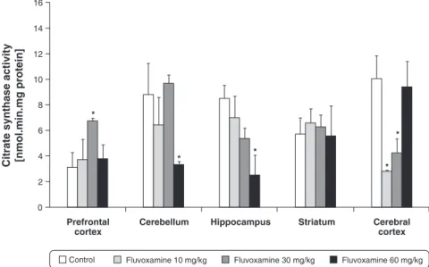
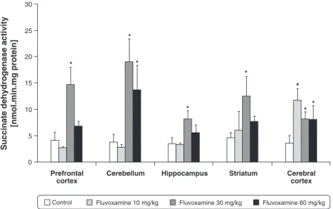
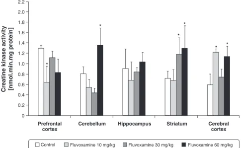
synthase activity was decreased in the cerebellum and hippocampus (60 mg/kg) and cerebral cortex (10 and 30 mg/kg) and increased in the prefrontal cortex (30 mg/kg) (Figure 1). Succinate dehydrogenase activity was increased in the prefrontal cortex, hippocampus and striatum (30 mg/kg), cerebellum (30 and 60 mg/kg), and cerebral cortex (at all doses) (Figure 2). Malate dehy-drogenase activity was decreased in the prefrontal cortex (10 mg/kg) and striatum (at all doses) (Figure 3). Complex I activity was decreased in the prefrontal cortex, hippocampus, and striatum (10 mg/kg); however, com-plex I activity was increased in the prefrontal cortex in the 30 mg/kg group (Figure 4A). Complex II activity was increased in the prefrontal cortex and cerebellum (30 mg/ kg) and cerebral cortex (10 mg/kg) (Figure 4B). Complex II-III activity was decreased in the prefrontal cortex (10 mg/kg) and cerebellum (20 mg/kg) (Figure 4C). Complex IV activity was decreased in the prefrontal cortex (10 and 30 mg/kg), hippocampus (30 and 60 mg/kg), and cerebral cortex (60 mg/kg) (Figure 4D). Finally, creatine kinase activity was increased in the cerebellum (60 mg/kg), striatum (30 and 60 mg/kg), and cerebral cortex (10 and 30 mg/kg); however, it was decreased in prefrontal cortex in the 10 mg/kg group (Figure 5).
Discussion
Mitochondria presumably produce much of the ATP essential for the excitability and survival of neurons and the protein phosphorylation reactions that mediate synaptic signaling and related long-term changes in neuronal structure and function. Evidence shows that patients with psychiatric disorders (depression, bipolar disorder, and schizophrenia) exhibit mitochondrial abnormalities at the structural, molecular, and functional levels.22 Indeed, Gardner et al.23 showed a significant decrease in mitochondrial ATP production rates and
mitochondrial enzyme ratios in muscle of major depres-sive disorder patients. Considering that life stressors may contribute to the development of depression, chronic stress has been used as an animal model of depression. In this scenario, it has been reported that activity of brain Na+, K+-ATPase and respiratory chain complexes I, III, and IV is inhibited after chronic variable stress in rats and that complexes I-III and II-III of the mitochondrial respiratory chain are inhibited in the rat brain after chronic stress.24An abnormal cellular energy state can lead to alterations in neuronal function, plasticity, and brain circuitry, and thereby affect cognition.25
The action of various therapeutic agents on mitochon-dria is relatively unknown. Evidence suggests that mitochondrial dysfunctions are implicated in etiology of drug-induced toxicities.26However, there is relatively little information about associations between antidepressant-induced changes in mitochondrial functions and thera-peutic or side effects of these drugs. Our findings showed that fluvoxamine alters the activity of energy metabolism enzymes in the brain, although this influence varied depending on the dose, brain region, and enzyme evaluated. Furthermore, different regions of the central nervous system can respond distinctly,27 and the
Figure 2 Effect of prolonged administration of fluvoxamine on succinate dehydrogenase activity in the rat prefrontal cortex, hippocampus, striatum, cerebellum, and cerebral cortex. Data were analyzed by one-way analysis of variance followed by Tukey test when F results were significant.*Different from control, p,0.05.
activities of enzymes involved in energy metabolism were analyzed in different brain regions, which in part represent different cell types, indicating heterogeneity in terms of physiological and metabolic characteristics.28-30 Our data are consistent with previous studies that showed antidepressants might cause impairment in mitochondrial function.31 Recent studies showed that citalopram and escitalopram decreased the activity of respiratory chain complexes.32 Dykens et al.33 showed that antidepressants induce mitochondrial dysfunction and cytotoxicity. In this context, a recent study showed that tricyclic antidepressants can modulate mitochondrial functions indirectly through a decrease in nitric oxide production.34More recently, Hroudova´ & Fisar35showed that amitriptyline, fluoxetine, and tianeptine are potent partial inhibitors of energized mitochondrial respiration.
The reasons for the region-specific effect of fluvox-amine on the activity of energy metabolism enzymes are unclear, but studies have demonstrated that fluvoxamine has different effects in specific brain areas. Muck-Seler
et al.36 reported that fluvoxamine administration
de-creased 5-HT synthesis rates in serotonergic cell bodies in the raphe magnus, with a trend toward decrease in the dorsal and median raphe nuclei. On the other hand, 5-HT synthesis rates were increased in the hippo-campus, substantia nigra, and hypothalamus, and re-mained unchanged in the caudate-putamen and nucleus accumbens.
that mitochondrial dysfunction induces activation of gene expression through the transcription factor cyclic adeno-sine monophosphate (cAMP) response element-binding protein (CREB) via the cAMP signaling pathway and that effects of long-term treatment with antidepressants are linked to the activation of the cAMP/protein kinase A/ CREB/brain-derived neurotrophic factor pathway,39it can be speculated that antidepressant-induced mitochondrial dysfunction could be involved in early biochemical processes leading to changes in neuroplasticity. In this context, Abdel-Razaq et al.40 suggest that the weak antimitochondrial actions of antidepressants could pro-vide a potentially protective preconditioning effect, in which antidepressant-induced mitochondrial dysfunction below the threshold of injury results in subsequent protection.
However, it is well known that the mitochondrial oxidative phosphorylation system generates reactive oxygen species (ROS).41Complex I plays a major role in controlling oxidative phosphorylation, and its abnormal activity can lead to defects in energy metabolism and thereby to changes in neuronal activity. This unique feature explains the mitochondrion’s great vulnerability to lipophilic molecules.41 Inhibition of complex III usually results in the generation of ROS as a consequence of the intrinsic characteristics of the electron-transfer process to this complex from reduced ubiquinone (UQ).42 To
maintain homeostasis, mitochondria possess a number of compartmentalized proteolytic systems that are cap-able of degrading transient or abnormal proteins. Within the mitochondrial matrix, the ATP-dependent Lon pro-tease has been suggested to perform this vital function. The increased susceptibility of Lon protease inactivation in comparison with electron transport chain inhibition raises the possibility that Lon protease dysfunction may be an early event in the pathogenesis of mitochondrial disorders associated with elevated ROS production. This
may result in a self-amplifying cycle of oxidative damage to the mitochondria as a result of aconitase oxidation and the release of unbound iron ions, leading to impaired respiration and cellular degeneration.43Thus, the poten-tially protective preconditioning caused by antidepres-sant-induced mitochondrial dysfunction below the threshold of injury can be associated with adverse effects through the formation of ROS, since said mitochondrial dysfunction generates ROS, which in turn are associated with a reduction in antioxidant defenses that can lead to oxidative stress.
In conclusion, based on the hypothesis that metabolic impairments might be implicated in the pathophysiology of depression, and on previous studies that have demonstrated that some antidepressants induce mito-chondrial dysfunction, we suggest that an increase in complex II, succinate dehydrogenase, and creatine kinase activities is associated with the therapeutic effects of antidepressants, and that a decrease in citrate synthase, malate dehydrogenase, and complexes I, II-III, and IV activities could be related to adverse effects of pharmacotherapy, but long-term molecular adaptations cannot be ruled out. In addition, we demonstrated that these changes varied according to brain structure or biochemical analysis and were not dose-dependent.
Acknowledgements
This study was supported by grants from Conselho Nacional de Pesquisa e Desenvolvimento (CNPq), Fundac¸a˜o de Apoio a` Pesquisa Cientı´fica e Tecnolo´gica do Estado de Santa Catarina (FAPESC), and Universidade do Extremo Sul Catarinense (UNESC).
Disclosure
The authors report no conflicts of interest.
References
1 Kessler RC, Walters EE. Epidemiology of DSM-III-R major depres-sion and minor depresdepres-sion among adolescents and young adults in the National Comorbidity Survey. Depress Anxiety. 1998;7:3-14. 2 Kiss JP. Theory of active antidepressants: a nonsynaptic approach
to the treatment of depression. Neurochem Int. 2008;52:34-9. 3 Zhang X, Beaulieu JM, Sotnikova TD, Gainetdinov RR, Caron MG.
Triptophan hydroxylase-2 controls brain serotonin synthesis. Science. 2004;305:217.
4 Pacher P, Kohegyi E, Kecskemeti V, Furst S. Current trends in the development of new antidepressants. Curr Med Chem. 2001;8:89-100.
5 Skolnick P. Beyond monoamine-based therapies: clues to new approaches. J Clin Psychiatry. 2002;63:19-23.
6 Charney DS. Monoamine dysfunction and the pathophysiology and treatment of depression. J Clin Psychiatry. 1998;59:11-4.
7 Mundo E, Rouillon F, Figuera ML, Stigler M. Fluvoxamine in obsessive-compulsive disorder: similar efficacy but superior toler-ability in comparison with clomipramine. Hum Psychopharmacol. 2001;16:461-8.
8 Potter WZ, Rudorfer MV, Manji H. The pharmacologic treatment of depression. N Engl J Med. 1991;325:633-42.
9 Tretter L, Mayer-Takacs D, Adam-Vizi V. The effect of bovine serum albumin on the membrane potential and reactive oxygen species generation in succinate-supported isolated brain mitochondria. Neurochem Int. 2007;50:139-47.
10 Dykens JA, Will Y. The significance of mitochondrial toxicity testing in drug development. Drug Discov Today. 2007;12:777-85. 11 Calabrese V, Scapagnini G, Giuffrida Stella AM, Bates TE, Clark JB.
Mitochondrial involvement in brain function and dysfunction: rele-vance to aging, neurodegenerative disorders and longevity. Neurochem Res. 2001;26:739-64.
12 Kelly DP, Gordon JI, Alpers R, Strauss AW. The tissue-specific expression and developmental Regulation of two nuclear genes encoding rat mitochondrial proteins. Medium chain acyl-CoA dehydrogenase and mitochondrial malate dehydrogenase. J Biol Chem. 1989;264:18921-5.
13 Bessman SP, Carpenter CL. The creatine-creatine phosphate energy shuttle. Annu Rev Biochem. 1985;54:831-62.
14 Maurel S, De Vry J, Schreiber R. Comparison of the effects of the selective serotonin-reuptake inhibitors fluoxetine, paroxetine, citalo-pram and fluvoxamine in alcohol-preferring cAA rats. Alcohol. 1999;17:195-201.
15 Lowry OH, Rosebough NG, Farr AL, Randall RJ. Protein measure-ment with the Folin phenol reagent. J Biol Chem. 1951;193:265-75. 16 Srere PA. Citrate synthase. Methods Enzymol. 1969;13:3-11. 17 Kitto GB. Intra- and extramitochondrial malate dehydrogenases from
chicken and tuna heart. Methods Enzymol. 1969;13:107-16. 18 Fischer JC, Ruitenbeek W, Berden JA, Trijbels JM, Veerkamp JH,
Stadhouders AM, et al. Differential investigation of the capacity of succinate oxidation in human skeletal muscle. Clin Chim Acta. 1985;153:23-36.
19 Cassina A, Radi R. Differential inhibitory Aation of nitric oxide and peroxynitrite on mitochondrial electron transport. Arch Biochem Biophys. 1996;328:309-16.
20 Rustin P, Chretien D, Bourgeron T, Gerard B, Rotig A, Saudubray JM, et al. Biochemical and molecular investigations in respiratory chain deficiencies. Clin Chim Acta. 1994;228:35-51.
21 Hughes BP. A method for estimation of serum creatine kinase and its use in comparing creatine kinase and aldolase activity in normal and pathologic sera. Clin Chim Acta. 1962;7:597-603.
22 Shao L, Martin MV, Watson SJ, Schatzberg A, Akil H, Myers RM, et al. Mitochondrial involvement in psychiatric disorders. Ann Med. 2008;40:281-95.
23 Gardner A, Johansson A, Wibom R, Nennesmo I, von Do¨beln U, Hagenfeldt L, et al. Alterations of mitochondrial function and correlations with personality traits in selected major depressive disorder patients. J Affect Disord. 2003;76:55-68.
24 Madrigal JL, Olivenza R, Moro M, Lizasoain I, Lorenzo P, Rodrigo J, et al. Glutathione depletion, lipid peroxidation and mitochondrial dysfunction are induced by chronic stress in rat brain. Neuropsychopharmacology. 2001;24:420-9.
25 Barrett SL, Kelly C, Bell R, King DJ. Gender influences the detection of spatial working memory deficits in bipolar disorder. Bipolar Disord. 2008;10:647-54.
26 Maurer I, Moller HJ. Inhibition of complex I by neuroleptics in normal human brain cortex parallels the extrapyramidal toxicity of neuro-leptics. Mol Cell Biochem. 1997;174:255-9.
27 Sullivan PG, Rabchevsky AG, Waldmeier PC, Springer JE. Mitochondrial permeability transition in CNS trauma: cause or effect of neuronal cell death? J Neurosci Res. 2005;79:231-9.
28 Lai YL, Rodarte JR, Hyatt RE. Effect of body position on lung emptying in recumbent anesthetized dogs. J Appl Physiol Respir Environ Exerc Physiol. 1977;43:983-7.
29 Sims DE. Recent advances in pericyte biology implications for health and disease. Can J Cardiol. 1991;7:431-43.
30 Sonnewald U, Hertz L, Schousboe A. Mitochondrial heterogeneity in the brain at the cellular level. J Cereb Blood Flow Metabol. 1998;18:231-7.
31 Mattson MP, Gleichmann M, Cheng A. Mitochondria in neuroplas-ticity and neurological disorders. Neuron. 2008;60:748-66. 32 Goncalves CL, Rezin GT, Ferreira GK, Jeremias IC, Cardoso MR,
Carvalho-Silva M, et al. Differential effects of escitalopram admin-istration on cortical and subcortical brain regions metabolic para-meters of Wistar rats. Acta Neuropsychiatr. 2012;24:147-54. 33 Dykens JA, Jamieson JD, Marroquin LD, Nadanaciva S, Xu JJ, Dunn
MC, et al. In vitro assessment of mitochondrial dysfunction and cytotoxicity of nefazodone, trazodone, and buspirone. Toxicol Sci. 2008;103:335-45.
34 Hwang J, Zheng LT, Ock J, Lee MG, Kim SH, Lee HW, et al. Inhibition of glial inflammatory activation and neurotoxicity by tricyclic antidepressants. Neuropharmacology. 2008;55:826-34.
35 Hroudova´ J, Fisˇar Z. In vitro inhibition of mitochondrial respiratory rate by antidepressants. Toxicol Lett. 2012;213:345-52.
36 Muck-Seler D, Pivac N, Diksic M. Acute treatment with fluvoxamine elevates rat brain serotonin synthesis in some terminal regions: an autoradiographic study. Nucl Med Biol. 2012;39:1053-7.
37 Szewczyk A, Wojtczak L. Mitochondria as a pharmacological target. Pharmacol. Rev. 2002;54:101-27.
38 D’Sa C, Duman RS. Antidepressants and neuroplasticity. Bipolar Disord. 2002;4:183-94.
39 Calabrese V, Cornelius C, Dinkova-Kostova AT, Calabrese EJ, Mattson MP. Cellular stress responses, the hormesis paradigm, and vitagenes: novel targets for therapeutic intervention in neurodegen-erative disorders. Antioxid Redox Signal. 2010;13:1763-811. 40 Abdel-Razaq W, Kendall DA, Bates TE. The effects of
antidepres-sants on mitochondrial function in a model cell system and isolated mitochondria. Neurochem Res. 2011;36:327-38.
41 Adam-Vizi V. Production of reactive oxygen species in brain mitochondria: contribution by electron transport chain and non-electron transport chain sources. Antioxid Redox Signal. 2005;7:1140-9.
42 Pathak RU, Davey GP. Complex I and energy thresholds in the brain. Biochim Biophys Acta. 2008;1777:777-82.


