J Bras Pneumol. 2010;36(3):372-391
Cell profile of BAL fluid in children and
adolescents with and without lung disease*
Celularidade do líquido de LBA em crianças e adolescentes saudáveis e com doenças pulmonares
Isabela Furtado de Mendonça Picinin, Paulo Augusto Moreira Camargos, Christophe Marguet
Abstract
The objective of this study was to review the literature on bronchoalveolar lavage fluid cell profiles in healthy children and adolescents, as well as on the use of BAL as a diagnostic and follow-up tool for lung disease patients in this age bracket. To that end, we used the Medline database, compiling studies published between 1989 and 2009 employing the following MeSH descriptors (with Boolean operators) as search terms: bronchoalveolar lavage AND cytology OR cell AND child. In healthy children, the cell profile includes alveolar macrophages (> 80%), lymphocytes (approximately 10%), neutrophils (approximately 2%) and eosinophils (< 1%). The profile varies depending on the disease under study. The number of neutrophils is greater in wheezing children, especially in non-atopic children, as well as in those with pulmonary infectious and inflammatory profiles, including cystic fibrosis and interstitial lung disease. Eosinophil counts are elevated in children/adolescents with asthma and can reach high levels in those with allergic bronchopulmonary aspergillosis or eosinophilic syndromes. In a heterogenous group of diseases, the number of lymphocytes can increase. Evaluation of the BAL fluid cell profile, when used in conjunction with clinical and imaging findings, has proven to be an essential tool in the investigation of various lung diseases. Less invasive than transbronchial and open lung biopsies, BAL has great clinical value. Further studies adopting standard international protocols should be carried out. Such studies should involve various age groups and settings in order to obtain reference values for BAL fluid cell profiles, which are necessary for a more accurate interpretation of findings in children and adolescents with lung diseases.
Keywords: Bronchoalveolar lavage; Bronchoalveolar lavage fluid/cytology; Child; Adolescent.
Resumo
Este estudo teve como objetivo rever a literatura existente sobre a celularidade do LBA em crianças e adolescentes saudáveis, bem como sobre sua utilização como método propedêutico e de acompanhamento nas afecções pulmonares neste grupo etário. Para tanto, utilizamos o banco de dados médico Medline com a seleção de artigos publicados entre 1989 e 2009 utilizando os seguintes descritores MeSH com operadores boolianos: bronchoalveolar lavage AND cytology OR cell AND child. Em crianças saudáveis, a celularidade é composta por macrófagos alveolares (> 80%), linfócitos (cerca de 10%), neutrófilos (cerca de 2%) e eosinófilos (< 1%). O perfil celular sofre alterações de acordo com a doença estudada. Ocorre uma elevação no número de neutrófilos em sibilantes, especialmente os não atópicos, bem como em indivíduos com quadros infecciosos e inflamatórios pulmonares, incluindo fibrose cística e doenças intersticiais pulmonares. Os eosinófilos se elevam em crianças/adolescentes com asma e podem atingir níveis acentuados na aspergilose broncopulmonar alérgica e nas síndromes hipereosinofílicas. A elevação dos linfócitos pode ocorrer em um grupo heterogêneo de doenças. Conclui-se que a celularidade do líquido de LBA, juntamente com dados clínicos e de imagem, tem se mostrado um instrumento essencial de investigação de diversas afecções pulmonares. O LBA possui uma grande utilidade clínica e é menos invasivo que a biópsia pulmonar transbrônquica e a céu aberto. Estudos sobre a celularidade normal do líquido de LBA utilizando-se protocolos internacionalmente padronizados e em diversas faixas etárias para a verificação de valores de referência são necessários para a interpretação mais acurada de resultados em crianças e adolescentes com pneumopatias.
Descritores: Lavagem broncoalveolar; Líquido da lavagem broncoalveolar/citologia; Criança; Adolescente.
* Study carried out at the Universidade Federal de Minas Gerais – UFMG, Federal University of Minas Gerais – Belo Horizonte, Brazil, and the University of Rouen, Rouen, France.
Correspondence to: Paulo Camargos. Departamento de Pediatria, Faculdade de Medicina da UFMG, Avenida Professor Alfredo Balena, 190, sala 267, CEP 30130-100, Belo Horizonte, MG, Brasil.
Tel 55 31 3409-9773. Fax 55 31 3409-9664. E-mail: pcamargs@medicina.ufmg.br or pauloamcamargos@gmail.com Financial support: None.
Submitted: 9 February 2010. Accepted, after review: 25 February 2010.
of proteins and analysis of inflammatory mediators.(3)
The material should be processed by an experienced professional following standardized procedures, in accordance with international recommendations that include the following(3): storage of the material at 4°C until the sample is analyzed in order to optimize cell viability; filtering of the material using sterile gauze to remove mucus particles (except in samples that will be sent for culture); performance of total and differential cytology after cytocentrifugation (in Citospin® centrifuges; Shandon Inc., Pittsburgh, PA, USA), with Giemsa staining. Other special staining methods can be used according to the clinical and laboratory suspicion. The sample used for microbiological study should be analyzed as fast as possible to avoid contamination and hinder the identification of agents.(3)
In accordance with the guidelines of the European Respiratory Society (ERS),(3) the BAL fluid can be technically acceptable when the volume recovered is more than 40% of the volume instilled and contains few epithelial cells, as we will discuss later.
Procedure safety
Various studies carried out with pediatric patients and even neonates showed that BAL is a safe procedure, presenting low rates of complications, which are usually minor and self-limited.(5-7) The most frequent complications are cough and transient bronchospasm (in less than 7% of the cases), as well as an isolated peak of fever after the procedure (in approximately 15% of the cases).(8)
It is recommended, however, that the procedure be performed by an experienced team specifically trained for treating possible emergencies, in an appropriate location, equipped with HR and oxygen saturation monitors, as well as with life support equipment. Appropriate cleaning and disinfection of the bronchoscope are essential to prevent patient contamination and avoid incorrect interpretation of the laboratory results.(3)
Indications
The evaluation of cellularity is one of the principal indications for BAL, in an attempt to obtain additional data (essential for the diagnosis
Introduction
With the development of fiberoptic bronchoscopy (FB) in 1965, BAL was introduced into clinical practice for adults. In the past 20 years, FB has been progressively used in pediatric clinics, and the use of BAL has been disseminated to a wide array of fields, including neonatology.(1)
The analysis of the cellular components of the BAL fluid has proven useful in the diagnosis and evaluation of the response to treatment in a vast number of lung diseases.(1,2) The determination of reference values in children without lung diseases is based in a few studies carried out in Europe, Australia and Korea,(3,4) and no similar publications are available until now in Latin-American countries, including Brazil.
Currently, BAL is well established as an extremely important procedure to obtain biological material from the lungs and has been widely used for diagnostic, therapeutic and research purposes.(1) Even when performed in high-risk children, the rate of complications, which are generally minor and self-limited, is minimal.(5,6) BAL correlates with lung biopsy by precluding the need for the latter in a vast number of diseases. Therefore, it is designated by some specialists “liquid biopsy”. Immunophenotyping of the T lymphocytes found in the BAL fluid has proven an important diagnostic tool for several childhood lung diseases.
Technical aspects
In children and adolescents, BAL is habitually performed with a flexible bronchoscope, the diameter of which can range from 2.8 to 4.9 mm. The procedure can be performed under general anesthesia with spontaneous breathing, although many centers opt for the use of local anesthesia and conscious sedation.(3)
The BAL procedure is performed by instilling isotonic saline solution through the suction channel of the flexible fiberoptic bronchoscope, with a volume that varies according to the weight of the child (generally 3 mL/kg), divided into three equal aliquots.(3)
374 Picinin IFM, Camargos PAM, Marguet C
J Bras Pneumol. 2010;36(3):372-391
airway infections in the two years preceding the study, history of bronchial hyperresponsiveness or history of atopy were excluded. Later, two groups of authors(1,11) analyzed children investigated for the diagnosis of laryngeal stridor without lung or systemic diseases, with normal physical examination and chest X-rays. Another group of authors(9) described BAL cell profiles in 16 children and adolescents without lung diseases, investigated for the diagnosis of stridor or suspicion of foreign body, who presented normal physical examination, chest X-rays and pulmonary function.
More recently, one group of authors(4) performed BAL in 10 children with no history of acute or chronic lung disease, submitted to elective surgery. These children comprised the control group for the study of BAL cell profiles in post-viral bronchiolitis obliterans.
None of the authors previously cited documented relevant clinical complications, including bleeding, cough or wheezing, during or after the procedure. In one of the aforementioned studies,(11) the authors reported that few children presented a slight and transient drop in oxygen saturation (in levels that were not reported) while performing the BAL, but this drop was not associated with changes in the HR.
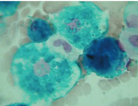
Table 1 describes the results for BAL cell profiles found by the aforementioned authors, as well as some characteristics of the studied population.
Although the normal cell profile values have been found to vary in the studies carried out, it has been observed that alveolar macrophages are the predominant cell type in the normal BAL fluid, accounting for over 80% of the cells found. According to the cell subtype and depending on the stimulus these cells are exposed to, they can play a role in phagocytosis, inflammatory activity and, especially, anti-inflammatory activity in the lungs.(12) The presence of these cells ensures the alveolar origin of the recovered material and the correct interpretation of the cell profile. However, the presence of a large number of epithelial cells reveals bronchial, non-alveolar material, which impedes or hinders the interpretation of total and differential cell distribution.
Lymphocytes are the second most common cell type in the normal BAL fluid of pediatric patients, with values ranging from 8-16% of the cells. These cells significantly contribute to and follow up of various lung diseases) from the
alveolar environment, as will subsequently be discussed. The analysis of cytokines, as well as of pro- and anti-inflammatory molecules, in the BAL fluid is also the focus of increasing interest, both in clinical practice and research.
Other indications, which are not the objective of the present article, include the identification of infectious agents (when there is suspicion of lung infection and when other forms of identification were not possible or inconclusive) and the therapeutic removal of airway materials, observed, for instance, in alveolar proteinosis and lipoid pneumonia.(3)
Reference values for cell profiles:
ethical and operational aspects
Although BAL is well tolerated in children and plays a key role in clinical practice and research, the determination of reference values in healthy children is difficult due to the ethical limitations of using healthy volunteers, as frequently observed in research with adults.(9)
Because of the ethical and operational issues involved, the determination of reference values for children can be based on data obtained from individuals submitted to elective surgery (usually orthopedic, otorhinolaryngologic or urogenital procedures) or bronchoscopy to clarify laryngeal stridor, after written informed consent has been obtained from the parents or legal guardians. Care should be taken to select patients without a history of asthma, atopy, acute respiratory infections or chronic lung diseases, so that the selected individuals can be considered representative of children without lung diseases.(3)
Cell profiles in healthy children and adolescents
The epithelial cells of bronchial origin that are found in the BAL fluid, as well as squamous cells from the upper airways, are not included in BAL differential cell counts;(12) however, they should ideally be mentioned in the cytological report, in order to allow the evaluation of the reliability of the analyzed sample by the clinician. The same should be observed for red blood cells, which might be found even in the normal BAL fluid, due to minor trauma caused by the contact of the bronchoscope with the bronchial mucosa.
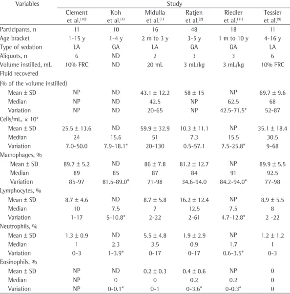
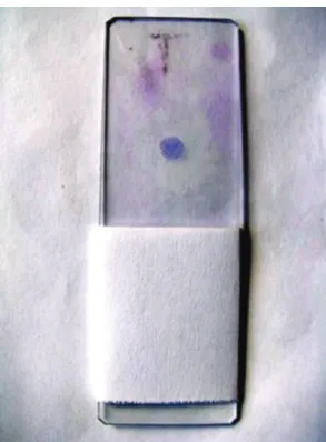
Figures 1 and 2 reveal, respectively, the macroscopic and microscopic aspect of a slide with normal BAL fluid after cytocentrifugation. the defense against tumors and infections, as
well as to the modulation of inflammation in the lungs.(12)
Neutrophils are inflammatory cells that are also found at a lower percentage in the normal BAL fluid, accounting for approximately 2% of the cells. Their primary function is phagocytosis and the elimination of invasive pathogens, and they play a key role in the pulmonary inflammatory process.(12) The increase in the number of these cells is common in diseases of different pathogenesis.
Eosinophils are not habitually found in the BAL fluid, or are found at very low percentages, accounting for less than 1% of the cells.(12)
Table 1 - Total and differential BAL cell profiles in studies involving children without lung diseases.
Variables Study
Clement et al.(10)
Koh et al.(4)
Midulla et al.(1)
Ratjen et al.(2)
Riedler et al.(11)
Tessier et al.(9)
Participants, n 11 10 16 48 18 11
Age bracket 1-15 y 1-4 y 2 m to 3 y 3-5 y 1 m to 10 y 4-16 y
Type of sedation LA GA LA GA GA LA
Aliquots, n 6 ND 2 3 3 6
Volume instilled, mL 10% FRC ND 20 mL 3 mL/kg 3 mL/kg 10% FRC
Fluid recovered
(% of the volume instilled)
Mean ± SD NP ND 43.1 ± 12.2 58 ± 15 NP 69.7 ± 9.6
Median NP ND 42.5 NP 62.5 68
Variation NP ND 20-65 NP 42.5-71.5* 52-87
Cells/mL, × 104
Mean ± SD 25.5 ± 13.6 ND 59.9 ± 32.9 10.3 ± 11.1 NP 35.1 ± 18.4
Median 24 15.6 51 7.3 15.5 30.5
Variation 7.0-50.0 7.9-18.1* 20-130 0.5-57.1 7.5-25.8* 9-68
Macrophages, %
Mean ± SD 89.7 ± 5.2 ND 86 ± 7.8 81.2 ± 12.7 NP 89.9 ± 5.5
Median 89 85 87 84 91 92.5
Variation 85-97 81.5-89.0* 71-98 34.6-94.0 84.2-94.0* 77-98
Lymphocytes, %
Mean ± SD 8.7 ± 4.6 ND 8.7 ± 5.8 16.2 ± 12.4 NP 8.9 ± 5.5
Median 10 7.5 7 12.5 7.5 8
Variation 1-17 5-10.8* 2-22 2-61 4.7-12.8* 2 -22
Neutrophils, %
Mean ± SD 1.3 ± 0.9 ND 5.5 ± 4.8 1.9 ± 2.9 NP 1.2 ± 1.2
Median 1 2.3 3.5 0.9 1.7 1
Variation 0-3 1-3.9* 0-17 0-17 0.6-3.5* 0-3
Eosinophils, %
Mean ± SD NP ND 0.2 ± 0.3 0.4 ± 0.6 NP 0
Median NP 0 0 0.2 0.2 0
Variation NP 0-0.1* 0-1 0-3.6* 0-0.3* 0
376 Picinin IFM, Camargos PAM, Marguet C
J Bras Pneumol. 2010;36(3):372-391
childhood, which are different from those found in adults.
Studies reveal that the number of eosinophils in the BAL fluid is higher in children with atopic asthma than in wheezing children with non-atopic asthma (p < 0.01).(13,14) These findings indicate that there is eosinophilic inflammation of the airways in atopic wheezing children, which might aid in differentiating
Usefulness of the analysis of BAL
cell profiles for the diagnosis of lung
diseases
Various diseases involve a deviation from the normal BAL cell profiles. Cytological studies can contribute to the correct diagnosis of these diseases, as well as to a better understanding of the pathophysiological mechanisms involved and of the response to treatment. However, it should be borne in mind that BAL cell profiles should not be interpreted in isolation, but in conjunction with data obtained from a detailed clinical history, a thorough physical examination and a careful evaluation of the available laboratory tests and imaging studies.
In general, infectious (pneumonia, bacterial bronchitis), suppurative (bronchiectasis) or pulmonary inflammatory processes (interstitial lung diseases, collagenosis, bronchopulmonary dysplasia, cystic fibrosis and even asthma) lead to greater recruitment of neutrophils to the lungs, which leads to an increase in the number of these cells in the BAL fluid.(3,12) For instance, in patients with active bacterial infection in the lungs, neutrophil values can range from 25-95% of the total of cells.(12) The increase in the differential counts of these cells is also seen in interstitial pneumonia.
Diseases with different etiologies cause the increase in the number of lymphocytes in the BAL fluid. Some examples are sarcoidosis (lymphocytes can correspond to 20-50% of the cells), hypersensitivity pneumonia, Crohn’s disease and some interstitial diseases.(3,12)
An eosinophilic infiltrate can be seen in other situations, including asthma, reaction to drugs, allergic bronchopulmonary aspergillosis and eosinophilic syndromes, such as the Churg-Strauss syndrome, idiopathic hypereosinophilic syndrome and chronic eosinophilic pneumonia.(3)
Next, aspects related to BAL cell profiles in some diseases and conditions of great clinical and epidemiological importance are discussed. Some of these data are synthesized in Table 2.
Asthma and wheezing
The study of BAL cell profiles has contributed to a better understanding of the pathophysiological and immunological mechanisms involved in asthma and wheezing in
Figure 1 - Slide with BAL fluid sample after cytocentrifugation (macroscopic aspect). The purple circle corresponds to the site where the cells are fixed.
proportion of CD8 lymphocytes (59% vs. 40%) was higher and, consequently, the CD4/CD8 ratio (0.266 vs. 0.455) was lower in children with asthma than in wheezing infants (p = 0.02).
Some authors support the theory that the greater presence of eosinophils is associated with allergic sensitization in predisposed individuals but not necessarily with persistent or more severe symptoms.(14,23)
There is no doubt that these findings provide essential information to guide therapeutic strategies and contribute to a better understanding of the various phenotypes involved in the disease. However, the clinical follow up of the investigated children and the objective evaluation of the response to treatment are indispensable for the critical analysis of the significance of the findings.(15)
Future studies and new strategies are required in order to identify the sequence of inflammatory processes involved in the development of asthma and wheezing.(22) In this sense, FB and BAL are essential investigation tools.
Cystic fibrosis
The BAL procedure has been increasingly used to monitor children with cystic fibrosis, being used for the early detection of microorganisms, for the monitoring of the inflammatory response and for the evaluation of therapeutic strategies.
One group of authors(15) studied the BAL fluid of 10 children and adolescents with cystic fibrosis and found that they presented an increased number of total cells that was proportional to the increased number of neutrophils (p = 0.004) and alveolar macrophages (p = 0.05) when compared with the other groups studied. High percentages of eosinophils have been found in children diagnosed with allergic bronchopulmonary aspergillosis, a condition that affects approximately 5% of the children with cystic fibrosis.
The BAL fluid has also contributed to the understanding of the impact of infectious agents in the pulmonary inflammatory process of children with cystic fibrosis. Studies(24,25) have revealed that children with positive cultures for Pseudomonas aeruginosa, especially mucoid strains, present significant higher total cell counts and a higher percentage of neutrophils and inflammatory cytokines than do individuals with negative cultures or positive cultures for patients with transient wheezing from patients
who will actually develop asthma. One study(15) concluded that the increase in the number of eosinophils in the BAL fluid is clearly present in asthma patients (aged 4-15 years, with a history of recurrent wheezing and dyspnea attacks, showing confirmed reversibility of airway obstruction after using β2 agonists) but is rare in wheezing infants (aged 5-43 months, with at least 3 episodes of wheezing or cough, associated with viral infection). This increase was identified in 64% of the children with asthma and in only 27% of the wheezing infants (p = 0.04). The number of eosinophils seems to differentiate allergic wheezing infants from nonallergic wheezing infants, suggesting that the eosinophilic inflammation starts at a very early stage of allergic asthma.(13) However, other authors(16) did not find significant differences between a group of 47 atopic wheezers and a group of 19 non-atopic wheezers regarding the presence of eosinophils in the BAL fluid. This same study found a normal number of eosinophils and a significantly higher number of neutrophils in children younger than 3 years with severe recurrent wheezing.
The number of neutrophils in the BAL fluid tends to increase in individuals with positive microbiological culture.(13,17) However, various authors have also confirmed the participation of neutrophils in the bronchial inflammation found in asthma patients and, especially, in wheezing infants,(15-19) regardless of the presence of bacterial infection. Various studies(14,15,18,20) have associated the increase in the number of neutrophils with greater severity of symptoms.
378
P
ic
in
in
IFM
, C
am
ar
gos
P
A
M
, M
ar
gu
et
C
J
B
ra
s P
n
eu
m
ol
. 2010;
36(
3)
:372-39
1
Table 2 - BAL cell profiles in some diseases and conditions of clinical and/or epidemiological importance.
Study Disease studied Participants Age Total cellsa Macrophages Lymphocytes Neutrophils Eosinophils
n % Cells* % Cellsa % Cellsa % Cellsa
Najafi
et al.(13)b
Asthma/Wheezing 39
Atopic 21 3 (0.3-11.9) y 177 (7-1,030) 39 (5-81) 62 (1-560) 12 (3-51) 22 (1-181) 39 (2-91) 92 (0-808) 3 (0-19) 4 (0-68)
Non-atopic 18 1.0 (0.3-8.8) y 245 (9-1,804) 24 (7-90) 58 (2-527) 10 (1-34) 19 (0-136) 56 (0-83) 92 (0-1,257) 0 (0-3) 0 (0-13)
Positive culture 16 2.1 (0.4-10.7) y 460 (9-1,804) 17 (5-62) 52 (2-560) 7 (1-34) 25 (3-112) 73 (19-91) 342 (3-1,257) 0 (0-4) 0 (0-29)
Negative culture 23 2.1 (0.3-11.9) y 126 (7-952) 47 (10-90) 79 (1-517) 12 (2-51) 18 (0-181) 22 (0-71) 19 (0-646) 2 (0-19) 2 (0-68)
Just
et al.(14)c
Asthma/Wheezing 79
Wheezing infants 21 15 ± 7.2 m 292.9 ± 142.4 235.3 ± 123.4 38.7 ± 38.9 18.8 ± 51.4 0 ± 0.2
Children with asthma 58 92.9 ± 56.0 m 244.7 ± 148.7 178.4 ± 118.3 41.9 ± 38.7 13.3 ± 39.9 6.4 ± 11.9
Atopic 38 273.4 ± 170.7 199.6 ± 140.9 43.9 ± 41.9 13.0 ± 25.6 8.4 ± 13.5
Non-atopic 41 251.3 ± 130.3 191.8 ± 107.3 41.7 ± 36.9 17.5 ± 56.9 0.4 ± 1.5
Positive culture 13 240.0 ± 109.8 195.0 ± 83.9 28.3 ± 17.3 9.8 ± 9.3 6.5 ± 15.6
Negative culture 66 261.2 ± 154.6 193.3 ± 128.2 43.6 ± 41.0 15.8 ± 46.8 4.4 ± 9.4
Episodic asthma 45 267.7 ± 154.6 212.5 ± 120.4 44.6 ± 42.8 6.1 ± 6.5 4.4 ± 10.1
Persistent asthma 34 246.1 ± 141.3 171.0 ± 122.0 35.6 ± 32.2 26.4 ± 63.7 5.3 ± 11.4
Using ICs 60 239.0 ± 137.5 178.9 ± 118.2 34.7 ± 31.4 17.8 ± 48.8 3.8 ± 9.2
Not using ICs 19 313.9 ± 167.3 239.8 ± 123.5 61.0 ± 51.4 5.2 ± 6.4 7.7 ± 13.8
Le Bourgeois Wheezing infants 83 10 (7-13) m 400 (272-582) 87 (75-92) 812 (230-438) 5.0 (3.0-8.8) 19.4
(9.6-37.7)
4 (2-10) 12.4 (5.4-39) 0 (0-0) 0 (0-0)
et al.(16)d
de Blic et al.(20)d
Asthma 28
Minimal symptoms 15 13.3
(10.1-15.0) a
130 (100-150) 72 (50-85) 4.6 (3.7-8) 3.2 (1.3-6) 1.0 (0.7-2.4)
Maximal symptoms 13 10.4 (9.6-12.5) a 125 (115-175) 42 (28-54) 3.5 (2.2-4.4) 4.0 (3.0-14.4) 1.0 (0.4-6.5)
Marguet et al.(15)d
Asthma/Wheezing
Wheezing infants 26 24 (5-48) m 705 (291-930) 252 (8-440) 25 (6-92) 39 (9-278) 0 (0-5)
Children with asthma 14 85 (50-180) m 521 (400-700) 342 (217-490) 29 (20-56) 16 (5-719) 13 (0-47)
Chronic cough 12 51.7 (10-143) m 630 (403-986) 434 (227-599) 25 (20-69) 41 (8-57) 0 (0-4)
Cystic fibrosis 10 78.5 (32-187) m 1.015
(692-1,460)
625 (381-756) 68 (46-102) 265 (168-448) 23 (0-81)
y: years; m: months; ICs: inhaled corticosteroids. aData expressed as cells × 103/mL.bData expressed as median (variation). cData expressed as mean ± SD. dData expressed as median (interquartile
is suspicion of pulmonary hemorrhage and hemosiderosis.(29)
The diagnosis of pulmonary histiocytosis can be documented (in the BAL fluid) with the use of monoclonal antibodies revealing the presence of more than 5% CD1a+ cells.(3,30) This finding, associated with clinical and radiological findings, can contribute to reduce the indication for lung biopsy in a significant number of cases. (30) Studies in adults with histiocytosis revealed a
tendency towards a slight increase in the number of eosinophils, but future studies are needed in order to confirm this finding, especially in children.(30)
Alveolar proteinosis is characterized by intra-alveolar accumulation of material of lipoprotein origin, similar to surfactant. This resemblance suggests that there is a defect in the resorption (or excessive production) of surfactant by type II pneumocytes or by alveolar macrophages. The BAL fluid is an essential diagnostic tool when there is recovery of opaque fluid or when there is identification of periodic acid-Schiff-positive material within alveolar macrophages. Cytological analyses of BAL fluid do not reveal a specific pattern, although hypercellularity and lymphocytosis are frequently observed.(31)
The investigation of lipid-laden alveolar macrophages (LLAM) is classically cited as an aid in the diagnosis of aspiration associated with diseases that originate in the gastrointestinal tract, such as the gastroesophageal reflux disease (GERD).(32,33) It is considered that, when there is clinical suspicion, a LLAM index higher than 165 is useful for the diagnosis of aspiration, with sensitivity of 98.6% and specificity of 78.0%.(32) However, this index has been greatly questioned because of its low specificity and because of the large variability in the cut-off values cited in different studies.(32) One group of authors(34) reported that more sensitive tests, such as pH-metry, should remain as the gold standard for the diagnosis of GERD and proposed that the identification of lipid-laden macrophages should be reserved for children with no definitive diagnosis and no improvement with the proposed therapy. However, this investigation is not needed when there is strong evidence of aspiration, such as in the presence of tracheoesophageal fistula, identified by means of radiology or endoscopy, or in severe neurological conditions, since no other pathogenic agents (p < 0.01). Similarly,
individuals in which Staphylococcus aureus was isolated presented higher counts of neutrophils (p < 0.0001) and inflammatory markers (p = 0.003) than did individuals whose cultures were negative for this agent. Children coinfected with P. aeruginosa and S. aureus presented the highest rate of inflammatory markers.(24) Another group of authors(26) also showed that factors associated with bacterial infection and host defense mechanisms contribute more significantly to the inflammatory response mediated by neutrophils than does the innate deregulation caused by the mutation of the gene that encodes the cystic fibrosis transmembrane conductance regulator protein. In addition, these authors(26) reported that the early detection and appropriate treatment of the infections appears to be the best strategy to prevent irreversible lung injury due to inflammation. However, another group of authors(27) argued that more than one mechanism might be associated with airway remodeling in young children with cystic fibrosis, regardless of the presence of inflammatory markers.
Cell profiles in the BAL fluid also play an important role in evaluating the effectiveness of the therapeutic strategies. One group of authors(28) observed that, in children treated with inhaled DNase, the increase in the number of neutrophils in the BAL fluid throughout a three-year-follow-up period was less pronounced (p < 0.01) than was that in patients who had not been treated with inhaled DNase (p < 0.005). These authors suggested that the treatment had a positive effect in the airway inflammation of patients with cystic fibrosis.(28)
Interstitial diseases
380 Picinin IFM, Camargos PAM, Marguet C
J Bras Pneumol. 2010;36(3):372-391
hypereosinophilic syndrome, this value can be higher than 75%.(40)
Figures 3 and 4 illustrate the use of the BAL fluid for the diagnosis of lung diseases, depicting the radiologic image (Figure 3) and the BAL fluid (Figure 4) that aided in the diagnosis of pulmonary hemosiderosis in a child with a history of hemoptysis and iron-deficiency anemia.
Postinfectious bronchiolitis obliterans
Studies carried out with children presenting postinfectious bronchiolitis obliterans (BO) revealed that there is an increase in the number of neutrophils and CD8+ lymphocytes, which causes a drop in the CD4/CD8 ratio.(4,41) One group of authors(4) compared 12 children who developed postinfectious BO caused by the measles virus with 10 control children without lung disease and observed that the number of neutrophils was considerably higher in the first group (16% vs. 2.3%; p < 0.05) and that the CD4/CD8 ratio was lower in the study group than in the control group (0.41 vs. 0.65; p < 0.01). Another group of authors(41) studied 11 children with postinfectious BO and reported a drop in the CD4/CD8 ratio (median = 0.45; interquartile range: 0.4-0.6) and a sharp increase in the number of neutrophils (median = 50%; interquartile range: 1-66%). This increase in the recruitment of neutrophils is common in inflammatory pulmonary diseases and, in the specific case of BO, seems to have an effect in lung injury after the initial infection, as well as in the clinical course of the disease and in the decline in pulmonary function.(41)
Chronic cough
The BAL profile in children with chronic cough (i.e., presence of cough for over six months with no associated wheezing and absence of alternative diagnosis, such as immunodeficiency, cystic fibrosis or ciliary dyskinesia) has also been studied in an attempt to understand its mechanisms and how it is associated with asthma. Studies suggest that the pathophysiology of cough in these children is different from that in children with asthma and that the association between chronic cough and asthma is indeed controversial.(15,42) One study(42) showed that children with chronic cough, regardless of the atopic status, presented additional information will be provided in these
cases.
A large proportion of LLAM is observed in cases of lipoid pneumonia, generally secondary to mineral oil aspiration. In these cases, multiple BALs have been described as a valid treatment, since they remove lipid-laden cells, which are involved in alveolar and interstitial fibrosis. (35,36) Serial evaluation of BAL cell profiles allows
a more objective evaluation of the treatment outcomes. In a recent study,(35) it was reported that, after multiple BALs (4-10 procedures), the number of LLAM dropped from 1,179 to 265 cells (p < 0.004), and the mean total cell counts dropped from 1,574 cells/mm3 to 363 cells/mm3, a value considered normal (p < 0.003).
Although lung biopsy is considered the gold standard, the presence of hemosiderin-laden macrophages (HLM) in the BAL fluid is considered crucial for the diagnosis of idiopathic pulmonary hemosiderosis when there are findings suggestive of the disease, such as hemoptysis, interstitial infiltrate in chest X-rays and iron-deficiency anemia.(37,38) One group of authors(38) reviewed data from BAL cell profiles in children to correlate the presence of hemosiderosis with HLM and concluded that the presence of 36% HLM among the total of macrophages corresponded to a sensitivity of 100% and a specificity of 96% for the diagnosis of the disease.
in the BAL fluid of control children without the disease (22 × 106 cells/100 mL and 10 × 106 cells/100 mL vs. 10 × 106 cells/100 mL and < 1 × 10⁶ cells/100 mL, respectively; p < 0.02). In addition, another study(46) revealed that there seems to be a preferential recruitment of a specific type of CD4+ T lymphocytes to sites of active tuberculosis infection and that the specific identification of these cells in the BAL fluid can hasten the diagnostic process and aid in the distinction between patients with tuberculosis and patients with other diseases.(46)
Complications after lung and bone
marrow transplantation
The monitoring of BAL cell profiles can aid in the diagnosis of complications after lung transplantation. One study involving adolescents and young adults submitted to lung transplantation showed that an increase in the number of neutrophils (> 40%) can indicate a percentage of neutrophils in the BAL fluid
that was higher than the reference values reported in the literature and that this higher percentage might be related to an underlying inflammatory process. Few atopic children and no non-atopic children with persistent cough presented eosinophilic inflammation typical of asthma, which might predict their poor response to the use of inhaled corticosteroids. (42) Although the microbiological examination results were negative in all studied patients, the authors associated the neutrophilia with a possible infection undiagnosed by the available methods; to the presence of GERD, detected by pH-metry in 9 patients; or even to the exposure to environmental factors, such as tobacco, which were not investigated in the study.(42)
The most common etiologic diagnosis in 108 patients with chronic cough studied by one group of authors(43) was protracted bacterial bronchitis (defined as productive cough, positive BAL fluid culture and response to antibiotic therapy with the resolution of the cough up to two weeks after the end of the treatment), which was identified by the study of the BAL fluid in 39.8% of the cases. The median percentage of neutrophils found in the BAL fluid of this group (in relation to the total number of cells) was 40%. Diseases habitually described in association with chronic cough, such as asthma, GERD and upper airway disorders, were found in less than 10% of the patients of this study, based on the diagnostic criteria used.
Tuberculosis
The BAL cell profile can help to understand the role of the inflammatory cells in the pathogenesis of some infectious diseases, such as tuberculosis, contributing to the selection of diagnostic and therapeutic strategies. Recent evidence suggests that neutrophils are more abundant than macrophages in the BAL fluid of individuals infected by Mycobacterium tuberculosis (78.8 ± 5.8% and 11.8 ± 4.1%, respectively) and that more bacilli are found within neutrophils than within macrophages (65.1 ± 14.4% and 28.3 ± 1.6%). These findings suggest that neutrophils work as active replication sites for the bacillus in the initial stages of the disease.(44) In another study,(45) the mean number of lymphocytes and eosinophils found in the BAL fluid of children with tuberculosis was higher than that found
Figure 3 - Chest X-ray revealing diffuse pulmonary infiltrate.
382 Picinin IFM, Camargos PAM, Marguet C
J Bras Pneumol. 2010;36(3):372-391
Mechanical ventilation and
non-bronchoscopic BAL
Serial BAL can aid in the study of the progress of the inflammatory process in respiratory disturbances, especially in intubated children, and in the diagnosis of pneumonia associated with mechanical ventilation (MV). In addition, it enables the collection of samples for culture and the identification of causative agents. However, it should be borne in mind that MV itself is a risk factor for lung injury. Therefore, it is difficult to obtain a control group of “normal” children who can be compared with a study group of ill, intubated children.(23)
Non-bronchoscopic BAL is a valid strategy to evaluate bronchial inflammation and obtain culture samples in intubated children, especially in premature neonates (who require an ultrafine flexible bronchoscope of 2.2 mm). The technique consists in introducing a catheter in the tracheal tube and advancing it until it enters the bronchial tree. Then a volume of saline solution based on the weight of the child is instilled and immediately aspirated with syringes, following practically the same guidelines cited for the performance of BAL via FB. Some advantages of this technique are that the catheter can be passed even through endotracheal tubes of small diameter (< 4 mm), that the procedure can be easily performed next to the bed of the patient, that there is low discomfort for the patient and that the cost is low. The main disadvantage is that, without direct visualization provided by FB, it is impossible to predict from which area of the lung the sample will be collected, although it is expected that samples from the right lung will predominate because of anatomical characteristics. Non-bronchoscopic BAL can be performed with a protected catheter (double lumen) or a conventional catheter. The protected catheter has the advantage of avoiding contamination of the sample with upper airway flora or secretion.(51) Some studies have confirmed the reproducibility of culture results(51) and of differential cell counts(52) in different samples obtained with this technique.
Final considerations
Since BAL cell profiles can present similar patterns in diseases of various etiologies, this procedure should be analyzed in conjunction with progression to BO, whereas lymphocytosis (>
20%) and/or eosinophilia (> 5%), accompanied by a drop in the CD4/CD8 ratio, with no evidence of lung infection, can suggest acute rejection. (47) One group of authors(48) concluded that BAL
cell profiles can also be useful for recognizing pulmonary graft-versus-host disease (GVHD) in children with respiratory symptoms submitted to bone marrow transplantation and for recognizing GVHD in other organs, by revealing lymphocytosis (> 18%) and severe epithelial cell atypia.
Immunodeficiency and neoplasia
In diseases that lead to primary or acquired immunosuppression, alterations in BAL cell profiles are generally associated with bacterial infections. According to one group of authors,(49) when there is strong clinical suspicion but specific isolation is not possible, the presence of neutrophilia (> 30%), lymphocytosis (> 12%) or an increase in the number of cells can provide indirect evidence of the presence of the infectious agent or can inform decisions regarding empirical antibiotic therapy.
Another group of authors(50) studied 26 patients presenting immunodeficiencies (including AIDS, leukemia and cancer) and pulmonary infiltrate and concluded that differential cell counts in the BAL fluid of children with positive culture for fungi or bacteria showed a higher percentage of neutrophils than did the counts of control individuals (31.4% vs. 0.3%; p < 0.001) or the counts of immunocompromised individuals with negative culture for fungi or bacteria (31.4% vs. 0.8%; p = 0.003). However, the percentage of lymphocytes was higher in uninfected immunocompromised patients than in infected immunocompromised patients (14.5% vs. 4.3%; p = 0.62) or control individuals (14.5% vs. 1.8%; p = 0.008).
cytology and cytokine expression profiles by alveolar cells in pediatric sarcoidosis. Chest. 1996;109(6):1430-8. 10. Clement A, Chadelat K, Masliah J, Housset B, Sardet
A, Grimfeld A, et al. A controlled study of oxygen metabolite release by alveolar macrophages from children with interstitial lung disease. Am Rev Respir Dis. 1987;136(6):1424-8.
11. Riedler J, Grigg J, Stone C, Tauro G, Robertson CF. Bronchoalveolar lavage cellularity in healthy children. Am J Respir Crit Care Med. 1995;152(1):163-8. 12. Barnes PJ. Biology and assessment of airway
inflammation. In: Chernick V, Boat TF, Wilmott RW, Bush A, editors. Kendig’s Disorders of Respiratory Tract in Children. Philadelphia: Saunders/Elsevier; 2006. p. 65-74.
13. Najafi N, Demanet C, Dab I, De Waele M, Malfroot A. Differential cytology of bronchoalveolar lavage fluid in asthmatic children. Pediatr Pulmonol. 2003;35(4):302-8.
14. Just J, Fournier L, Momas I, Zambetti C, Sahraoui F, Grimfeld A. Clinical significance of bronchoalveolar eosinophils in childhood asthma. J Allergy Clin Immunol. 2002;110(1):42-4.
15. Marguet C, Jouen-Boedes F, Dean TP, Warner JO. Bronchoalveolar cell profiles in children with asthma, infantile wheeze, chronic cough, or cystic fibrosis. Am J Respir Crit Care Med. 1999;159(5 Pt 1):1533-40. 16. Le Bourgeois M, Goncalves M, Le Clainche L, Benoist
MR, Fournet JC, Scheinmann P, et al. Bronchoalveolar cells in children < 3 years old with severe recurrent wheezing. Chest. 2002;122(3):791-7.
17. Schellhase DE, Fawcett DD, Schutze GE, Lensing SY, Tryka AF. Clinical utility of flexible bronchoscopy and bronchoalveolar lavage in young children with recurrent wheezing. J Pediatr. 1998;132(2):312-8.
18. Barbato A, Panizzolo C, Gheno M, Sainati L, Favero E, Faggian D, et al. Bronchoalveolar lavage in asthmatic children: evidence of neutrophil activation in mild-to-moderate persistent asthma. Pediatr Allergy Immunol. 2001;12(2):73-7.
19. Krawiec ME, Westcott JY, Chu HW, Balzar S, Trudeau JB, Schwartz LB, et al. Persistent wheezing in very young children is associated with lower respiratory inflammation. Am J Respir Crit Care Med. 2001;163(6):1338-43. 20. de Blic J, Tillie-Leblond I, Tonnel AB, Jaubert F,
Scheinmann P, Gosset P. Difficult asthma in children: an analysis of airway inflammation. J Allergy Clin Immunol. 2004;113(1):94-100.
21. Cloutier MM. Neutrophils or eosinophils in young children with wheezing: which comes first? Chest. 2002;122(3):761-3.
22. Bush A. How early do airway inflammation and remodeling occur? Allergol Int. 2008;57(1):11-9. 23. Gaston B. Rethinking doctrine: bronchitis, eosinophils,
and bronchoscopy in pediatric asthma. J Allergy Clin Immunol. 2002;110(1):24-5.
24. Sagel SD, Gibson RL, Emerson J, McNamara S, Burns JL, Wagener JS, et al. Impact of Pseudomonas and Staphylococcus infection on inflammation and clinical status in young children with cystic fibrosis. J Pediatr. 2009;154(2):183-8.
25. Douglas TA, Brennan S, Gard S, Berry L, Gangell C, Stick SM, et al. Acquisition and eradication of P. aeruginosa in young children with cystic fibrosis. Eur Respir J. 2009;33(2):305-11.
clinical, laboratory and radiological findings, in order to allow appropriate diagnosis and follow up of lung diseases in childhood and adolescence. Because BAL has low complication rates, it can be performed even in immunocompromised children or in children under MV, thereby allowing better understanding of the pulmonary inflammatory process and precluding the need for lung biopsy in a considerable number of cases.
However, future studies on BAL cell profiles in children without lung diseases and in different age brackets are needed. Such studies should adopt protocols that are similar to the ones already used in order to determine reference values, essential for a more accurate interpretation of findings in children with lung diseases.
Further studies involving BAL in children and adolescents who are normal and in those with lung diseases will allow a better understanding of and a more effective approach to lung diseases in this age bracket, as well as providing new perspectives on treatment and research.
References
1. Midulla F, Villani A, Merolla R, Bjermer L, Sandstrom T, Ronchetti R. Bronchoalveolar lavage studies in children without parenchymal lung disease: cellular constituents and protein levels. Pediatr Pulmonol. 1995;20(2):112-8. 2. Ratjen F, Bredendiek M, Brendel M, Meltzer J, Costabel
U. Differential cytology of bronchoalveolar lavage fluid in normal children. Eur Respir J. 1994;7(10):1865-70. 3. de Blic J, Midulla F, Barbato A, Clement A, Dab I, Eber
E, et al. Bronchoalveolar lavage in children. ERS Task Force on bronchoalveolar lavage in children. European Respiratory Society. Eur Respir J. 2000;15(1):217-31. 4. Koh YY, Jung da E, Koh JY, Kim JY, Yoo Y, Kim
CK. Bronchoalveolar cellularity and interleukin-8 levels in measles bronchiolitis obliterans. Chest. 2007;131(5):1454-60.
5. Ahrens P, Pabelick C, Schledt U, Behne M, Zielen S. Safety aspects of bronchoalveolar lavage in risk patients in childhood--continuous end-expiratory pCO2 monitoring [Article in German]. Pneumologie. 1998;52(3):157-60. 6. Birriel JA Jr, Adams JA, Saldana MA, Mavunda
K, Goldfinger S, Vernon D, et al. Role of flexible bronchoscopy and bronchoalveolar lavage in the diagnosis of pediatric acquired immunodeficiency syndrome-related pulmonary disease. Pediatrics. 1991;87(6):897-9.
7. Pue CA, Pacht ER. Complications of fiberoptic bronchoscopy at a university hospital. Chest. 1995;107(2):430-2.
8. de Blic J, Marchac V, Scheinmann P. Complications of flexible bronchoscopy in children: prospective study of 1,328 procedures. Eur Respir J. 2002;20(5):1271-6. 9. Tessier V, Chadelat K, Baculard A, Housset B, Clement
384 Picinin IFM, Camargos PAM, Marguet C
J Bras Pneumol. 2010;36(3):372-391
40. Jeong YJ, Kim KI, Seo IJ, Lee CH, Lee KN, Kim KN, et al. Eosinophilic lung diseases: a clinical, radiologic, and pathologic overview. Radiographics. 2007;27(3):617-37; discussion 637-9.
41. Cazzato S, Poletti V, Bernardi F, Loroni L, Bertelli L, Colonna S, et al. Airway inflammation and lung function decline in childhood post-infectious bronchiolitis obliterans. Pediatr Pulmonol. 2008;43(4):381-90. 42. Ferreira Fde A, Filho LV, Rodrigues JC, Bush A, Haslam
PL. Comparison of atopic and nonatopic children with chronic cough: bronchoalveolar lavage cell profile. Pediatr Pulmonol. 2007;42(10):857-63.
43. Marchant JM, Masters IB, Taylor SM, Cox NC, Seymour GJ, Chang AB. Evaluation and outcome of young children with chronic cough. Chest. 2006;129(5):1132-41.
44. Eum SY, Kong JH, Hong MS, Lee YJ, Kim JH, Hwang SH, et al. Neutrophils are the predominant infected phagocytic cells in the airways of patients with active pulmonary TB. Chest. 2010;137(1):122-8.
45. Swaminathan S, Venkatesan P, Sankaran K, Prabhakar R, Vijayan VK, Somu N, et al. Cellular profile of bronchoalveolar lavage fluid in pulmonary tuberculosis. Arch Dis Child. 1995;73(2):182.
46. Nemeth J, Winkler HM, Zwick RH, Rumetshofer R, Schenk P, Burghuber OC, et al. Recruitment of Mycobacterium tuberculosis specific CD4+ T cells to the site of infection for diagnosis of active tuberculosis. J Intern Med. 2009;265(1):163-8.
47. Reynaud-Gaubert M, Thomas P, Gregoire R, Badier M, Cau P, Sampol J, et al. Clinical utility of bronchoalveolar lavage cell phenotype analyses in the postoperative monitoring of lung transplant recipients. Eur J Cardiothorac Surg. 2002;21(1):60-6.
48. Rochat I, Posfay-Barbe KM, Kumar N, Pache JC, Kaiser L, Ozsahin H, et al. Bronchoalveolar cytology for diagnosing pulmonary GVHD after bone marrow transplant in children. Pediatr Pulmonol. 2008;43(7):697-702. 49. Efrati O, Gonik U, Bielorai B, Modan-Moses D, Neumann
Y, Szeinberg A, et al. Fiberoptic bronchoscopy and bronchoalveolar lavage for the evaluation of pulmonary disease in children with primary immunodeficiency and cancer. Pediatr Blood Cancer. 2007;48(3):324-9. 50. Ratjen F, Costabel U, Havers W. Differential cytology
of bronchoalveolar lavage fluid in immunosuppressed children with pulmonary infiltrates. Arch Dis Child. 1996;74(6):507-11.
51. Gauvin F, Lacroix J, Guertin MC, Proulx F, Farrell CA, Moghrabi A, et al. Reproducibility of blind protected bronchoalveolar lavage in mechanically ventilated children. Am J Respir Crit Care Med. 2002;165(12):1618-23.
52. Warke TJ, Kamath S, Fitch PS, Brown V, Shields MD, Ennis M. The repeatability of nonbronchoscopic bronchoalveolar lavage differential cell counts. Eur Respir J. 2001;18(6):1009-12.
26. Armstrong DS, Hook SM, Jamsen KM, Nixon GM, Carzino R, Carlin JB, et al. Lower airway inflammation in infants with cystic fibrosis detected by newborn screening. Pediatr Pulmonol. 2005;40(6):500-10. 27. Hilliard TN, Regamey N, Shute JK, Nicholson AG, Alton
EW, Bush A, et al. Airway remodelling in children with cystic fibrosis. Thorax. 2007;62(12):1074-80.
28. Paul K, Rietschel E, Ballmann M, Griese M, Worlitzsch D, Shute J, et al. Effect of treatment with dornase alpha on airway inflammation in patients with cystic fibrosis. Am J Respir Crit Care Med. 2004;169(6):719-25.
29. Hilman BC, Amaro-Galvez R. Diagnosis of interstitial lung disease in children. Paediatr Respir Rev. 2004;5(2):101-7.
30. Takizawa Y, Taniuchi N, Ghazizadeh M, Enomoto T, Sato M, Jin E, et al. Bronchoalveolar lavage fluid analysis provides diagnostic information on pulmonary Langerhans cell histiocytosis. J Nippon Med Sch. 2009;76(2):84-92.
31. de Blic J. Pulmonary alveolar proteinosis in children. Paediatr Respir Rev. 2004;5(4):316-22.
32. Furuya ME, Moreno-Córdova V, Ramírez-Figueroa JL, Vargas MH, Ramón-García G, Ramírez-San Juan DH. Cutoff value of lipid-laden alveolar macrophages for diagnosing aspiration in infants and children. Pediatr Pulmonol. 2007;42(5):452-7.
33. Ahrens P, Noll C, Kitz R, Willigens P, Zielen S, Hofmann D. Lipid-laden alveolar macrophages (LLAM): a useful marker of silent aspiration in children. Pediatr Pulmonol. 1999;28(2):83-8.
34. Pérez-Tarazona S, Andreu JA, Cortell-Aznar I, Vila-Carbó JJ. Pulmonary aspiration and lipid-laden alveolar macrophages. Pediatr Pulmonol. 2008;43(6):620-1; author reply 622-3.
35. Sias SM, Daltro PA, Marchiori E, Ferreira AS, Caetano RL, Silva CS, et al. Clinic and radiological improvement of lipoid pneumonia with multiple bronchoalveolar lavages. Pediatr Pulmonol. 2009;44(4):309-15. 36. Sias SM, Ferreira AS, Daltro PA, Caetano RL, Moreira
Jda S, Quirico-Santos T. Evolution of exogenous lipoid pneumonia in children: clinical aspects, radiological aspects and the role of bronchoalveolar lavage. J Bras Pneumol. 2009;35(9):839-45.
37. Kabra SK, Bhargava S, Lodha R, Satyavani A, Walia M. Idiopathic pulmonary hemosiderosis: clinical profile and follow up of 26 children. Indian Pediatr. 2007;44(5):333-8.
38. Salih ZN, Akhter A, Akhter J. Specificity and sensitivity of hemosiderin-laden macrophages in routine bronchoalveolar lavage in children. Arch Pathol Lab Med. 2006;130(11):1684-6.
About the authors
Isabela Furtado de Mendonça Picinin
Professor. University José do Rosário Vellano School of Medicine, Belo Horizonte, Brazil.
Paulo Augusto Moreira Camargos
Full Professor. Department of Pediatrics, Universidade Federal de Minas Gerais – UFMG, Federal University of Minas Gerais – School of Medicine, Belo Horizonte, Brazil.
Christophe Marguet