Use of virtual bronchoscopy in children with
suspected foreign body aspiration*
Uso da broncoscopia virtual em pacientes pediátricos com suspeita de aspiração de corpo estranho
Tiago Neves Veras, Gilberto Hornburg,
Adrian Maurício Stockler Schner, Leonardo Araújo Pinto
Abstract
Foreign body aspiration (FBA) into the tracheobronchial tree is a common problem in children, especially in those under three years of age. Preliminary radiological evaluation reveals normal chest X-rays in nearly 30% of such patients. Tomography-generated virtual bronchoscopy (VB) can facilitate the early diagnosis and rapid management of these cases. The definitive treatment is the removal of the foreign body by means of rigid bronchoscopy under general anesthesia. The objective of this study was to describe the use of VB in two patients with suspicion of FBA, as well as to review the literature regarding this topic. The two patients presented with sudden onset of respiratory symptoms and history of cough or choking with foods before these symptoms. Both patients were submitted to VB. In both cases, we detected an endobronchial foreign body, which was then removed by conventional rigid bronchoscopy in one of the cases. Only recently developed, VB is a noninvasive imaging method that has potential for use in detecting foreign bodies in the airways of children. In select cases, VB can indicate the exact location of the foreign body and even preclude the need to submit patients to rigid bronchoscopy in the absence of a foreign body.
Keywords: Bronchoscopy; Pediatrics; Respiratory aspiration.
Resumo
A aspiração de corpo estranho (ACE) para o trato respiratório é um problema comum em pacientes pediátricos, em especial abaixo dos três anos de idade. Na avaliação radiológica inicial, cerca de 30% dos pacientes apresentam radiograma de tórax normal. A tomografia com broncoscopia virtual (BV) pode auxiliar no diagnóstico precoce desse quadro e seu pronto manejo. O tratamento definitivo se dá com a retirada do corpo estranho através de broncoscopia rígida e mediante anestesia geral. O objetivo deste trabalho foi descrever o uso da BV na abordagem de dois pacientes com suspeita de ACE e realizar uma revisão da literatura sobre este tópico. Os dois pacientes tiveram início súbito de sintomas respiratórios e relato de tosse ou engasgo com alimentos antecedendo o quadro. Os pacientes foram submetidos à BV, e foi detectada a presença de corpo estranho endobrônquico em ambos os casos, com remoção posterior por broncoscopia rígida convencional em um caso. A BV é um método não-invasivo recente e com potencial para detectar a presença de corpo estranho na via respiratória em crianças. Em casos selecionados, BV pode auxiliar na localização correta do corpo estranho e até mesmo evitar o procedimento de broncoscopia rígida na ausência de corpo estranho.
Descritores: Broncoscopia; Pediatria; Aspiração respiratória.
* Study conducted at the Hospital Dona Helena – HDH, Dona Helena Hospital – Joinville, Brazil.
Correspondence to: Tiago Neves Veras. Rua Três Barras, 539, casa 01, Saguaçu, CEP 89221-430, Joinville, SC, Brasil. Tel 55 47 3027-1113. E-mail: tnveras@pneumoped.com.br
Financial Support: None.
the tissue studied. The thickness of the slice was 1 mm, with a 1.4 pitch, at 120 kVp, with 45 mA and exposure time of 3.53 s. The patients were sedated with chloral hydrate at 15%, at a dose of 50 mg/kg. The rigid bronchoscope used for the FB removal in one of the patients (Case 1) had a diameter of 3.5 mm (Karl Storz Instruments, Tuttlingen, Germany), and the child was sedated with propofol during the procedure.
Case 1
A 17-month-old male patient presented to the pediatric emergency room with breathless-ness after choking during a meal 30 min prior
Introduction
Foreign body aspiration (FBA) into the trache-obronchial tree is a frequent and serious cause of respiratory distress and visits to the pediatrics emergency, principally in patients under 3 years
of age.(1,2) Sudden onset of cyanosis, cough and
wheezing are the principal symptoms described
for this condition.(2) However, in other situations,
there is scarcity of clinical signs, either due to spontaneous improvement of the patients or due to failure, on the part of family members, to report episodes of suffocation or choking.
The radiological manifestation depends on the size, location and nature of the foreign body (FB) aspirated. Chest X-ray can show an unspe-cific infiltrate, atelectasis, areas of unilateral or bilateral hyperinflation or even parenchymal
consolidations and bronchiectasis.(3) Most FBs
are not radiopaque, and approximately one third of the children admitted present normal chest X-ray results.(4,5)
Serious respiratory sequelae, such as recur-rent pulmonary infections, bronchiectasis and destruction of the previously healthy lung paren-chyma, can result from FBA. Therefore, early diagnosis and intervention are fundamental for
better management of this condition.(6) Definite
treatment is conducted by bronchoscopy, an invasive procedure, which, although simple, is not free of complications and risks. Virtual bron-choscopy (VB) is a relatively new, non-invasive, procedure that provides a three-dimensional view of the internal walls of the tracheobron-chial tree through the reconstruction of axial images.
The objective of this study was to describe the use of the VB technique associated with low-dose multidetector CT (MDCT) in two pedi-atric patients with suspected FBA, as well as to conduct a systematic review on the subject, using PubMed and SciELO as data sources, in addition to the Brazilian literature related to the theme.
Case reports
The device used to perform the examina-tions was a 64-slice multidetector computed tomography scanner (SOMATOM Sensation 64; Siemens, Erlangen, Germany), equipped with Care Dose 4D, a device which modulates the radiation emitted depending on the thickness of
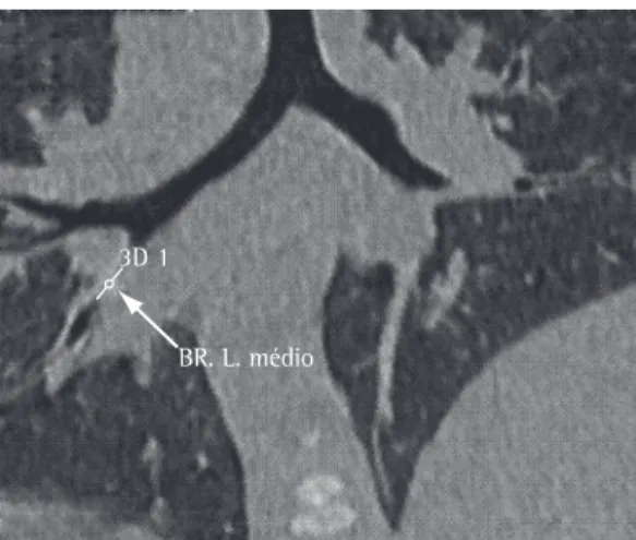
3D 1
BR. L. médio
Figure 1 - CT scan of the chest in a patient with suspected FBA. Organic foreign body in topography of middle lobe (arrow).
Chest X-ray evidenced right lung hyperinfla-tion, without consolidation or pleural effusion. The trachea and main bronchi were normal, bilaterally, in the VB. However, in the topography of the right main bronchus, next to the carina, there was evidence of the presence of an image with well-defined, irregular borders obliterating the bronchial lumen (Figure 3). The patient was not submitted to rigid bronchoscopy, since the guardian refused to give consent, despite having been informed of the risk of future respiratory complications.
Discussion
In children, principally those in the 6- to 24-month age bracket, FBA is a relevant cause of breathlessness. In patients suspected of having this condition, after the survey of the history and the performance of meticulous clin-ical examination, radiologclin-ical evaluation is the first step in the investigation. The most common radiographic findings are air trapping, atelectasis
and shadows suggestive of a radiopaque FB.(7,8)
However, those findings are nonspecific and have low accuracy.
One group of authors reported that chest X-ray has a sensitivity of 68% and a specificity
of 67% for the diagnosis of FBA.(3) In a study
with similar characteristics, an 85% sensitivity
and a 68% specificity were demonstrated.(2) In
other studies, the conclusion reached was that the absence of radiological alterations does
not exclude a diagnosis of FBA.(9,10) In order
to increase radiological accuracy, fluoroscopy imaging can be conducted in inhalation and
exhalation, in lateral decubitus.(8) However,
conventional bronchoscopy cannot be ruled out based exclusively on these findings, principally in patients in whom there is a high suspicion of FBA.
In the cases described in our study, it was possible to identify the presence of FB in the tracheobronchial tree of the patients using VB associated with MDCT. Images in the axial plane as well as those created through formatting allowed the diagnosis of FBA. Rigid bronchos-copy remains the gold standard for the diagnosis and for the removal of FB in the tracheobron-chial tree; however, it is an invasive procedure, which requires general anesthesia and can cause complications.(11,12)
to the medical visit. The child was previously healthy, with no history of asthma or fever.
Physical examination revealed that the patient was in good general health, he presented an
SpO2 of 96% on room air, mild subcostal
retrac-tion, tachypneic (respiratory rate = 46 breaths/ min) and pulmonary auscultation with diffuse inspiratory and expiratory wheezing.
Chest X-ray evidenced interstitial infiltrate in the right hilar region. The trachea and main bronchi were normal in the VB. However, complete obstruction of the lumen of the intermediate bronchus was observed (Figures 1 and 2). The subjacent lung parenchyma was normal. The patient was submitted to rigid bronchoscopy with the removal of an organic FB (grain of rice) and subsequent local bronchial hygiene, without complications.
Case 2
A 14-month-old female patient was referred to the emergency room after reportedly wheezing for 2 months following the ingestion of peanut offered by the mother. The patient had used an oral corticosteroid on three previous occasions, for seven days each time, in combination with a nebulized bronchodilator. Physical examination revealed that the patient was in good general
health, presented an SpO2 of 97% on room air,
Cushingoid appearance, normal respiration and pulmonary auscultation with wheezing located in the right hemithorax. The rest of the exami-nation was unremarkable.
FBA. The correct location of the FB, the possi-bility of visualizing the lung parenchyma and the possibility of conducting the examination without anesthesia make this method viable for this condition. This study demonstrated the use of this new method in two cases with different evolutions. In addition, the perform-ance of conventional bronchoscopy with general anesthesia can be avoided in cases when VB does not show the presence of FB in the airway.
References
1. Koşucu P, Ahmetoğlu A, Koramaz I, Orhan F, Ozdemir O, Dinç H, et al. Low-dose MDCT and virtual bronchoscopy in pediatric patients with foreign body aspiration. AJR Am J Roentgenol. 2004;183(6):1771-7.
2. Haliloglu M, Ciftci AO, Oto A, Gumus B, Tanyel FC, Senocak ME, et al. CT virtual bronchoscopy in the evaluation of children with suspected foreign body aspiration. Eur J Radiol. 2003;48(2):188-92.
3. Svedström E, Puhakka H, Kero P. How accurate is chest radiography in the diagnosis of tracheobronchial foreign bodies in children? Pediatr Radiol. 1989;19(8):520-2. 4. Sodhi KS, Saxena AK, Singh M, Rao KL, Khandelwal
N. CT virtual bronchoscopy: new non invasive tool in pediatric patients with foreign body aspiration. Indian J Pediatr. 2008;75(5):511-3.
5. Fraga Ade M, Reis MC, Zambon MP, Toro IC, Ribeiro JD, Baracat EC. Foreign body aspiration in children: clinical aspects, radiological aspects and bronchoscopic treatment. J Bras Pneumol. 2008;34(2):74-82. 6. Burke AJ, Vining DJ, McGuirt WF Jr, Postma G, Browne
JD. Evaluation of airway obstruction using virtual endoscopy. Laryngoscope. 2000;110(1):23-9.
7. Honnef D, Wildberger JE, Das M, Hohl C, Mahnken AH, Barker M, et al. Value of virtual tracheobronchoscopy and bronchography from 16-slice multidetector-row spiral computed tomography for assessment of suspected tracheobronchial stenosis in children. Eur Radiol. 2006;16(8):1684-91.
8. Adaletli I, Kurugoglu S, Ulus S, Ozer H, Elicevik M, Kantarci F, et al. Utilization of low-dose multidetector CT and virtual bronchoscopy in children with suspected foreign body aspiration. Pediatr Radiol. 2007;37(1):33-40. 9. Zerella JT, Dimler M, McGill LC, Pippus KJ. Foreign
body aspiration in children: value of radiography and complications of bronchoscopy. J Pediatr Surg. 1998;33(11):1651-4.
10. Hartnick CJ, Chung S, Emery KH, Myer CM 3rd. Imaging case study of the month. Pediatric virtual bronchoscopy. Ann Otol Rhinol Laryngol. 2002;111(3 Pt 1):281-3. 11. Konen E, Katz M, Rozenman J, Ben-Shlush A, Itzchak
Y, Szeinberg A. Virtual bronchoscopy in children: early clinical experience. AJR Am J Roentgenol. 1998;171(6):1699-702.
12. Ozkurt H, Bahadir E, Ucgul A, Altuna C, Basak M, Cevizci NM, et al. Comparison of multidetector computed tomography-virtual bronchoscopy and conventional bronchoscopy in children with suspected foreign body aspiration. Emerg Radiol. Epub 2008 Nov 19.
The modality of VB associated with MDCT is an imaging method capable of identifying the presence of FB and the associated parenchymal alterations. The greatest advantages over conven-tional tomography are in the increased speed in performing the examination and the possibility of thinner slices, with better spatial resolution
for the 3D reconstruction.(13,14) However, the
principal disadvantage of this procedure is the exposure to radiation, which can be minimized by avoiding the excessive use of tomography scans and reducing the tube current (in mA) to the minimum possible, and the need for the sedation of the patient. Typically, the minimal limit accepted for a good quality image is 30 mA and 80 kVp.
One group of authors investigated the use of VB in pediatric patients with suspected FBA and concluded that it can correctly identify the
posi-tion of the FB in the respiratory tree.(8) The authors
demonstrated that, when the VB result is normal, without evidence of endobronchial obstruction, the use of conventional bronchoscopy was not superior in providing relevant additional infor-mation. In another study, when images obtained by means of VB and conventional bronchoscopy in patients with suspected FBA were compared,
similar results were found.(2) In a recent study
comparing VB and conventional bronchoscopy, the estimated sensitivity and specificity of VB
were 96% and 58%, respectively.(12)
Another group of authors recently published a study comparing VB and rigid bronchoscopy in
patients with suspected FBA,(15) suggesting that
the use of this new technique can shorten the time to the performance of bronchoscopy and accurately locate the FB. Although VB does not remove the FB, it can provide the precise location of the latter and can be useful in the prepara-tion prior to the removal of the FB by means of rigid device. A possible disadvantage of the use of VB is the false-positive examination results. The presence of purulent endobronchial secre-tion or protrusion of some tumor mass towards
the bronchial lumen must be ruled out.(8)
In a recent review, the importance of the development of respiratory endoscopy emer-gency centers was highlighted in order to optimize the treatment of patients suspected of
having this condition.(16)
15. Cevizci N, Dokucu AI, Baskin D, Karadağ CA, Sever N, Yalçin M, et al. Virtual bronchoscopy as a dynamic modality in the diagnosis and treatment of suspected foreign body aspiration. Eur J Pediatr Surg. 2008;18(6):398-401.
16. de Lima AG, dos Santos NA, Rocha ER, Toro IF. Bronchoscopy for foreign body removal: where is the delay? J Bras Pneumol. 2008;34(11):956-8.
13. Sorantin E, Geiger B, Lindbichler F, Eber E, Schimpl G. CT-based virtual tracheobronchoscopy in children--comparison with axial CT and multiplanar reconstruction: preliminary results. Pediatr Radiol. 2002;32(1):8-15. 14. Heyer CM, Nuesslein TG, Jung D, Peters SA, Lemburg
SP, Rieger CH, et al. Tracheobronchial anomalies and stenoses: detection with low-dose multidetector CT with virtual tracheobronchoscopy--comparison with flexible tracheobronchoscopy. Radiology. 2007;242(2):542-9.
About the authors
Tiago Neves Veras
Pediatric Pulmonologist. Hospital Jeser Amarante Filho – HIJAF, Jeser Amarante Filho Hospital – Joinville, Brazil.
Gilberto Hornburg
Radiologist. Hospital Dona Helena – HDH, Dona Helena Hospital – Joinville, Brazil.
Adrian Maurício Stockler Schner
Thoracic Surgeon. Hospital Jeser Amarante Filho – HIJAF, Jeser Amarante Filho Hospital – Joinville, Brazil.
Leonardo Araújo Pinto