Current concepts in multiparametric magnetic resonance
imaging for active surveillance of prostate cancer
Alexandre Cavalcante,I,* Pu´blio Cesar C. Viana,IGiuliano B. Guglielmetti,IIJose´ Pontes Junior,I Henrique Nonemacher,IMauricio D. Cordeiro,IRegis Otaviano F. Bezerra,I Rafael F. Coelho,I William Carlos NahasII
IInstituto do Cancer do Estado de Sao Paulo (ICESP), Hospital das Clinicas HCFMUSP, Faculdade de Medicina, Universidade de Sao Paulo, Sao Paulo, SP, BR. IIGrupo de Uro-Oncologia, Departamento de Urologia, Faculdade de Medicina FMUSP, Universidade de Sao Paulo, Sao Paulo, SP, BR.
Cavalcante A, Viana PC, Guglielmetti GB, Pontes Junior J, Nonemacher H, Cordeiro MD, et al. Current concepts in multiparametric magnetic resonance imaging for active surveillance of prostate cancer. Clinics. 2018;73(suppl 1):e464s
*Corresponding author. Email: [email protected]
Active surveillance (AS) is a possible new approach for men with very low-risk and low-risk prostate cancer (PCa) and, less frequently, those with intermediate-risk PCa as a way to reduce overtreatment of clinically indolent disease (1-3). The very low likelihood of progression of this disease to unfavorable outcomes in appropriately selected men has demonstrated that AS can be used with good outcomes (4,5). High cancer-specific survival rates have been described, reaching 94.3% and 99.9% at the University of Toronto and Johns Hopkins Hospital, respectively (4,5), during 15 years of follow-up. Data from Sweden and the United States have shown cancer-specific survival rates of 60% and 64%, respectively, in patients who participated in AS after 5 years (6,7), and 55% cancer-specific survival has been reported in Canada after 15 years (4).
Despite good outcomes with AS, the poorly defined criteria for intervention and the lack of standardized surveillance protocols are important limitations. In all protocols for AS around the world, systematic transrectal ultrasound (TRUS)-guided biopsy is the tool used to establish a diagnosis of PCa (8), but the morbidity associated with this method is well known and can range from local symptoms, such as hematuria and acute urinary retention, to systemic symptoms, such as bacteremia and sepsis. Each new biopsy greatly increases the complication rates (9).
Another issues related to AS is the underestimation of the real extent of the disease and aggressiveness based only on standard biopsy (SB) data. Approximately thirty percent of patients with low-risk PCa on initial biopsy have a Gleason score upgrade when treated by radical prostatectomy; thus, SB is associated with substantial misclassification that may lead to inappropriate selection of patients for AS and postpone-ment of the necessary treatpostpone-ment (10). Therefore, distinction of patients who have low-risk cancers and those who need
immediate treatment is a serious challenge. In this scenario, multiparametric magnetic resonance imaging (mpMRI) has appeared as a promising imaging modality to help to better select and manage patient candidates for AS (11-13).
Currently, many studies confirmed that mpMRI is a use-ful instrument for detecting clinically significant PCa. The sensitivity is 86% for identifying tumors greater than 0.5 cm3 in size (14) and 80% for detecting index tumors (15). How-ever, the precise role of mpMRI in AS is not yet well established. Defining the role of mpMRI during the initial diagnosis and follow-up of patients undergoing AS is the next step.
mpMRI
In recent years, rapid technological advances have taken place. Thus, the acquisition of high quality images has advanced the current knowledge about the findings of PCa on mpMRI. Exam standardization and a high level of reader training have made mpMRI an important tool in PCa man-agement in daily practice. Usually, the exam is performed by a 3-Tesla scanner; however, a 1.5-Tesla magnet with an endorectal coil can be used to perform diffusion weighted imaging (DWI), T2-weighted imaging (T2WI) and dynamic contrast-enhanced MRI (DCE-MRI) (16).
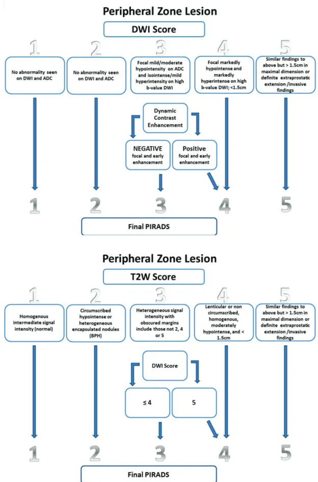
The famous Prostate Imaging Reporting and Data System version 2 (PI-RADS), which is frequently used in the descrip-tion of PCa, was developed to standardize MRI reporting. PI-RADS can be used to generate a map of the prostate to define the area of interest (17). Suspicious lesions are graded on a 5-point scale based on features suggestive of malig-nancy, and the probability of clinically significant PCa is related to a higher PI-RADS grade. The lesion with highest PI-RADS category if there is more than one lesion or the largest lesion when the lesions have same PI-RADS grade is considered the index lesion.
Certain characteristics of the parameters of T2WI, DWI and DCE-MRI aid in disease diagnosis. Currently, the use of DCE-MRI has low applicability. DCE-MRI is used only for lesions in in situ irradiated glands in which an enhancing nodule is strongly suggestive of PCa or to separate PI-RADS 3 and 4 lesions in the peripheral zone. Greer et al. demon-strated a high positive predictive value for PI-RADSX3 and X4 lesions (85% and 90%, respectively) to detect clinically
significant cancer with PI-RADS 2.0 (18) (Figure 1).
DOI:10.6061/clinics/2018/e464s
Copyright&2018CLINICS–This is an Open Access article distributed under the terms of the Creative Commons License (http://creativecommons.org/licenses/by/ 4.0/) which permits unrestricted use, distribution, and reproduction in any medium or format, provided the original work is properly cited.
No potential conflict of interest was reported.
Received for publication onDecember 22, 2017.Accepted for publi-cation onAugust 28, 2018
Candidate selection for AS and the role of mpMRI
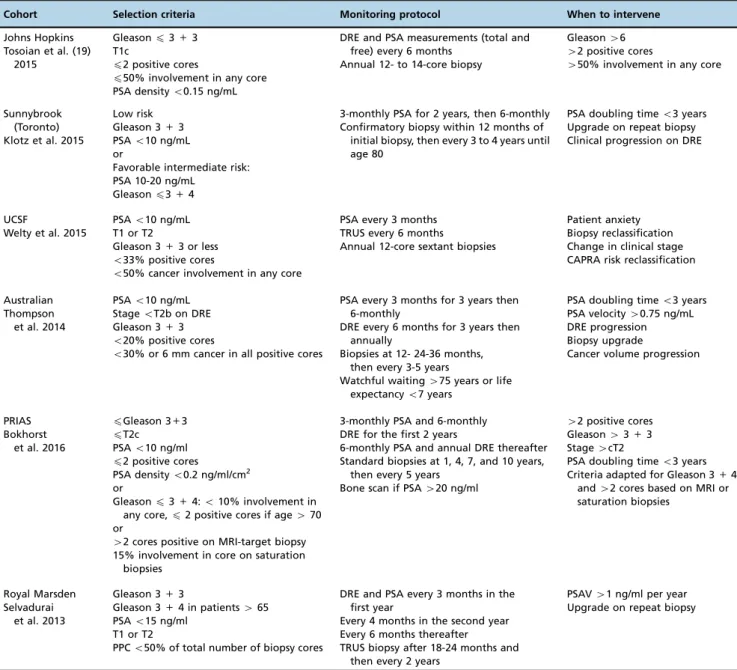
There are several AS protocols described in the literature with different inclusion criteria, and the lack of uniformity among these protocols poses the first challenge in clinical practice (Table 1). The most frequently used criteria to include patients in AS are Gleason score p3+3, prostate-specific
antigen (PSA)p10 ng/mL, clinical stagepT2, maximum of
2 positives cores or p50% per core. Two important
excep-tions are the groups from Memorial Sloan-Kettering Cancer Center who admitted patients with up to three involved cores and the group from Toronto who admitted patients with Gleason scores up to 7 (3+4) and with PSA levels up to 15 ng/mL. When the patients do not fulfill all these criteria, they cannot be included in AS because are deemed to have clinically significant disease. The problem with AS is that conventional prostate biopsy undersamples roughly one-third of patients when compared with prostatectomy specimens (20). mpMRI can yield important details about the disease
since this modality detects with higher accurately tumors with Gleason score higher than 6 and has a higher sensitivity than computed tomography for detecting extracapsular extension thus decreasing the undergrading and under-staging rates. However, in the majority of AS programs, mpMRI findings are not adopted as initial inclusion criteria. Currently, mpMRI has helped in the early detection and staging of PCa. Many studies have demonstrated that the incorporation of mpMRI findings during the initial diag-nosis of PCa has better accuracy than TRUS findings (21,22). If MRI shows a suspected lesion, these patients must undergo image-guided fusion biopsies. There are three possible modalities that can be used to perform this procedure: in-bore MR fusion, cognitive fusion, and finally, MR-US software fusion biopsy (16). Overall cancer detec-tion is significantly higher when MR-US software is used to generate the fusion image (48.1%) than when cognitive fusion alone is used (34.6%) (p=0.04) (23).
Table 1-Active surveillance protocols. Inclusion criteria, monitoring and intervention criteria.
Cohort Selection criteria Monitoring protocol When to intervene
Johns Hopkins Tosoian et al. (19)
2015
Gleasonp3+3 T1c
p2 positive cores
p50% involvement in any core PSA densityo0.15 ng/mL
DRE and PSA measurements (total and free) every 6 months
Annual 12- to 14-core biopsy
Gleason46 42 positive cores
450% involvement in any core
Sunnybrook (Toronto) Klotz et al. 2015
Low risk Gleason 3+3 PSAo10 ng/mL or
Favorable intermediate risk: PSA 10-20 ng/mL
Gleasonp3+4
3-monthly PSA for 2 years, then 6-monthly Confirmatory biopsy within 12 months of
initial biopsy, then every 3 to 4 years until age 80
PSA doubling timeo3 years Upgrade on repeat biopsy Clinical progression on DRE
UCSF
Welty et al. 2015
PSAo10 ng/mL T1 or T2
Gleason 3+3 or less
o33% positive cores
o50% cancer involvement in any core
PSA every 3 months TRUS every 6 months
Annual 12-core sextant biopsies
Patient anxiety Biopsy reclassification Change in clinical stage CAPRA risk reclassification
Australian Thompson et al. 2014
PSAo10 ng/mL StageoT2b on DRE Gleason 3+3
o20% positive cores
o30% or 6 mm cancer in all positive cores
PSA every 3 months for 3 years then 6-monthly
DRE every 6 months for 3 years then annually
Biopsies at 12- 24-36 months, then every 3-5 years
Watchful waiting475 years or life expectancyo7 years
PSA doubling timeo3 years PSA velocity40.75 ng/mL DRE progression Biopsy upgrade
Cancer volume progression
PRIAS Bokhorst
et al. 2016
pGleason 3+3
pT2c PSAo10 ng/ml
p2 positive cores
PSA densityo0.2 ng/ml/cm2
or
Gleasonp3+4:o10% involvement in
any core,p2 positive cores if age470 or
42 cores positive on MRI-target biopsy 15% involvement in core on saturation
biopsies
3-monthly PSA and 6-monthly DRE for the first 2 years
6-monthly PSA and annual DRE thereafter Standard biopsies at 1, 4, 7, and 10 years,
then every 5 years Bone scan if PSA420 ng/ml
42 positive cores Gleason43+3 Stage4cT2
PSA doubling timeo3 years Criteria adapted for Gleason 3+4
and42 cores based on MRI or
saturation biopsies
Royal Marsden Selvadurai
et al. 2013
Gleason 3+3
Gleason 3+4 in patients465 PSAo15 ng/ml
T1 or T2
PPCo50% of total number of biopsy cores
DRE and PSA every 3 months in the first year
Every 4 months in the second year Every 6 months thereafter
TRUS biopsy after 18-24 months and then every 2 years
PSAV41 ng/ml per year
Siddiqui et al. (24) recruited 1003 men with PCa who underwent SB and targeted biopsy. This prospective study showed that targeted biopsy had increased accuracy for high-risk cancer, diagnosing 30% more cases (173 vs. 122 cases, po0.001) and decreased accuracy for low-risk cancer,
diagn-osing 17 fewer cases (213vs.258 cases,po0.001). In a second
analysis evaluating targeted biopsy combined with SB, an increase in the diagnosis of low-risk PCa with an additional 103 cases (22%) was observed. Among 170 patients who underwent radical prostatectomy and were diagnosed with whole-gland disease after pathologic analysis, the preopera-tive targeted biopsy demonstrated better predicpreopera-tive ability to differentiate low-risk, intermediate or high-risk tumors than the two approaches used together or SB alone (0.73, 0.67 and 0.59, respectively,po0.05) (24).
Pessoa et al. (25) recently published a prospective cohort study involving 105 patients on AS who underwent mpMRI examinations. Reclassification rates among patients with PI-RADS 1, 2, 3, 4, and 5 were 0%, 23.1%, 9.1%, 74.5%, and 100%, respectively. The overall reclassification rate was 55.2%, and the most frequent criterion change was Gleason score classification (44.8%). Approximately 60% of the population was PI-RADS 4 and 5 on mpMRI, and in this group (PI-RADS grade 4 and 5), the rate of reclassification was 85.71%. The sensitivity, specificity, positive predictive value, and negative predictive value of mpMRI for disease reclassification were 92.5%, 76%, 81%, and 90.5%, respectively. These findings are comparable to what Lee et al. (26) found at the Cleveland Clinic on prostate specimens from patients who underwent radical prostatectomy for low-risk disease.
Other similar studies have been recently published. Da Rosa et al. (27) from the University of Toronto, in a prospective study, reported upgrading in 19 out of 72 patients on AS with a Gleason scoreX7. Seven of these upgraded patients were
detected only on MR-fusion-targeted biopsy, two were detected only on TRUS-guided systematic biopsy alone, and 10 were detected using both techniques. The negative predictive value for significant cancer approached 100% among men with no suspicious lesions identified using mpMRI. Current data from Memorial Sloan-Kettering (28) have been reported regarding the reclassification rate of 206 men with low-risk PCa enrolled in AS who underwent MR-fusion-targeted biopsy and systematic prostate biopsy. MRI identified suspicious lesions in 66% of patients, reinforcing that MRI findings have the potential to alter the management of low-risk PCa patients. In a retrospective study, Vargas et al. (13) found that 20% (79 of 388) of patients with clinically low-risk PCa had Gleason scores upgraded on confirmatory biopsy. These patients underwent MRI scans between the initial and confirmatory biopsies. MRI scoresp2 had a high negative predictive value
(0.96-1.0) for upgrading on confirmatory biopsy, while a score of 5 were highly sensitive for upgrading on confirma-tory biopsy (0.87-0.98).
In summary, the use of mpMRI as initial screening exam for PCa allows better stratification of the disease. The sen-sitivity and predictive negative value of mpMRI are high, both of which are important features for screening tests. MRI fusion biopsy of target lesions has higher predictive negative than TRUS-guided biopsy (29).
Role of mpMRI during follow-up of patients on AS
Once patients are selected to undergo AS, they are moni-tored with PSA exams, digital rectal exams (DREs) and
standard prostate biopsies. Sometimes patients are followed for many years and may require a considerable number of biopsies. However, prostate biopsy is an invasive technique and is associated with morbidity. The most common compli-cations are pain, hematuria, hematospermia, erectile dys-function, and infections. Thus, some recent studies have demonstrated that mpMRI is a promising alternative to decrease biopsies and thus prevent the complications related to this method.
The true role of mpMRI for monitoring patients on AS is not fully established; however, some studies suggest that serial MRI scans improve prediction of pathological progres-sion compared to clinicopathological variables alone, and stable MRI findings are associated with pathological stabil-ity (30-32). In the largest published retrospective study, Frye et al. (33) analyzed 166 men with PCa on AS in whom mpMRI-evident lesions were monitored, and fusion-guided biopsy was properly indicated. The mean follow-up was 25.5 months, and pathologic progression was observed in 29.5% of patients. Targeted fusion biopsy identified progres-sion in 44.9% of patients, and systematic biopsy detected progression in 30.6% (p=0.03). Progression was observed 26% more frequently with fusion biopsy than with systema-tic biopsy, and in this series, progression on mpMRI was shown to be the sole predictor of pathological progression in patients on AS (p=0.013). The analysis of pathologic progres-sion on mpMRI in a cohort study demonstrated a negative predictive value of 81%, a sensitivity of 77.6%, a positive predictive value of 35% and a specificity of 40.5%. These authors concluded that as imaging and technology evolve, the number or frequency of biopsies in men on AS may potentially be reduced (33).
In their cohort, Felker et al. (30), found that mpMRI had low sensitivity (37%) but high specificity (90%) for predicting pathologic progression. The main changes were maximum cancer core length (MCCL)X3 mm at baseline biopsy and
prostate-specific antigen density (PSAD) X0.15 ng/ml at
follow-up biopsy. Overall, 19 patients had pathologic pro-gression: 9 patients (47%) were identified on targeted biopsy, 7 (37%) on systematic biopsy, and 3 (16%) on systematic and targeted biopsy, highlighting the importance of targeted biopsies in patients on AS. In a similar study, Rosenkrantz et al. (32) analyzed and compared the changes in prostate index lesions identified on serial mpMRI with the follow-up biopsy results of patients on AS. A total of 55 patients were analyzed with median follow-up of 14 months; these authors observed that MRI had a high specificity (76% to 90%) for predicting positive follow-up biopsy results but a low sen-sitivity (23% to 35%).
In 206 men selected for AS, Recabal et al. (28) noted that 66% had regions of interest on MRI, and 35% were upgraded. They also found that when MRI-targeted biopsy was per-formed, the increase in detection of higher grade cancer was 23%; furthermore, the higher the PI-RADS score on MRI was, the greater the likelihood of finding a high-grade tumor (po0.0001).
Tran et al. (35) evaluated the utility of mpMRI fusion biopsy for predicting disease progression in patients on AS compared with systematic biopsy. They showed that 83 men (40%) experienced upgrading including 49 (24%) who under-went systematic sampling, 30 (14%) who underunder-went MRI-targeted core biopsies, and 4 (2%) who underwent both. Seven patients (9%) exhibited major upgrading with sys-tematic biopsy among those with negative results on MRI-US fusion biopsy.
Cost-effectiveness analysis of mpMRI during AS
An important point for deciding whether a method should be used routinely in the investigation and follow-up of a disease is whether this method has clinical applicability and feasibility from an economic point of view. However, this analysis becomes difficult since the cost of a specific exam and its particularities, such as hospitalization, treatment of complications and protocols for institutional execution of the exams, are extremely variable around the world. A few studies have attempted to analyze the cost-effectiveness of mpMRI in patients on AS and the results have varied. The methods and parameters used to evaluate cost-effectiveness in these studies were not uniform; thus, making comparisons of the results is not possible. The main points evaluated in the cost-effectiveness analysis studies were the estimated cost of the examination and the quality of life assessed by quality-adjusted life years (QALYs). QUALYs is a generic measure used in economic evaluation to assess the value for money of medical interventions; one QUALY is equal to to one year in perfect health and 0 QUALY indicates death.
European studies have had divergent results about the cost-effectiveness of MRI for AS (36-38). In these studies, the costs of MRI and TRUS were similar. Nicholson et al. (36) showed that the use of MRI to follow-up patients with PCa on AS was not cost-effective when compared with other strategies. Two other studies demonstrated divergent results, stating that MRI-based strategies were cost-effective (37,38). Mowatt et al. (38) suggested that the use of MRI in the patients studied was d7,685 below the threshold stipulated by QUALY (d20,000 vs d12,315) calculated in order to be considered cost-effective in the UK. In a study conducted in the Netherlands, de Rooij et al. (37) also showed the cost-effectiveness of strategies based on MRI.
In an Australian study, Gordon et al. (39) analyzed the survival, total number of biopsies, healthcare costs, insignif-icant and signifinsignif-icant cancer and QUALYs in patients in 3 different scenarios: patients diagnosed by TRUS without MRI and referred for radiotherapy, surgery or AS; patients who underwent TRUS guided by MRI and referred for radiotherapy, surgery or AS; and patients who underwent TRUS guided by MRI who all underwent AS. After separately analyzing the cost of the exam and QUALY, it was not possible to state that an MRI was cost-effective in patients with PCa in the current scenario of AS. However, it is believed that if MRI is able to include more patients on AS thus postponing radical treatment (surgery or radiotherapy), this tool can become cost-effective. A recent study entitled the ProstateMR Imaging Study (PROMIS) was conducted to analyze the cost of diagnosing clinically significant (CS) cancer by comparing MRI with TRUS findings in the UK (40). The results showed that MRI as the first examination detected more CS cancer per pound than TRUS (sensitivity =0.95 [95% confidence interval (CI) 0.92-0.98]vs0.91 [95% CI
0.86-0.94]) and was also cost-effective (ICER=d7,076 [h8350 / QALY gained]). Even though the PROMIS study did not aim to evaluate cost-effectiveness in AS patients, we can infer that this study has indirect applicability in the AS scenario, since diagnosing patients with CS cancer earlier can lead to exclusion or inclusion of these patients in AS programs.
Due to the wide divergence between studies in the anal-ysis of the cost of MRI compared to TRUS or the cost-effectiveness of these techniques used during AS, we cannot extrapolate the results of one study to a location other than where the study was conducted. In addition to cost variability among exams, hospitalizations and procedures worldwide, the lack of uniformity in the protocols used for AS make it difficult to standardize cost-effectiveness studies. Thus, at this time, it is not possible to make final statements about the cost-effectiveness of MRI in the AS scenario. Individualized studies for the given reality in which the use of mpMRI is desired to be evaluated seem to be the best option for a more reliable analysis of cost-effectiveness.
Current data show that mpMRI is an adjunctive power-ful tool that can assist in the selection of patients for AS, increasing the diagnostic accuracy of prostate biopsy. The integration of mpMRI data and the combination of targeted biopsy and systematic biopsy improve detection of higher grade disease thus decreasing incorrect patient selection for AS. Therefore, we believe that MRI findings should be used as additional criteria for inclusion of patients in future pro-tocols for AS. The role of MRI in the follow-up of patients on AS is not yet well established. Recent data support that MRI should not be used as an autonomous tool to follow-up and trigger biopsies because upgrading also occur in areas outside the targeted biopsy, but improved definition and standardization of radiological progression for patients on AS is expected.
’ REFERENCES
1. Heidenreich A, Bastian PJ, Bellmunt J, Bolla M, Joniau S, van der Kwast T, et al. European Association of Urology. EAU guidelines on prostate can-cer. part 1: screening, diagnosis, and local treatment with curative intent-update 2013. Eur Urol. 2014;65(1):124-37, http://dx.doi.org/10.1016/ j.eururo.2013.09.046.
2. Mohler JL, Armstrong AJ, Bahnson RR, D’Amico AV, Davis BJ, Eastham JA, et al. Prostate Cancer, Version 1.2016. J Natl Compr Canc Netw. 2016;14(1):19-30, http://dx.doi.org/10.6004/jnccn.2016.0004.
3. Klotz L, Zhang L, Lam A, Nam R, Mamedov A, Loblaw A. Clinical results of long-term follow-up of a large, active surveillance cohort with localized prostate cancer. J Clin Oncol. 2010;28(1):126-31, http://dx.doi.org/ 10.1200/JCO.2009.24.2180.
4. Klotz L, Vesprini D, Sethukavalan P, Jethava V, Zhang L, Jain S, et al. Long-term follow-up of a large active surveillance cohort of patients with prostate cancer. J Clin Oncol. 2015;33(3):272-7, http://dx.doi.org/10.1200/ JCO.2014.55.1192.
5. Tosoian JJ, Mamawala M, Epstein JI, Landis P, Wolf S, Trock BJ, et al. Intermediate and Longer-Term Outcomes From a Prospective Active-Surveillance Program for Favorable-Risk Prostate Cancer. J Clin Oncol. 2015;33(30):3379-85, http://dx.doi.org/10.1200/JCO.2015.62.5764. 6. Loeb S, Folkvaljon Y, Makarov DV, Bratt O, Bill-Axelson A, Stattin P.
Five-year nationwide follow-up study of active surveillance for prostate cancer. Eur Urol. 2015;67(2):233-8, http://dx.doi.org/10.1016/j.eururo.2014.06.010. 7. Welty CJ, Cowan JE, Nguyen H, Shinohara K, Perez N, Greene KL, et al. Extended followup and risk factors for disease reclassification in a large active surveillance cohort for localized prostate cancer. J Urol. 2015;193(3): 807-11, http://dx.doi.org/10.1016/j.juro.2014.09.094.
8. Dall’Era MA, Albertsen PC, Bangma C, Carroll PR, Carter HB, Cooper-berg MR, et al. Active surveillance for prostate cancer: a systematic review of the literature. Eur Urol. 2012;62(6):976-83, http://dx.doi.org/10.1016/ j.eururo.2012.05.072.
10. Lellig E, Gratzke C, Kretschmer A, Stief C. Final pathohistology after radical prostatectomy in patients eligible for active surveillance (AS). World J Urol. 2015;33(7):917-22, http://dx.doi.org/10.1007/s00345-015-1604-6. 11. Hamoen EH, de Rooij M, Witjes JA, Barentsz JO, Rovers MM. Use of
the Prostate Imaging Reporting and Data System (PI-RADS) for pro-state cancer detection with multiparametric magnetic resonance imaging: a diagnostic meta-analysis. Eur Urol. 2015;67(6):1112-21, http://dx.doi. org/10.1016/j.eururo.2014.10.033.
12. Vargas HA, Akin O, Franiel T, Mazaheri Y, Zheng J, Moskowitz C, et al. Diffusion-weighted endorectal MR imaging at 3 T for prostate cancer: tumor detection and assessment of aggressiveness. Radiology. 2011;259(3): 775-84, http://dx.doi.org/10.1148/radiol.11102066.
13. Vargas HA, Akin O, Afaq A, Goldman D, Zheng J, Moskowitz CS, et al. Magnetic resonance imaging for predicting prostate biopsy findings in patients considered for active surveillance of clinically low risk prostate cancer. J Urol. 2012;188(5):1732-8, http://dx.doi.org/10.1016/j.juro.2012. 07.024.
14. Puech P, Potiron E, Lemaitre L, Leroy X, Haber GP, Crouzet S, et al. Dynamic contrast-enhanced-magnetic resonance imaging evaluation of intraprostatic prostate cancer: correlation with radical prostatectomy specimens. Urology. 2009;74(5):1094-9, http://dx.doi.org/10.1016/j.urology. 2009.04.102.
15. Le JD, Tan N, Shkolyar E, Lu DY, Kwan L, Marks LS, et al. Multifocality and prostate cancer detection by multiparametric magnetic resonance imaging: correlation with whole-mount histopathology. Eur Urol. 2015; 67(3):569-76, http://dx.doi.org/10.1016/j.eururo.2014.08.079.
16. Nassiri N, Natarajan S, Margolis DJ, Marks LS. Targeted Prostate Biopsy: Lessons Learned Midst the Evolution of a Disruptive Technology. Urology. 2015;86(3):432-8, http://dx.doi.org/10.1016/j.urology.2015.07.001. 17. Weinreb JC, Barentsz JO, Choyke PL, Cornud F, Haider MA, Macura KJ,
et al. PI-RADS Prostate Imaging Reporting and Data System: 2015, Version 2. Eur Urol. 2016;69(1):16-40, http://dx.doi.org/10.1016/j.eururo. 2015.08.052.
18. Greer MD, Brown AM, Shih JH, Summers RM, Marko J, Law YM, et al. Accuracy and agreement of PIRADSv2 for prostate cancer mpMRI: A multireader study. J Magn Reson Imaging. 2017;45(2):579-85, http://dx. doi.org/10.1002/jmri.25372.
19. Thompson JE, Hayen A, Landau A, Haynes AM, Kalapara A, Ischia J, et al. Medium-term oncological outcomes for extended vs saturation biopsy and transrectal vs transperineal biopsy in active surveillance for prostate cancer. BJU Int. 2015;115(6):884-91, http://dx.doi.org/10.1111/ bju.12858.
20. Conti SL, Dall’era M, Fradet V, Cowan JE, Simko J, Carroll PR. Patholo-gical outcomes of candidates for active surveillance of prostate cancer. J Urol. 2009;181(4):1628-33, http://dx.doi.org/10.1016/j.juro.2008.11.107. 21. Meng X, Rosenkrantz AB, Mendhiratta N, Fenstermaker M, Huang R, Wysock JS, et al. Relationship Between Prebiopsy Multiparametric Mag-netic Resonance Imaging (MRI), Biopsy Indication, and MRI-ultrasound Fusion-targeted Prostate Biopsy Outcomes. Eur Urol. 2016;69(3):512-7, http://dx.doi.org/10.1016/j.eururo.2015.06.005.
22. NiMhurchu E, O’Kelly F, Murphy IG, Lavelle LP, Collins CD, Lennon G, et al. Predictive value of PI-RADS classification in MRI-directed trans-rectal ultrasound guided prostate biopsy. Clin Radiol. 2016;71(4):375-80, http://dx.doi.org/10.1016/j.crad.2016.01.001.
23. Oberlin DT, Casalino DD, Miller FH, Matulewicz RS, Perry KT, Nadler RB, et al. Diagnostic value of guided biopsies: fusion and cognitive-registration magnetic resonance imaging versus conventional ultrasound biopsy of the prostate. Urology. 2016;92:75-9, http://dx.doi.org/10.1016/ j.urology.2016.02.041.
24. Siddiqui MM, Rais-Bahrami S, Turkbey B, George AK, Rothwax J, Shakir N, et al. Comparison of MR/ultrasound fusion-guided biopsy with ultrasound-guided biopsy for the diagnosis of prostate cancer. JAMA. 2015;313(4):390-7, http://dx.doi.org/10.1001/jama.2014.17942. 25. Pessoa RR, Viana PC, Mattedi RL, Guglielmetti GB, Cordeiro MD, Coelho
RF, et al. Value of 3-Tesla multiparametric magnetic resonance imaging and targeted biopsy for improved risk stratification in patients considered for active surveillance. BJU Int. 2017;119(4):535-42, http://dx.doi.org/ 10.1111/bju.13624.
26. Lee MC, Dong F, Stephenson AJ, Jones JS, Magi-Galluzzi C, Klein EA. The Epstein criteria predict for organ-confined but not insignificant disease
and a high likelihood of cure at radical prostatectomy. Eur Urol. 2010; 58(1):90-5, http://dx.doi.org/10.1016/j.eururo.2009.10.025.
27. Da Rosa MR, Milot L, Sugar L, Vesprini D, Chung H, Loblaw A, et al. A prospective comparison of MRI-US fused targeted biopsy versus sys-tematic ultrasound-guided biopsy for detecting clinically significant pro-state cancer in patients on active surveillance. J Magn Reson Imaging. 2015;41(1):220-5, http://dx.doi.org/10.1002/jmri.24710.
28. Recabal P, Assel M, Sjoberg DD, Lee D, Laudone VP, Touijer K, et al. The efficacy of multiparametric magnetic resonance imaging and magnetic resonance imaging targeted biopsy in risk classification for patients with prostate cancer on active surveillance. J Urol. 2016;196(2):374-81, http:// dx.doi.org/10.1016/j.juro.2016.02.084.
29. Pokorny MR, de Rooij M, Duncan E, Schröder FH, Parkinson R, Barentsz JO, et al. Prospective study of diagnostic accuracy comparing prostate cancer detection by transrectal ultrasound-guided biopsy versus magnetic resonance (MR) imaging with subsequent MR-guided biopsy in men without previous prostate biopsies. Eur Urol. 2014;66(1):22-9, http://dx. doi.org/10.1016/j.eururo.2014.03.002.
30. Felker ER, Wu J, Natarajan S, Margolis DJ, Raman SS, Huang J, et al. Serial magnetic resonance imaging in active surveillance of prostate cancer: incremental value. J Urol. 2016;195(5):1421-7, http://dx.doi.org/10.1016/ j.juro.2015.11.055.
31. Walton Diaz A, Shakir NA, George AK, Rais-Bahrami S, Turkbey B, Rothwax JT, et al. Use of serial multiparametric magnetic resonance imag-ing in the management of patients with prostate cancer on active surveil-lance. Urol Oncol. 2015;33(5):202.e1-202.e7, http://dx.doi.org/10.1016/ j.urolonc.2015.01.023.
32. Rosenkrantz AB, Prabhu V, Sigmund EE, Babb JS, Deng FM, Taneja SS. Utility of diffusional kurtosis imaging as a marker of adverse pathologic outcomes among prostate cancer active surveillance candidates under-going radical prostatectomy. AJR Am J Roentgenol. 2013;201(4):840-6, http://dx.doi.org/10.2214/AJR.12.10397.
33. Frye TP, George AK, Kilchevsky A, Maruf M, Siddiqui MM, Kongnyuy M, et al. Magnetic resonance imaging-transrectal ultrasound guided fusion biopsy to detect progression in patients with existing lesions on active surveillance for low and intermediate risk prostate cancer. J Urol. 2017; 197(3 Pt 1):640-6, http://dx.doi.org/10.1016/j.juro.2016.08.109. 34. Morgan VA, Riches SF, Thomas K, Vanas N, Parker C, Giles S, et al.
Diffusion-weighted magnetic resonance imaging for monitoring prostate cancer progression in patients managed by active surveillance. Br J Radiol. 2011;84(997):31-7, http://dx.doi.org/10.1259/bjr/14556365. 35. Tran GN, Leapman MS, Nguyen HG, Cowan JE, Shinohara K, Westphalen
AC, et al. Magnetic resonance imaging-ultrasound fusion biopsy during prostate cancer active surveillance. Eur Urol. 2017;72(2):275-81, http://dx. doi.org/10.1016/j.eururo.2016.08.023.
36. Nicholson A, Mahon J, Boland A, Beale S, Dwan K, Fleeman N, et al. The clinical effectiveness and cost-effectiveness of the PROGENSAs
prostate cancer antigen 3 assay and the Prostate Health Index in the diagnosis of prostate cancer: a systematic review and economic evaluation. Health Technol Assess. 2015;19(87):1-191, http://dx.doi.org/10.3310/hta19870. 37. de Rooij M, Crienen S, Witjes JA, Barentsz JO, Rovers MM, Grutters JP.
Cost-effectiveness of magnetic resonance (MR) imaging and MR-guided targeted biopsy versus systematic transrectal ultrasound-guided biopsy in diagnosing prostate cancer: a modelling study from a health care perspective. Eur Urol. 2014;66(3):430-6, http://dx.doi.org/10.1016/j.eururo.2013.12.012. 38. Mowatt G, Scotland G, Boachie C, Cruickshank M, Ford JA, Fraser C,
et al. The diagnostic accuracy and cost-effectiveness of magnetic resonance spectroscopy and enhanced magnetic resonance imaging techniques in aiding the localisation of prostate abnormalities for biopsy: a systematic review and economic evaluation. Health Technol Assess. 2013;17(20): 1-281, http://dx.doi.org/10.3310/hta17200.
39. Gordon LG, James R, Tuffaha HW, Lowe A, Yaxley J. Cost-effectiveness analysis of multiparametric MRI with increased active surveillance for low-risk prostate cancer in Australia. J Magn Reson Imaging. 2017;45(5): 1304-1315, http://dx.doi.org/http://dx.doi.org/10.1002/jmri.25504. 40. Faria R, Soares MO, Spackman E, Ahmed HU, Brown LC, Kaplan R, et al.