Full paper published online: August 31, 2009 ISSN 1678-9199.
BIOACTIVITIES OF EXTRACTS FROM THE MARINE SPONGE Halichondria
panicea
Purushottama GB (1), Venkateshvaran K (1), Pani Prasad K (2), Nalini P (2)
(1) Laboratory of Aquatic Biotoxinology, Central Institute of Fisheries Education,
Mumbai, India; (2) Department of Aquatic Animal Health Management, Central
Institute of Fisheries Education, Mumbai, India.
ABSTRACT: In the present study, we screened the biological activity of extracts from the marine sponge Halichondria panicea collected in the Arabian Sea. Crude toxin was obtained by methanol, chloroform-methanol (2:1) and aqueous extraction. Subsequently, the protein concentration of each crude extract was determined. The impact of both sponge methanolic and aqueous extracts was found to increase activities of Na+-K+ ATP-ase and Mg++ ATP-ase. In the case of chloroform-methanol extract, higher concentrations increased acetylcholine esterase (AchE) activity. The methanolic and chloroform-methanol extracts exhibited hemolytic activity on chicken and human erythrocytes, whereas the aqueous extract failed to do so. Methanol and aqueous extracts produced an immunostimulating effect and all extracts revealed angiogenic activity. The aqueous extract yielded nine bands by SDS-PAGE on 12% gel.
KEY WORDS: Halichondria panicea, phagocytosis, neuromodulation, hemolysis,
angiogenesis.
CONFLICTS OF INTEREST: There is no conflict.
CORRESPONDENCE TO:
K. VENKATESHVARAN, Aquatic Biotoxinology Laboratory, Central Institute of
Fisheries Education, Fisheries University Road, Seven Bungalows, Andheri (W),
Versova, Mumbai, 400061, India. Phone: +91 22 2636 1446 / 7. Fax: +91 22 2636
INTRODUCTION
Sponges are simple multicellular sessile animals with no true tissue layers or organs
(1). They populate tropical and subtropical benthic marine habitats but are also found
at higher latitudes and even in freshwater lakes and streams (2). As sessile filter
feeders, they pump large volumes of water through a specialized canal system,
known as its aquiferous system (1). Marine sponges are a rich source of structurally
novel and biologically active metabolites (3). Over 60% of potentially useful bioactive
compounds discovered so far from living organisms have been obtained from marine
fauna, 70% of which comes from sponges (4). There are approximately 15,000
different species of sponges throughout the globe, 150 of which occur in freshwater,
but only about 17 present commercial value for traditional use, including the cosmetic
industry. A total of 486 sponge species have been found in Indian waters (5).
The sponge class Demospongiae is known for producing the largest number and
diversity of secondary metabolites isolated from marine invertebrates (6). There is a
worldwide interest in marine natural products as one of the few de novo sources of
drug discovery (7). However, the bioactive potential of compounds from Indian
sponges has been little studied. Therefore, the present study comprises an initial
effort to assess the bioactivity of secondary metabolites from the marine subtidal
sponge Halichondria panicea (Demospongiae class, Halichondrida order,
Halichondriidae famaily).
MATERIALS AND METHODS
Sponge Collection
The sponge Halichondria panicea was collected from subtidal areas of the Arabian
Sea at Mumbai, India (latitude 18.55 N and longitude 72.54 E). Animals were taken
alive to the laboratory in seawater and immediately frozen at –20°C until use.
Preparation of Sponge Extracts
Crude toxin was extracted following the method of Braekman et al. (8) with some
modifications. To 10 g of dried sponge sample, 200 mL of methanol and
chloroform-methanol (2:1) as well as deionized water were added and kept standing for five
hours. Solvents were then removed, by squeezing sponge samples, and filtered
through Whatman filter paper number 1. The remaining solvent was evaporated at
compound was finally dried in a vacuum desiccator and stored at 4°C in a refrigerator
for further use as crude-methanolic or chloroform-methanolic extracts. The aqueous
extract of sponges was prepared by squeezing sand-free specimens in triple distilled
water. The resultant solution was filtered and dialyzed by using Sigma (USA) dialysis
membrane-500 (average flat width: 24.26 mm; average diameter: 14.3 mm;
approximate capacity: 1.61 mL/cm) against D-glucose to remove excessive water.
Then, the supernatant obtained was lyophilized (FreeZone® Freeze Dry Systems,
Labconco, USA) and stored at 4°C in a refrigerator for subsequent use as crude
aqueous extract.
Partial Purification
Crude extracts were fractionated using a locally made DEAE-cellulose column (29 x
2.3 cm). Ten adsorbed fractions (designated F1 to F 10), 15 mL each, were
submitted to step-gradient elution of 0.1 to 1.0 M NaCl in phosphate buffered saline
(PBS) solution, pH 7.4. Moreover, five 15-mL unabsorbed fractions were also eluted
with phosphate buffer, pH 7.4. They were all stored at 4°C for later use.
Protein Estimation
Protein content from crude extracts and fractions was estimated (9).
Neuromodulatory Activity
P2 fraction (mitochondrial nerve endings) from male mouse (20 ± 2 g) brain was
prepared (10). The protein content of the enzyme source was estimated as
previously described.
ATP-ase assays for Na+-K+ ATP-ase activity and Mg++ ATP-ase activity were
conducted following the inorganic phosphate method (11). Control experiments were
also run simultaneously with 100 μL of triple distilled water instead of
extracts/fractions. Enzyme activity was expressed as micromoles of inorganic
phosphate per miligram per hour.
AChE inhibitory activity was calorimetrically measured (12). Briefly, to 3 mL of 0.1 M
phosphate buffer, pH 8.0, contained in each tube, 0.1 mL of enzyme source (2% w/v
homogenate) was added and stirred. Then, 100 μL of 0.01 DNTB (0 5-5-dithiobis-2
measured at 412 nm. The reaction was started by adding 20 μL of acetylthiocholine
iodide (ATCI) (0.075 M) as substrate to each tube; then, the reaction was allowed to
continue for 15 minutes at room temperature. The developed color was
spectrophotometrically measured at 412 nm and the inhibitory activity was
calculated.
Hemolytic Activity
The hemolysis of crude toxin on chicken and human red blood cells (RBC) was
tested by a micro-hemolytic method (13). Chicken and human blood were obtained,
respectively, from a nearby slaughterhouse and Sheena Clinic, Andheri, Mumbai,
India using EDTA solution (2.7 g in 100 mL of distilled water) as an anticoagulant at
5% of the blood volume and brought to the laboratory. A 1% erythrocyte suspension
was prepared by adding 99 mL of PBS, pH 7.4, to 1 mL of packed RBC (14). The
micro-hemolytic test was performed on 96-well U-bottom microtiter plates. Serial
two-fold dilutions of the extracts were carried out in 100 μL of PBS, pH 7.4. In controls, to
3% RBC suspension, 100 μL of distilled water was added, which served as positive
control and an equal amount of PBS, pH 7.4, was negative control. The plate was
gently shaken and allowed to stand for two hours at room temperature; then the
results were recorded. Uniform red color suspension in the wells was considered
positive hemolysis while sedimentation on the bottom was considered lack of
hemolysis.
Phagocytosis
Immunomodulatory activity was analyzed by a slide method through in vitro
phagocytosis of Candida albicans by polymorphonuclear cells (PMN) (15).
Chorioallantoic Membrane (CAM) Assay
Nine-day-old fertilized chicken eggs were selected and small holes of 1.0 cm2 were
made in their shells. Next, air was sucked out from the eggs to bring their
membranes down (16). Through each opening, a sterile disc of methylcellulose
containing concentration of 40 μg and 80 μg/mL of crude extracts was placed inside
the egg at the junction of two blood vessels. Subsequently, the opening was resealed
holes were then reopened and vessel formation was observed in terms of number
and caliber, and finally compared with eggs containing discs without any lyophilized
crude extract. Positive controls were also maintained in the same conditions.
SDS-PAGE
Crude extracts were submitted to electrophoresis in 12% polyacrylamide slab gels to
analyze their protein profile (17).
RESULTS
Preparation of Sponge Extract
From an initial 10-g sample of dried sponge tissue, methanolic and
chloroform-methanol (2:1) extraction, yielded, respectively 1.970 and 0.520 g of crude toxin,
while aqueous extraction yielded 3.0 g.
Protein Estimation
The crude protein contents were found to be 0.096 and 0.192 mg/mL, respectively, in
methanolic and chloroform-methanol extracts, and 0.124 mg/mL in aqueous extract.
It ranged from below detectable levels to 0.014 mg/mL in methanolic extract fractions
and from below detectable levels to 0.002 mg/mL in aqueous extract.
Neuromodulatory Activity
Na+-K+ ATP-ase activity was inhibited by methanolic (4.07 to 354.24%),
chloroform-methanol (319.79 to 242.61%) and aqueous (271.59 to 272.06%) extracts. However,
in all three cases, the activity was enhanced at doses from 250 to 1,000 μg.
The methanolic extract elevated Mg++ ATP-ase activity by 285.0% at 250 μg/mL
concentration, but inhibited it by up to 135.62% at 750 μg/mL and, finally, at 1,000
μg/mL concentration augmented the activity again. The chloroform-methanol extract
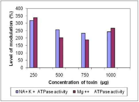
elevated Mg++ ATP-ase activity by 338.64% at a lower concentration of 250 μg/mL;
however, it inhibited the activity by up to 188.70% at 750 μg/ mL, and at 1,000 μg/ mL
increased the activity. The aqueous extract elevated Mg++ ATP-ase activity up to
355.87 and 355.64%, respectively, at concentrations of 250 and 500 μg/mL, but
inhibited the activity, up to 307.45%, at higher concentrations (Figures 1 to 3).
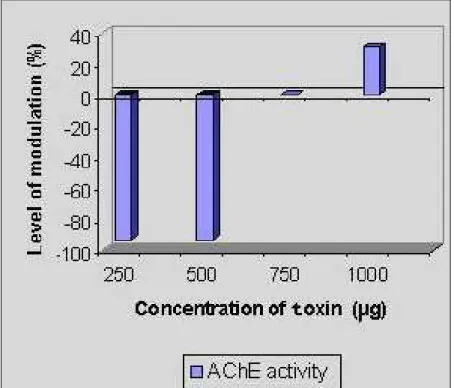
Methanolic extract inhibited AChE activity up to –93.10% at 250 μg and 500 μg/mL,
similar trend was found in the chloroform-methanol extract, in which inhibition was
observed at the lower concentration of 250 μg/mL (–13.79%), while increase was
registered at higher concentrations of 500, 750 and 1,000 μg/mL (respectively, 17.24,
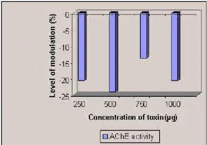
27.58 and 106.89%). In the aqueous extract, the inhibitory effect of –20.68% was
observed from lower to higher concentrations (Figures 4 to 6).
Figure 1. Modulation of Na+-K+ ATP-ase and Mg++ ATP-ase activity by the
methanolic extract of Halichondria panicea.
Figure 2. Modulation of Na+-K+ ATP-ase and Mg++ ATP-ase activity by the
Figure 3. Modulation of Na+-K+ ATP-ase and Mg++ ATP-ase activity by the aqueous
extract of Halichondria panicea.
Figure 4. Modulation of AChE activity by the methanolic extract of Halichondria
Figure 5. Modulation of AChE activity by the chloroform-methanol (2:1) extract of
Halichondria panicea.
Figure 6. Modulation of AChE activity by the aqueous extract of Halichondria
panicea.
Hemolytic Assay
In chicken blood, crude methanolic and chloroform-methanol extracts induced
pronounced hemolysis with specific hemolytic activity of 333.33 in each case. They
also lysed human blood, with respective specific hemolytic activities of 20.83 and
10.41. The aqueous extract failed to elicit hemolysis in either chicken or human
F6 fraction of the methanolic extract induced partial hemolysis in both chicken and
human blood (specific hemolytic activity 571.42). All other fractions failed to induce
hemolysis in chicken or human blood (Tables 1 and 2).
Table 1. Hemolytic activity of Halichondria panicea crude toxin at 5 mg/mL in chicken
and human erythrocytes
Blood
Chicken Human
Serial n.
Type of extract
Protein
(mg) Total
hemolysis (up to dilutions) Hemolytic titer Specific hemolytic activity (HT/mg) Total hemolysis (up to dilutions) Hemolytic titer Specific hemolytic activity (HT/mg)
1 Methanol 0.096 5 32 333.33 1 2 20.83
2
Chloroform- methanol
(2:1)
0.192 6 64 333.33 1 2 10.41
3 Aqueous 0.124 0 0 0 0 0 0
Table 2. Hemolytic activity of methanolic and aqueous fractions of Halichondria
panicea on chicken erythrocytes
Chicken
Methanolic Aqueous
Serial n. Type of solution Protein (mg) Total hemolysis (up to dilution) Hemolytic titer Specific hemolytic activity (HT/mg) Protein (mg) Total hemolysis (up to dilution) Hemolytic titer Specific hemolytic activity (HT/mg)
1 0.2 M ND ND ND ND ND ND ND ND
2 0.4 M ND ND ND ND ND ND ND ND
3 0.6 M 0.014 3 8 571.42 0.002 ND ND ND
4 0.8 M 0.006 ND ND ND ND ND ND ND
5 1.0 M 0.004 ND ND ND ND ND ND ND
Phagocytosis
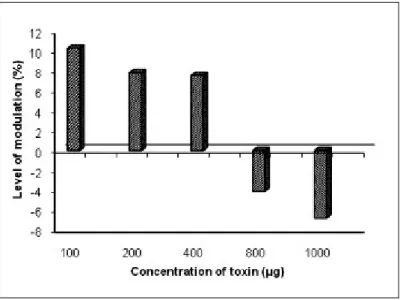
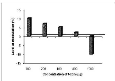
The methanol extract of Halichondria panicea exerted an immunostimulating effect of
7.57 to 10.30% magnitude at lower concentrations and a suppressive effect of 6.81%
at higher concentrations. The chloroform-methanol extract had a stimulative activity
of up to 12.27% at a lower concentration (100 μg/mL), but exerted a suppressive
activity from 1.91 to 8.24% at higher concentrations. The aqueous extract exhibited
immunostimulation ranging from 1.99 to 10.05% at concentrations up to 800 μg, but
showed immunosuppressive effects at 1,000 μg concentration (Figures 7 to 9).
Figure 7. Immunomodulation produced by the methanolic extract of Halichondria
panicea.
Figure 8. Immunomodulation produced by the chloroform-methanol (2:1) extract of
Figure 9. Immunomodulation produced by the aqueous extract of Halichondria
panicea.
Chorioallantoic Membrane (CAM) Assay
Results of the chorioallantoic membrane (CAM) assay are presented in Table 3. All
the extracts from Halichondria paincea showed angiogenic activity including increase
in thickness of blood vessels compared to the control group at 40 and 80 μg doses.
Table 3. Angiogenesis in 9-day-old chicken egg after treatment with methanolic,
chloroform-methanol (2:1) and aqueous extracts
Toxin concentration Extract type
40 µg/mL 80 µg/mL
1 Aqueous + +
2 Methanol ++ +++
3 Chloroform-methanol (2:1) ++ ++
+: slight; ++: moderate; +++: severe.
SDS-PAGE
By SDS-PAGE on 12% gel, crude toxins yielded nine bands in Halichondria panicea
aqueous extract, ranging from 14.3 to 116 kDa with three well-defined bands at 19.5,
Figure 10. SDS-PAGE of crude protein extracts of Halichondria panacea.
DISCUSSION
The amount of crude toxin extracted by lyophilization in the current study was higher
compared to those reported in earlier findings. Kerr et al. (18) obtained 1.67 g of
crude extract from 200 g of Epipolasis sp. and Kijjoa et al. (19) obtained 5.8 g of
crude extract from 490 g of fresh marine sponge Suberea praetensa.
The crude protein content of the various studied extracts could not be compared with
previous studies because similar data on protein contents of sponge toxins are not
available in the literature.
The present results on AChE activity of Halichondria panicea extracts correspond to
earlier findings of Elancheran (20), who reported elevated AChE activity due to
higher doses of tetrodotoxin. Wankhede (21) observed similar neuroinhibitory activity
by bile extracts of freshwater carps on the Na+-K+ ATP-ase enzyme system in
mammalian models The ATP-ase enzyme system is widely accepted as a structure
that employs part of the free energy from ATP hydrolysis for active transport of Na+
-K+. Lehinnger (22) found that metabolic stimulation provoked by ionic movement at
the membrane level results from a series of reactions that lead to accumulation of
ADP and Pi (inorganic phosphate), which play an important role in regulating
respiration. Augmented ADP concentration intensifies mitochondrial respiration that,
Schmitz et al. (23) reported the hemolytic activity of halitoxin from sponges of the
genus Haliclona. Similarly, Mebs et al. (24) observed hemagglutination and hemolytic
activity of aqueous extracts from 48 tropical sponge species. Malovrh et al. (25)
described cytotoxic and cytolytic activities of polymeric alkylpyridinium salts
(poly-APS) from the marine sponge Reniera sarai, which they inferred were due to their
detergent-like structure and behavior in aqueous solutions.
Indap and Pathare (26) reported hemolytic activity as HD50 at < 2 mg/mL in the
chloroform extract from Tethya sp. Moreover, Fusetani et al. (27) and Makarieva et
al. (28) reported that sterol derivatives from halichondriid sponges, namely halistanol
sulfates and sokotrasterol sulfates, possessed hemolytic activity. Abas et al. (4)
screened marine sponges from Malaysian waters and reported cytotoxic activity as
well as an effect on liver-metabolizing enzymes. Sepčićet al. (3) described significant
levels of hemolytic and hemagglutinating activities of Reniera sarai extracts
compared to the moderate hemolytic activity of extracts from Saracotragus
muscarum and Aplysina aerophoba.
Thus, Halichondria panicea appears to be a candidate for further studies on
analgesic or antineoplastic activities of its secondary metabolites in terms.
The present study revealed that the three crude extracts of Halichondria panicea had
immunostimulative effects at lower concentrations. However, at higher
concentrations, they exhibited immunosuppressive effects. This aspect requires
further studies in greater detail, since, according to Hudson and Hay (29),
immunomodulation by marine toxins has been a poorly studied subject.
Wound healing is a process that is fundamentally a connective tissue response. The
initial stage of this process involves an acute inflammatory phase followed by
synthesis of collagen and other extracellular macromolecules that are later
remodeled to form scar tissue (30). Extensive turnover (degradation and
biosynthesis) of the connective tissue is evident, requiring the action of proteolytic
enzymes such as collagenase and cathepsins.
Angiogenesis, the growth of new capillary blood vessels, is important in normal
processes such as embryo development, corpus luteum formation and wound
healing. It is also a component in pathological processes including chronic
inflammation, certain immune responses and neoplasia (31). Deo (32) had
demonstrated very similar angiogenesis activity by mucous extracts of two marine
SDS-PAGE analysis during the present study revealed medium-sized proteins in
crude toxin from the aqueous extract. Prominent bands indicated proteins of 19.5,
39.0 and 66.2 kDa in the aqueous extract of Halichondria panicea. Corresponding
data on SDS-PAGE is not available in the literature for comparison; further study on
the main compound responsible for the bioactivity of the marine sponge Halichondria
panicea will be necessary.
REFERENCES
1. Bergquist PR. Sponges. California: University of California Press; 1978. 268 p.
2. Hooper JNA, Van Soest RWM. Systema Porifera: a guide to the classification of
sponges. New York: Plenum Publishers; 2002. 1 vol. p. 721-3.
3. Sepčić K, Batista U, Vacelet J, Macek P, Turk T. Biological activities of aqueous
extracts from marine sponges and cytotoxic effects of 3-alkylpyridinium polymers
from Reniera sarai. Comp Biochem Physiol. 1997;117(1):47-53.
4. Abas HH, Zulfigar Y, Chan KL. Cytotoxicity and drug metabolism screening of
several marine sponges from Pulau Pasir, Kedah and Pulau Aur, Johor. Asean
Review of Biodiversity and Environmental Conservation (ARBEC). 1999.
5. Thomas PA. Porifera. In: Alfred JRB, Das AK, Sanyal AK, editors. Faunal diversity
in India. Calcutta: ENVIS Central Zoological Survey of India; 1998. p. 28-36.
6. Newbold RW, Jensen PR, Fenical W, Pawlik JR. Antimicrobial activity of
Caribbean sponge extracts. Aquat Microb Ecol. 1999;19:279-84.
7. McConnell OJ, Longley RE, Koehn FE. The discovery of marine natural products
with therapeutic potential. Biotechnology. 1994;26:109-74.
8. Braekman JC, Daloze D, Stoller C, Van Soest RWM. Chemotaxonomy of Agelas
(Porifera: Demospongiae). Biochem Syst Ecol.1992;20(5):417-31.
9. Peterson GL. A simplification of the protein assay method of Lowry et al. which is
more generally applicable. Anal Biochem. 1977;83(2):346-56.
10. Green DE, Lester RL, Ziegler DM. Studies on the mechanism of oxidative
phosphorylation. I. Preparation and properties of a phosphorylating electron transfer
practicle from beef heart mitochondria. Biochem Biophys Acta. 1957;23(3):516- 24.
11. Fiske CH, Subbarow YT. The colorimetric determination of phosphorus. J Biol
12. Ellman GL, Courtney KD, Andres V Jr, Feather-Stone RM. A new and rapid
colorimetric determination of acetylcholinesterase activity. Biochem Pharmacol.
1961;7:88-95.
13. Prasad KP, Venkateshvaran K. Microhemolytic assay. In: Venkateshvaran K,
Prasad KP, editors. Training manual on advance techniques in marine biotoxinology.
India: CAS in Fishery Science, Central Institute of Fisheries Education; 1997. p. 41-2.
14. Kwapinski JB. Methods of serological research. New York: John Wiley & Sons,
Inc.; 1965. 526 p.
15. Kulkarni SR, Karnade VS. Study of the immunostimulant activity of
naphthoquinone extract of leaves of Lawsonia alba Linn. Indian Drugs.
1998;35(7):427-34.
16. Lobb RR, Alderman EM, Fett JW. Induction of angiogenesis by bovine brain
derived class 1 heparin-binding growth factor. Biochemistry. 1985;24(19):4969-73.
17. Lamemli UK. Cleavage of structural proteins during the assembly of the head of
bacteriophage T4. Nature. 1970;227(5259):680-8.
18. Kerr RG, Foss C, Matsunaga S, Fusetani N. Isolation and structure elucidation of
epipolasterol and 22,23- dihydroepipolasterol from marine sponge Epipolasis sp.
Comp Biochem Physiol. 1997;117(4):561-3.
19. Kijjoa A, Watanadilok R, Sonchaeng P, Silva AM, Eaton G, Herz W.
11,17-dideoxyagelorin A and B, new bromotyrosine derivatives and analogs from the
marine sponge Suberea aff. praetensa. Z Naturforsch [C]. 2001;56(11-12):1116-9.
20. Elancheran P. Studies on toxins of certain marine fishes. [Dissertation]. Mumbai:
Central Institute of Fisheries Education; 1994. 155 p.
21. Wankhede M. Neuroinhibitory activity of fish bile and ovarian extracts of the
Horseshoe Crab. [Dissertation]. Mumbai: Central Institute of Fisheries Education;
1996. 58 p.
22. Lehninger AL. The mitochondrian. New York, Amsterdam: W. A. Benjamin; 1964.
132 p.
23. Schmitz FJ, Hollenbeak KH, Campbell DC. Marine natural products: halitoxin,
toxic complex of several marine sponges of genus Haliclona. J Org Chem.
1978;43(20):3916-22.
24. Mebs D, Weiler I, Heinke HF. Bioactive proteins from marine sponges: screening
of sponge extracts for hemagglutinating, hemolytic, ichthyotoxic and lethal properties
25. Malovrh P, Sepcić K, Turk T, Macek P. Characterization of hemolytic activity of
3-alkylpyridinium polymers from the marine sponge Reniera sarai. Comp Biochem
Physiol C Pharmacol Toxicol Endocrinol. 1990; 124(21):221-6.
26. Indap MM, Pathare SP. Cytotoxicity and bioactivity of some marine animals.
Indian J Mar Sci. 1998;27:433-7.
27. Fusetani N, Matsunaga S, Konosu S. Bioactive marine metabolites. II Halistanol
sulfate, an antimicrobial novel steroid sulfate from sponge Halichondria cf. moorei
Bergquist. Tetrahedron Lett. 1981;21:1985-8.
28. Makarieva TN, Shubina LK, Kalinovsky AI, Stonik VA, Elyakov GB. Steroids in
Porifera. II: Steroid derivatives from two sponges of the family Halichondriidae
sokotrasterol sulfate, a marine steroid with a new pattern of side chain alkylation.
Steroids. 1983;42(3):267-81.
29. Hudson L, Hay FC. Practical immunology. 3rd ed. Oxford: Blackwell Scientific
Publication; 1991. 507 p.
30. Chithra P, Sajithlal GB, Chandrakasan G. Influence of Aloe vera on collagen
characteristics in healing dermal wounds in rats. Mol Cell Biochem.
1998;181(1-2):71-6.
31. Taylor SL, Folkman J. Protamine is an inhibitor of angiogenesis. Nature.
1982;297:307-12.
32. Deo AD. Ichthyocrinotoxicity of Marine Catfishes of Mumbai Coast. [Dissertation].