Sheyla Farhayldes Souza DOMINGUES1; Luiz Viana DINIZ2; Sonia Helena Costa FURTADO3; Otavio Mitio OHASHI1; David RONDINA3; Lúcia Daniel Machado SILVA3.
ABSTRACT
The present study aimed to obtain quanti-qualitative data about the follicular ovarian population in Cebus apella females. Seven ovaries were obtained from 4 C. apella adult females. The ovaries were subjected to light microscopy. The number of preantral and antral follicles for each ovary was estimated using the Fractionator method. The preantral follicles were classified into primordial, transitional, primary and secondary follicles. Antral follicles were those that presented an antral cavity. All counted follicles were classified as normal or degenerated. The diameter of the follicles, oocytes and their nuclei were determined to accompany the follicular development. All results were represented as mean ± SE. The number of preantral follicles was 56,938 ± 21,888 and 49,133 ± 26,896 for the right and left ovaries, respectively. The percentage of normal follicles was 80 ± 4.95%. The follicular diameter ranged from 22 ± 0.5 Fm to 61.2 ± 4.0 Fm. Regarding the antral follicles, the number of normal and degenerate follicles per ovary were 60.0 ± 19.0 and 3 ± 1.8 follicles, respectively. The antral follicular diameter was 514.4 + 56.6 Fm. In conclusion, the information obtained in this study can be used as a parameter for subsequent in vivo or in vitro studies about folliculogenesis in non-human neotropical primates of the C. apella species.
KEY WORDS
Ovary, histological study, Cebus apella, ovarian follicles.
Estudo histológico de folículos ovarianos de
Cebus apella.
RESUMO
O objetivo do presente trabalho foi obter dados quantitativos e qualitativos da população folicular ovariana de fêmeas de Cebus apella. Foram obtidos 7 ovários de 4 fêmeas adultas de C. apella através de ovariectomia. Os ovários foram submetidos à preparação para histologia ótica de rotina. O número de folículos pré-antrais e antrais por ovário foi estimado utilizando o Método Fracionador. Os folículos pré-antrais foram classificados em primordial, transição, primário e secundário. Foram considerados folículos antrais todos aqueles que apresentavam uma cavidade antral. Todos os folículos contados foram classificados em normais ou degenerados. Com o intuito de acompanhar o desenvolvimento folicular, os diâmetros médios folicular, oocitário e do núcleo do oócito foram determinados. Todos os resultados foram apresentados em Média ± Erro Padrão. A população média de folículos pré-antrais foi de 56.938 ±
21.888 e 49.133 ± 26.896 para os ovários direito e esquerdo, respectivamente. A percentagem de folículos pré-antrais estimados normais foi de 80,00 ± 4,95 %. O diâmetro médio folicular variou de 22,0 ± 0,5 Fm a 61,2 ± 4,0 Fm. No tocante aos folículos antrais, a população média de folículos normais e degenerados por ovário foi de 60,0 ± 19,0 e 3
± 1,8 folículos, respectivamente. O diâmetro médio folicular foi de 514,4 ± 56,6 Fm. Para concluir, as informações obtidas neste trabalho poderão servir como parâmetro para posteriores estudos in vivo ou in vitro da foliculogênese de primatas não-humanos neotropicais da espécie C. apella.
PALAVRAS-CHAVE
Ovário, estudo histológico, Cebus apella, folículos ovarianos.
1 Faculdade de Medicina Veterinária-Universidade Federal do Pará, Belém - Brasil - fone: (91) 3721-1686, 3724-1777.
2 Parque Zoológico Sargento Prata, Fortaleza - Ceará, Brasil.
INTRODUCTION
In the last 30 years, the primatology has been receiving great attention, certainly because of the anatomical and physiological similarity with the human species. As studies develop, the fragility of most primates species has been found caused mainly by destruction of the natural primate habitat (Aurichio, 1995). In this context, captive breeding programs for endangered primates need to be developed (Hearn, 1994). However, even for those species that reproduce naturally, it is necessary to understand the processes involved in reproduction, before developing an artificial reproduction program with success (Asa, 1996).
At present, many reproductive biotechnology have been developed in domestic animals, which includes the manipulation of oocytes enclosed in preantral follicles, this process seeks to isolate, culture and/or conserve preantral follicles, to optimize the use of oocitary potential with high genetic value or from endangered species (Figueiredo et al., 1993). However, for successful development of this biotechnique, it is important to know histological aspects of the ovarian follicles from the species under study. The aim of this work was to obtain morphometric and histological data of the ovarian follicular population in capuchin monkey females (Cebus apella). In the future, these data will support other studies with manipulation of oocytes enclosed in preantral follicles from the Cebus apella species.
MATERIAL AND METHODS
1. Ovarian collect and histological treatment
Ovaries (n=7) from 4 adult C. apella females were used. All ovaries were obtained through ovariectomy. These females were part of the animal surplus from the Municipal Zoological Park (Fortaleza-Ceará, Brazil). The animals had been recovered by IBAMA (Brazilian Wildlife Police). The sterilization was part of the contraception management control of the C. apella population.
Each whole ovary was submitted to histological analysis. The ovaries were fixed in Carnoy and sectioned serially at a thickness of 7 mm. Each 10th section of ovarian tissue fragment was mounted and stained with periodic acid Schiff and hematoxylin (PAS-Hematoxylin).
2. Ovarian follicle classification
The preantral follicles were classified according to Lintern-Moore (1974), in: (i) primordial, that present a layer of flattened granular cells surrounding the oocyte; (ii) transitional follicle, in which the oocyte is surrounded by a layer of flattened and cuboidal granulosa cells; (iii) primary, that are formed by a layer of cuboidal granular cells, and
(iv) secondary, that possess two or more layers of cuboidal cells surrounding the oocyte. Antral follicles were considered those that presented an antral cavity replete of follicular fluid (Dellman & Brown, 1982).
3. Follicular population estimative
Each 50th section was evaluated to estimate the population of preantral follicles. Only follicles that presented a visible nucleus from the oocyte in the analyzed section were counted. The antral follicles were counted by analyzing each 10th histological section. The ovarian follicle populations were estimated using the Fractionator Method (Gundersen et al., 1988).
4. Qualitative analysis of follicular population
All follicles counted were classified as normal or degenerated for the qualitative evaluation. The normal follicles were those that presented the complete basal membrane, absence of pycnotic bodies in the oocyte nucleus, without signs of oocyte and/or granular degeneration (Bailey, 1976).
Follicular dimensions
The follicular dimensions were measured with a micrometric eye lens in an optical microscope (400X). The largest and the smallest diameters of the oocyte nucleus, oocyte and follicle were measured. The mean diameter of each structure was calculated. For this end, 10 preantral normal follicles of each category per ovary were measured. The antral follicular dimensions were also determined. The approximate diameter of the granulosa surface was estimated in all preantral follicles categories, subtracting the follicular diameter from the oocyte diameter. To accompany the follicular growth, the cell and layer number of the granulosa surface from 5 preantral follicles per ovary (n=7) were counted, totaling a number of 35 follicles per category.
5. Statistical data analysis
RESULTS
The presence of corpus luteum was observed in all C. apella females studied. Table 1 shows the total mean of preantral follicle population of the right and left ovaries from 4 adult C. apella females. A great variation in the parameters of both the right and left ovaries was observed. The mean number of preantral follicles present in situ was higher in the right ovary.
Table 2 shows the mean percentage of primordial, transition, primary and secondary follicles in 7 ovaries from 4 adult C. apella females. The largest percentage of follicles present in situ was the transition follicle, followed by the primordial, primary and secondary.
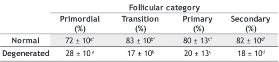
From the total number of follicles, 80 ± 4.95% was normal and 20 ± 4.95% was considerate degenerate. Table 3 shows the percentage of normal and degenerate follicles by follicular category. It was observed that the transition follicle has the grater percentage of normal follicles, followed by the secondary, primary and primordial. The percentage of normal follicles was significantly greater than the percentage of degenerate follicles in all categories.
Table 4 shows the follicular, oocyte, its nucleus and granulosa surface mean diameters from preantral follicles. The mean diameters of follicles and granulosa surface were statistically different from each other. There was no statistical difference between the transition and the primary follicle in relation to the oocyte diameter. The nuclear diameter of the secondary follicle was greater than all nuclear diameters from other preantral follicles. There was no statistical difference among the diameters of any preantral follicular parameters between the right and left ovaries.
Figure 1 shows the relationship between three morphometric values, i.e. follicular, oocitary and granulosa surface diameters in the largest cross-section, and oocyte nucleus diameter. The follicular development in C. apella is separated in two different phases. The primordial and transition follicles are in included the left graphs are . Until the 15 Fm nuclear diameter, the three follicular dimensions showed little change. There were significant (P < 0.001) linear relationships and the slopes of the regression lines possess low values, 1.17, 0.94, 0.22, respectively. The primary and secondary follicles are in the right graphs. In phase II there is fast growth in the three follicular measurements. There were significant linear relationships (P < 0.001). The slopes of the regression values are higher than in the previous phase.
The numbers of granulosa cells in relation to the preantral follicular category are shown in Table 5. The significant increase in the number of granulosa cells occurs among the different follicular categories, according to the follicular development.
The transformation from flattened to cuboidal granulosa cells in relation to the number of granulosa cells in the largest cross-section is shown in Figure 2. In the transition follicle, the numbers of flattened and cuboidal granular cells are 6 ± 0.35 and 7 ± 0.44, respectively. The cubic cells appear when the follicle presents approximately 11 cells in the granulosa cell layer. The flattened cells are present until the follicular 15-cell stage. The complete transformation from flattened to cuboidal cells was achieved at about the 16-cell stage.
Figure 3 (A, B, C and D) illustrates the different categories of the morphology of the preantral follicles analyzed in histological cross-section. Many polyovular follicles were found (Figure 3 E). The pellucid zone was first localized in the primary follicles. However, the pellucid zone just became easily shown in the secondary follicle with more than two layers of granulosa cells. The immature theca cells were already present in some secondary follicles with two layers of granulosa cells.
Figure 3 (F) shows an antral follicle in histological cross-section. The mean population of normal and degenerate follicles per ovary was 60.0 ± 19.0 and 3 l
a m i n
A Ovary
t h g i
R Left
I 34,500
-I
I 24,550 18,550
I I
I 47,650 26,100
V
I 121,050 102,750
) M E S ± ( n a e
M 56,938±21,888a 49,133±26,896b Table 1 - Preantral follicular mean ( ± SEM) population in the right and left ovaries from 4 adult C. apella females.
Among lines: a, b (P< 0.05).
Table 2 - Percentage of primordial, transition, primary and secondary ovarian follicles in 4 adult C. apella females.
y r o g e t a C r a l u c i l l o F l a i d r o m i r P ) % ( n o i t i s n a r T ) % ( y r a m i r P ) % ( y r a d n o c e S ) % ( 3 . 4 ± 0
3 a 60±4.8b 6±0.96c 4±0.67d
y r o g e t a c r a l u c i l l o F l a i d r o m i r P ) % ( n o i t i s n a r T ) % ( y r a m i r P ) % ( y r a d n o c e S ) % ( l a m r o
N 72±10a* 83±10b* 80±13c* 82±10d* d e t a r e n e g e
D 28±10a 17±10b 20±13c 18±10d Table 3 - Percentage of normal and degenerated preantral follicles from 4 adult C. apella.
Figure 2 - Relationship between granulosa cell number in the largest cross-section and its pattern of flattened and cuboidal cells distribution. Each point is the mean value of 35 follicles from 7 ovarian samples.
Figure 1 - Relationships between oocyte nuclear diameter with follicular, oocyte and granulosa layer diameters of 280 preantral follicles: b= slope of the regression line (y = a + bx).
Column A Column A Column A Column A Column A
Diameters from Primordial and Transitional follicles.
Column B Column B Column B Column B Column B
Diameters from Primary and Secondary follicles.
± 1.8, with the percentage of 95.24% and 4.76% respectively. The follicular, oocitary and oocyte nucleus mean diameter were 514.4 + 53 mm, 56.6 + 1.5 mm and 17.7 + 1.2 mm. The minimum and maximum diameters of the antral follicles were 262 mm and 983 mm, respectively.
DISCUSSION
The results of this study have shown an estimation of ovarian follicular population and data about the folliculogenesis in the C. apella female. The follicles have been classified as proposed by Lintern-Moore (1974). This classification considers a transitional follicular stage (category C/B), which we have called transitional follicle. This preantral follicle has characteristics of follicular
activation, with transformation of the flattened to cuboidal granulosa cells, characterizing that the follicle left the quiescent state (Erickson, 1986).
e l c i l l o
F Fol.Diam. Ooc.Diam. Nuc.Diam. Gran.Diam.
l a i d r o m i r P x a M -n i M 5 . 0 ± 1 . 2 2 a ) 7 4 -6 1 ( 4 . 0 ± 9 . 7 1 a ) 9 3 -2 1 ( 2 . 0 ± 6 . 0 1 a ) 5 1 -6 ( 2 . 0 ± 1 . 4 a ) 8 1 -2 ( n o i t i s n a r T x a M -n i M 0 . 5 ± 2 . 4 2 b ) 3 3 -7 1 ( 3 . 0 ± 9 . 8
1 bc
) 3 2 -2 1 ( 2 . 0 ± 0 . 1 1 a ) 6 1 -6 ( 2 . 0 ± 3 . 5 b ) 1 1 -2 ( y r a m i r P x a M -n i M 5 . 0 ± 3 . 7 2 c ) 8 3 -0 2 ( 3 . 0 ± 3 . 9 1 c ) 6 2 -2 1 ( 2 . 0 ± 0 . 1 1 a ) 6 1 -7 ( 3 . 0 ± 1 . 8 c ) 6 1 -4 ( y r a d n o c e S x a M -n i M 0 . 4 ± 2 . 1 6 d ) 3 9 1 -6 2 ( 2 . 1 ± 4 . 0 3 d ) 8 5 -8 1 ( 4 . 0 ± 7 . 3 1 b ) 4 2 -8 ( 0 . 3 ± 6 . 0 3 d ) 1 4 1 -7 (
Table 4 - Mean diameter (mm) of follicles, oocytes, their nuclei and the granulosa layer of preantral follicles from ovaries of 4 adult C. apella females.
Among lines: a, b, c, d (P< 0.05)
Table 5 - Granulosa cell number of preantral follicles (n=35) according to their category from C. apella.
Comparisons among columns: a, b, c, e, f, g (P<0,05). l a i d r o m i r
P Transition Primary Secondary s r e y a l l l e C
2 3 4 5
9 ±0.3a 13±0,5b 23±1.0c 64±1.6d 133±8.1e 240 ±14f 400±27g
Figure 3 - Illustration of ovarian follicles. A, B, C, D, E (x 400) and F (x 100). PR - Primordial, TR – Transitional, PA – Primary, SC – Secondary, PO – Polyovular, AN – Antral and TC – Thecal cells.
There was a statistical difference between the right and left ovaries for the follicular population in adult Cebus apella female. The right ovary had a larger follicular population compared to the left. Miller et al. (1999) found no difference between the left and right ovary for the number of follicles in the Pigtailed Monkey. In C. apella, a great number of follicles in the right ovary can suggest a lower functionality of this compared to the left ovary. Nagle et al. (1994) showed that the ovulation rate in the left ovary is 62.5% and 37.5% for the right ovary in the C. apella. In this study, the follicular ovarian population was found, without verifying the ovulation rate. In women controversy exists about the functionality of the ovaries. According to Potashnik et al. (1987), ovulation happens more frequently in the right ovary in cyclic women. However, more recently, Lass et al. (1997) observed that the ovulation rate is the same for both ovaries. Concerning the percentage of follicles by follicular category, in the present study a larger percentage of transitional follicles was found in relation to the other preantral follicles. This result is in agreement with the data presented by Block (1951), Gougeon & Chainy (1987) and Gougeon et al. (1994). These authors state that in adult woman, there is a larger proportion of preantral follicles that were activated and are growing.
The statistical difference found in the follicular diameter show that is possible to use it to aid in the classification of the different follicular categories. These results found in C. apella are similar to those found by Hulshof et al. (1994) for the bovine species. There was no statistical difference between the oocyte diameters of the transitional and primary follicles. This result suggests that the main responsible component for follicular growth in these stages is the granulosa surface.
The changes in the three follicular diameters observed in relation to the oocyte nucleus diameter, show that there are two phases of follicular growth. The first phase corresponds to transformation from flattened to cuboidal granulosa cells. At the second growth phase, there is an increase in the oocyte size and in the number of granulosa cells, with the increase in follicular diameter. These data are similar to those found by Gougeon & Chainy (1987). However, in the first phase, the small follicular growth is caused mainly by the increase in the oocyte size, while in the second phase of follicular growth, there is a larger participation of the granulosa cells.
The incidence of in situ polyovular follicles has been verified (with up to 4 oocytes). Forabosco et al. (1991) reported the presence of polyovular follicles in human ovaries during the neonatal period. According to these authors, the organogenesis of the ovary is not complete in humans at birth, and oogonias and follicles can be found in the same ovary. Such condition only happens close to birth in great primates, but it extends until the adult life in prosimians (Anand Kumar, 1974). In the present study, we did not find oogonias, but the presence of polyovular follicles in the ovaries of adult animals may suggest an incomplete organogenesis in the adult ovary.
The pellucid zone can be first visualized in the primary follicle in C. apella. This result is in agreement with Hirshfield (1991). According to this author, the pellucid zone begins to be formed soon after the primordial follicle begins growth. Regarding the theca, this does not appear until follicular growth is well underway. In the secondary follicles, a signal is generated and causes a stream of mesenchymal cells to migrate to the basal follicular membrane, where they become parallel to one another, forming a radial arrangement of fibroblast-like cells around the entire follicle. These cells will eventually develop into the theca intern and extern (Erickson, 1986). In the mouse, the first recognizable theca cells are reported to appear when the fully grown oocytes and the follicle have 2-3 layers of granulosa cells (Peters, 1969). In hamsters, the theca cells are not evident until the follicle reaches the 7-8 layers of granulosa cells stage (Roy and Greenwald, 1985). However, in female rats, a different theca is already evident in small follicles. These immature theca cells proliferate along with the granulosa cells during the follicular growth (Hirshfield, 1991).
In the present study, the antral follicles present in the ovaries were counted and measured, to provide more information about the folliculogensis in C. apella.
Comparing the results in C. apella with other primates, in Aotus trivirgatus (owl monkey) the diameter of the antral follicles can vary from 0.3 mm to 3 mm (Herting et al., 1976). Oerke (1995) reported that in Callitrichix jacchus, the preovulatory follicles diameter can vary from 2.1 to 3.2 mm. In women, the preovulatory follicle can reach 20 mm (Baker & Wai, 1976). In C. apella, the mean diameter of the preovulatory follicle can reach 10-12 mm (Nagle et al., 1980). The 983 Fm is not the maximum diameter that an antral follicle can reach in C. apella females and this diameter does not correspond to the final reached at follicular maturation. Regarding the quality of the antral follicles, a small number was degenerate, which may be because the antral follicles are more susceptible to follicular atresia in the final differentiation stage of the granulosa and theca cells than the antral follicles at the initial stage of development (Lussier et al., 1987).
Taking into consideration the results obtained in the present study, it can be concluded that the ovarian follicular population in adult C. apella females is variable. The number of preantral follicles is larger in the right ovary. The primordial follicle is the more susceptible to atresia. During the preantral stage, there are two phases of follicular development. In the first phase the oocitary growth has larger participation in the follicular growth, while in the second phase, the proliferation of the granulosa cells is predominant for the follicular growth. The follicular diameter can be used as a parameter to aid the classification of preantral follicles. Regarding the antral follicles, more studies are necessary to understand the folliculogenesis dynamics of this development stage in the C. apella species. The information obtained in this study can be used as a parameter for subsequent in vivo or in vitro studies about the folliculogenesis in neotropical primates of the C. apella species. The C. apella female may be used as an experimental model in subsequent studies on manipulation from oocytes of ovarian preantral follicles, to develop multiplication programs for endangered non-human primates in the future.
LITERATURE CITED
Aurichio, P. 1995. Primatas do Brasil. Terra Brasilis Editora, São Paulo, 168p.
Anand Kumar, T. C. 1974. Oogenesis in adult prossimians primates.
Contrib. Primatol., 3: 82-96.
Asa, C. S. 1996. Reproductive Physiology. In: Wild Mammals in Captivity. The University of Chicago Press. Chicago and London. 390 - 417pp
Bailey F.; Copenhaver W. M; Bunge, R. P; Bunge, M. B. 1976.
Histologia. Editora Edgard Blücher Ltda. São Paulo. 491 - 526pp Baker T. G; Wai, S. O. 1976. Developmental of the ovary and
oogenesis. Clin. Obstet. Gynecol. 3: 3- 26.
Cahill L. P; Mariana J.C.; Mauléon P. 1979. Total follicular populations in ewes of high and low ovulations rates. Journal of Reproduction and Fertility, 55: 27-36.
Driancourt, M. A; Cahill, L. P; Bindon B. M. 1985. Ovarian follicular populations and preovulatory enlargement in booroola and control merino ewes. Journal of Reproduction and Fertility, 10: 97-105.
Erickson, B. H. 1966. Development and senescence of the postnatal bovine ovary. J.Animal Science, 25: 800-805.
Erickson, B. H.; Reynolds, R. A; Murphree, R. L. 1976. Ovarian characteristics and reproductive performance of the aged cow.
Biology of Reproduction, 15: 555-560.
Erickson, G. F. 1986. An analysis of follicle development and ovum maturation. In: Seminars and Reproductive Endocrinology, San Diego – California, pp. 233 – 254. (seminários)
Figueiredo, J. R.; Hulshof, S. C. J; Van den Hurk, R. 1993. Development of a combined new mechanical and enzimatic method for the isolation of intact follicles from fetal, calf and adult bovine ovaries. Theriogenology, 40: 789-799.
Forabosco A.; Sforza C.; De Pol A.; Vizzoto L.; Marzona, L.; Ferrario, V. F. 1991. Morphometric study of the human neonatal ovary.
The Anatomical Record, 231:201-208.
Gougeon, A.; Chainy, G. B. N. 1987. Morphometric studies of small follicles in ovaries of women at different ages. Journal Reproduction and Fertility, 81: 433- 442.
Gougeon, A.; Ecochard, R.; Thalabard, J. C. 1994. Age-related changes of the population of human ovarian follicles: Increased in the disappearance rate of non-growing and early-growing follicles in aging women. Biology of Reproduction, 50: 653- 663.
Gundersen, H.; Bagger, P.; Bendtsen, T. 1988. The new sterological tools: dissector, fractionator, nucleator and point sampled intercepts and their use in pathological research and diagnosis.
AMPIS, 96: 857-881.
Hearn, J. 1994. New world primates for research in human reproductive health. American Journal of Primatology, 34: 11-17.
Herting, A.; Barton, B. R; Mackey, J. J. 1976. The female genital tract of the owl monkey (Aotus trivirgatus) with special reference to the ovary. Laboratory Animal Science, 26 (6): 1041- 1067.
Hirshfield, A. N. 1991. Development of follicles in the mammalian ovary. International Review of cytology. 124: 43 - 108 Hulshof, S. C. J; Figueiredo, J. R; Beckers, J. F; Bevers, M. M; Van den
Hurk, R. 1994. Isolation and characterization of preantral follicles from fetal bovine ovaries. Veterinary Quarterly, 16 ( 2): 78-80.
Lass, A.; Croucher, C.; Lawrie, H.; Margara, R.; Winston, R. M. L. 1997. Right or left ovary – which one is better? Human Reproduction, 12 (8): 1730-1731.
Lintern-Moore, S.; Peters, H; Moore, G. P. .M; Faber, M. 1974. Follicular development in the infant human ovary. Journal Reproductio and Fertility, 20: 773- 778.
Lussier, J. G; Matton, P.; Duffort, J. J. 1987. Growth rates of follicles in the ovary of the cow. JournalReproduction and Fertility, 81: 301-307.
Miller, P. B.; Charlenston, J.S.; Battaglia, D. E; Klein, N. A.; Soules, M. R. 1999. Morphometric analysis of primordial follicle number in pigtailed monkey ovaries: symmetry and relationship with age. Biology of Reproduction, 61: 553-556. Nagle, C. A.; Riarte, A.; Quiroga, S.; Azorero, R. M.; Carril, M.; Denari, J. H.; Rosner, J. M. 1980.Temporal Relantionship Between Follicular Development, Ovulation and Hormonal Profile in the Capuchin Monkey (Cebus apella). Biology of Reproduction, 23: 629- 635.
Nagle, C. A.; Digiano, L.; Paul, N.; Terlato, M.; Quiroga, S.; Mendizabal, A. F. 1994. Interovarian communication for the control of follicular growth and corpus luteun fuction in the cebus monkey. American Journal of Primatology, 34: 19 – 28. Oeke, A-K. 1995. Follicular development and corpus luteum formation as determined by ultrasonography in the marmoset monkey (Callithrix jacchus). Primate Report, 43: 25. Peters, H. 1969. The developmental of the mouse ovary from birth
to maturity. Acta Endocrinologica, 62: 98- 116.
Potashnik, G.; Inter, V.; Meizeneq, I.; Stenberg, M. 1987. Frequency, sequence and side of ovulation in women menstruating normally. Br. Med. Journal, 24: 294 – 319.
Roy, S. K.; Greenwald, G. S. 1985. An enzymatic method for dissociation of intact follicles from the hamster ovary: Histological and Quantitative Aspects. Biology of Reproduction, 32: 205-215.
Scaramuzzi, R. J; Adams, N. R, Baird, D. T; Campbell, B. K.; Downing, J. A.; Findlay, J. K.; Henderson, K. M.; Martín, G. B.; Macnatty, K. P.; Mcneilly, A. S.; Tsonis, C. G. 1993. A model for follicle selection and the determimation of ovulation rate in the ewe. Reprod. Fertil. Dev., 5: 459-478.