This Accepted Author Manuscript is copyrighted and published by Elsevier. It is posted here by agreement between Elsevier and University of Brasilia. Changes resulting from the publishing process - such as editing, corrections, structural formatting, and other quality control
mechanisms - may not be reflected in this version of the text. The definitive version of the text was subsequently published in [Small Ruminant Research, Volume 41, Issue 1, July 2001, Pages 61–69, Pages 809–822, doi:10.1016/S0921-4488(01)00196-1].You may download, copy and otherwise use the AAM for non-commercial purposes provided that your license is limited by the following restrictions:
(1) You may use this AAM for non-commercial purposes only under the terms of the CC-BY-NC-ND license.
(2) The integrity of the work and identification of the author, copyright owner, and publisher must be preserved in any copy.
(3) You must attribute this AAM in the following format: [agreed attribution language, including link to CC BY-NC-ND license + Digital Object Identifier link to the published journal article on Elsevier’s ScienceDirect® platform].
________________________________________________________________________
Este Manuscrito do Autor Aceito para Publicação (AAM) é protegido por direitos autorais e publicado pela Elsevier. Ele esta disponível neste Repositório, por acordo entre a Elsevier e a Universidade de Brasília. As alterações decorrentes do processo de publicação - como a edição, correção, formatação estrutural, e outros mecanismos de controle de qualidade - não estão refletidas nesta versão do texto. A versão definitiva do texto foi posteriormente publicado em [Small Ruminant Research, Volume 41, Número 1, Julho de 2001, Páginas 61–69,
doi:10.1016/S0921-4488(01)00196-1]. Você pode baixar, copiar e utilizar de outra forma o AAM para fins não comerciais , desde que sua licença seja limitada pelas seguintes restrições:
(1) Você pode usar este AAM para fins não comerciais apenas sob os termos da licença CC- BY- NC-ND.
(2) A integridade do trabalho e identificação do autor, detentor dos direitos autorais e editor deve ser preservado em qualquer cópia.
Light microscopical and ultrastructural characterization of goat preantral
follicles
Carolina M Lucci R.V Silva
C.A Carvalho R Figueiredo N Báo
Abstract
Goat ovarian preantral follicles were morphologically and ultrastructurally described in this work. Primordial follicles are oocytes surrounded by one layer of squamous or squamous-cuboidal granulosa cells; primary follicles have a single layer of squamous-cuboidal granulosa cells, and secondary follicles are oocytes surrounded by two or more layers of cuboidal granulosa cells. At all developmental stages a thick layer of glycoproteins, the basement membrane, surrounded the preantral follicles. The quiescent oocyte is spherical or oval and it has a large eccentrically located nucleus with a conspicuous nucleolus. The organelles were uniformly distributed in the cytoplasm. A large number of vesicles were spread throughout the cytoplasm in all the oocytes. The cytoplasm of oocytes also contains numerous rounded mitochondria besides the usual organelles. As the follicle develops, the mitochondria become elongated. The communication between the oocyte and the granulosa cells is apparently mediated through endocytosis as indicated by the abundant coated pits and vesicles noted in the cortical cytoplasm of the oocyte. The oocyte plasma membrane presented projections that penetrated between adjacent granulosa cells and a few short microvilli lying parallel to the oocyte surface. In secondary follicles, patches of zona pellucida material were observed. Overall, the results indicate that the morphological and ultrastructural organization of caprine preantral follicles resembles that of other mammals. However, some particularities were observed, and that may indicate species specific differences.
Keywords: Goat; Oocyte; Preantral follicle; Ultrastructure
1. Introduction
At birth, the mammalian ovary is endowed with hundreds of thousands of preantral
follicles, which represent the resting stockpile of germ cells (Saumande, 1991). However, very
little is known about the biology of these follicles and the events surrounding their growth or
atresia. Advances in this field will depend on in vitro systems which study preantral follicles.
During the past few years the possible exploitation of preantral follicles from domestic
animals, by isolation and culture, has received much attention (Figueiredo et al., 1994, Hulshof
et al., 1994, Jewgenow and Goritz, 1995, Nuttinck et al., 1996, Wandji et al., 1996, Lucci et al.,
1999 and Cecconi et al., 1999). However, in some studies, the results were not promising
possibly due to follicle damage induced by the isolation procedures (Van den Hurk et al., 1997
microscopic evaluation, but there is a great need for better viability analysis. With this
perspective in mind, the ultrastructural evaluation, after isolation, may provide more
information about the quality of the follicles and their oocytes, as it is capable of fine
discrimination of damage to membranes and organelles. In this way, a description of the
standard ultrastructure of preantral follicles in situ is very important as a model. Moreover, the
need for cell biological knowledge of preantral follicles is accentuated in order to develop
suitable culture systems.
Ultrastructural studies have been carried out on follicles and oocytes from farm
animals such as cows (Hyttel et al., 1986, Assey et al., 1994 and Van Wezel and Rodgers, 1996;
Fair et al., 1996, Fair et al., 1997a and Fair et al., 1997b) and ewes (Brand and Jong, 1973 and
Cran et al., 1980), mainly in tertiary and preovulatory follicles, and some differences among
species were demonstrated. However, little information about the ultrastructural aspects of
goat preantral follicles is available in the literature (Sharma et al., 1994). In order to increase
our knowledge of preantral follicle development in goat ovaries, the aim of the present study
is to describe the morphological and ultrastructural aspects of follicular development during
the preantral phases in the adult goat ovary. Oocyte and granulosa cells of primordial, primary
and secondary follicles were studied using light and transmission electron microscopy.
2. Materials and methods
Ovaries (n=6) from adult (1–3 years old) and non-pregnant goats of mixed breed were
collected at a slaughterhouse. The ovaries were removed, stripped of surrounding fat tissue
and ligaments, washed three times in phosphate buffered saline (PBS) and processed for light
and transmission electron microscopy. Primordial (n=162 and 6, respectively for light and
TEM), primary (n=162 and 5) and secondary (n=48 and 4) follicles were evaluated.
2.1. Light microscopy
The ovarian cortex was fixed by immersion for 24–48 h in 10% formaldehyde,
dehydrated in ethanol, and embedded in paraffin. Sections of 7 μm were stained with periodic acid Schiff (PAS) and haematoxylin. Preantral follicles were classified by their morphological
appearance, according to Hulshof et al. (1994). The diameters of the follicles and the oocytes
were determined when the nucleolus of the oocyte was observed (equatorial section), using
equatorial section were counted. Sections were examined and photographed using a Zeiss
Axiophot light microscope.
2.2. Transmission electron microscopy (TEM)
Small pieces of ovarian cortex were fixed during 3 h at room temperature in a solution
containing 2% paraformaldehyde and 2.5% glutaraldehyde in 0.1 M sodium cacodylate buffer
pH 7.2. After fixation, the specimens were rinsed in buffer and post-fixed in 1% osmium
tetroxide, 0.8% potassium ferricyanide and 5 mM CaCl2 in 0.1 M sodium cacodylate buffer for
1 h, followed by block staining in 0.5% uranyl acetate. Subsequently, the samples were
dehydrated in a series of ascending acetone concentrations (30–100%) and then embedded in
Spurr epoxy resin. Thin sections (70 nm) were contrasted with uranyl acetate and lead citrate,
and examined using a Jeol 100 C and a Zeiss 912 transmission electron microscopes. The
basement membrane thickness was estimated in all observed follicles (n=15) by measurement
in electron photomicrographs (10 000× final magnification) in three different regions (all of
them in the equatorial section).
2.3. Statistical analysis
The diameters of follicles and oocytes and the number of granulosa cells in the
equatorial section, as well as the basement membrane thickness, were compared between
primordial, primary and secondary follicles by Scheffe’s test (Stat View for Macintosh). Differences were considered statistically significant at 0.05 level.
3. Results
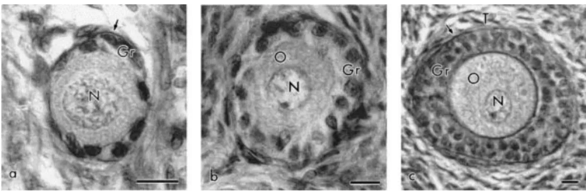
In this work, the preantral follicles were classified in three categories: primordial
follicles (oocytes surrounded by one layer of squamous or squamous-cuboidal granulosa cells
— Fig. 1a); primary follicles (with a single layer of cuboidal granulosa cells around the oocyte — Fig. 1b), and secondary follicles (oocytes surrounded by two or more layers of cuboidal granulosa cells — Fig. 1c). The mean (±S.E.M.) diameter of follicles and oocytes and the
number of granulosa cells at the equatorial section are presented in Table 1. At all
developmental stages a thick layer of glycoproteins, the basement membrane, surrounded the
histological sections. Its thickness in different follicles varied from 231 to 727 nm, but no
statistical differences were observed between follicular classes. The mean thickness of
basement membrane was 513±38 nm in preantral follicles in general.
Fig. 1. Light micrographs showing (a) primordial, (b) primary, and (c) secondary goat follicles. O: oocyte, N: nucleus of oocyte, Gr: granulosa cell, T: theca cells (arrow marks the basement membrane). Bar=10 μm.
Table 1.
Diameter of primordial, primary and secondary follicles and their oocytes and number of granulosa cells at the equatorial section (mean±S.E.M.)a
aValues with different letters in the same column are significantly different (Scheffe’s test; P<0.005).
3.1. Primordial follicles oocytes
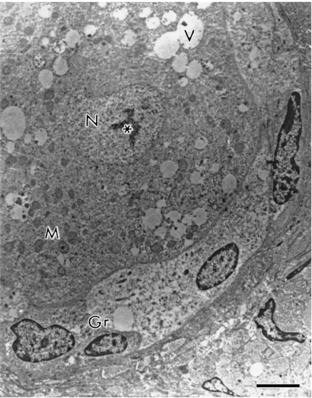
The quiescent oocytes in primordial follicles were spherical or oval and were
completely surrounded by 1–14 squamous or squamous-cuboidal granulosa cells at the
equatorial section. A large oocyte nucleus occupied a central or eccentric position showing a
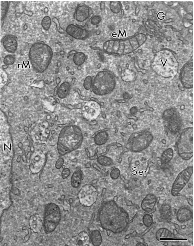
conspicuous nucleolus (Fig. 2). The organelles were uniformly distributed in the cytoplasm or,
sometimes, more closely to the nucleus. The mitochondria were the most evident organelle
and were predominantly round. These round mitochondria normally present few cristae often
arranged parallel close to the surface of the outer mitochondria membrane, leaving a very
large central area of moderately electron-dense inner matrix (Fig. 3). A small number of
elongated mitochondria were also observed in some cases. A few cistern of smooth
endoplasmic reticulum and Golgi apparatus were observed and a variable number of vesicles
stage, and the oocyte and granulosa cells appeared only juxtaposed, without any specific
junctions.
Fig. 3. Detail of cytoplasmic structures. N: nucleus of oocyte, V: vesicles, rM/eM: round or elongated mitochondria, G: Golgi apparatus, Ser: smooth endoplasmic reticulum. Bar=1 μm.
3.2. Primary follicles oocytes
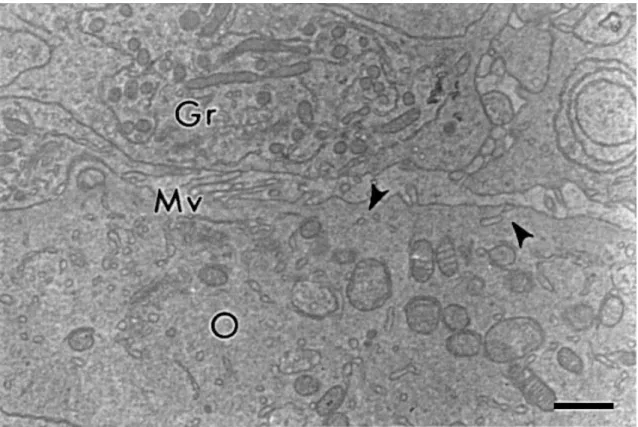
The oocytes in primary follicles appeared not to be quiescent any more. They were
surrounded by 5–26 cuboidal granulosa cells disposed in a single layer at the equatorial
section, with which they kept a narrow contact. The communication between the oocyte and
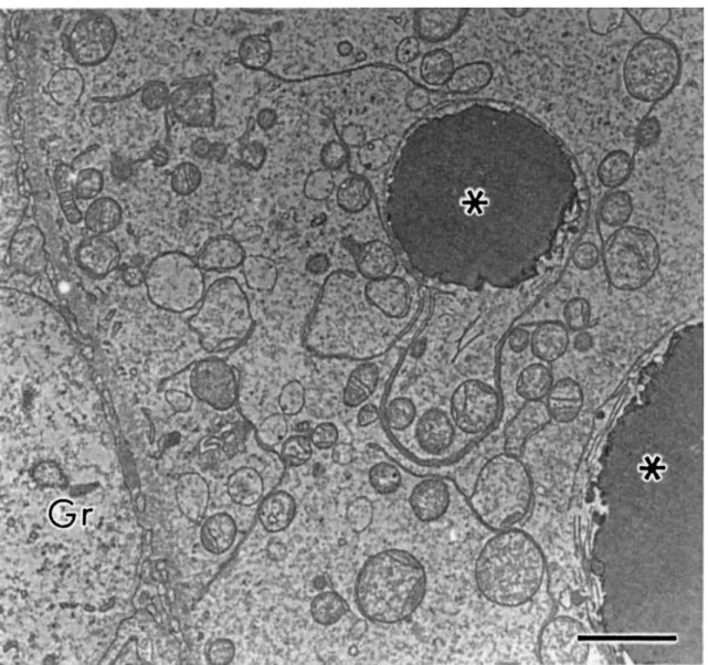
the granulosa cells is mediated through endocytosis as indicated by the abundant coated pits
projections that penetrated between adjacent granulosa cells and a few short bent microvilli
lying parallel to the oocyte surface (Fig. 4). As well as the primordial follicles oocytes, the
cytoplasm of oocytes in primary follicles also contains numerous rounded mitochondria
beyond the usual organelles. As the follicle develops the mitochondria take the elongated
shape (Fig. 3).
Fig. 4. Communication between oocyte and granulosa cells in a primary follicle. Gr: granulosa cell, O: oocyte, Mv: microvilli, arrowheads: coated pits. Bar=1 μm.
3.3. Secondary follicles oocytes
In these follicles, the oocytes were surrounded by 13–114 cuboidal granulosa cells
forming two or more layers at the equatorial section. The nucleus of the oocyte assumed an
eccentric position and the organelles started to move to the periphery. The number of smooth
endoplasmic reticulum increased and the vast majority of mitochondria were elongated. With
the development of the follicle, there appeared to be a slight increase in the number of
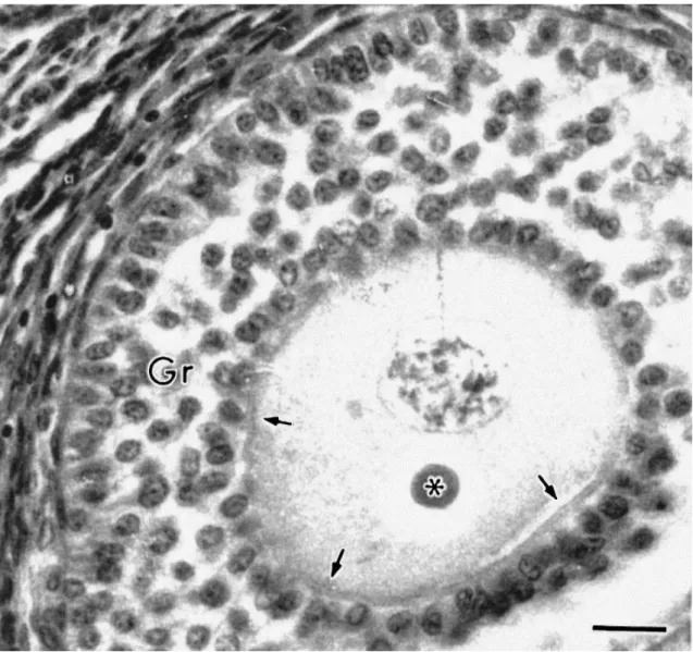
microvilli. In secondary follicles, patches of ZP material were observed and were usually
associated with erect oocyte microvilli (Fig. 5). Also in these follicles, electron-dense droplets
were commonly observed in the cytoplasm of both the oocyte and granulosa cells (Fig. 6).
These droplets could often be seen in histological sections and were strongly dyed by the
cells surrounding the follicle (Fig. 1c), outside the basement membrane. However, the theca
interna could only be clearly defined in follicles with four or more layers of granulosa cells.
Fig. 5. Forming zona pellucida in a secondary follicle. O: oocyte, Gr: granulosa cell, Mv: microvilli, ZP: zona pellucida. Bar=1 μm.
Fig. 7. Glycoprotein droplet (dyed by the Schiff’s reagent) in a histological section of a secondary follicle (∗). Arrows indicate the zona pellucida forming a complete ring around the oocyte. Gr: granulosa cell. Bar=20 μm.
3.4. Granulosa cells
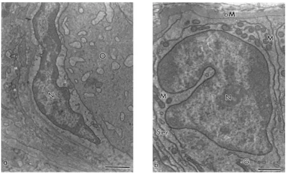
Granulosa cells were small, with high nuclear:cytoplasmic ratio. Squamous granulosa
cells had an elongated nucleus and few organelles in general. Cuboidal granulosa cells, on the
other hand, showed many elongated mitochondria, smooth and rough endoplasmic reticulum
and Golgi apparatus and some electron dense vesicles. Their nucleus showed an irregular
shape with loose chromatin and little peripheral agglomerates of dense chromatin (Fig. 8a and
b). In secondary follicles, the granulosa cells began to separate, forming wide irregular
intercellular spaces. At all follicular stages, granulosa cells rested on a basement membrane
Fig. 8. (a) Squamous and (b) cuboidal granulosa cells. N: nucleus, M: mitochondria, Ser: smooth endoplasmic reticulum, bM: basement membrane, G: Golgi apparatus, O: oocyte. Bar=1 μm.
4. Discussion
The results of this study highlight much information about the cell biology of ovarian
preantral follicles in the goat, which can be useful as a model to studies of integrity of
follicles/oocytes handled in vitro. Concerning the histological aspects, the mean diameter of
oocytes and follicles, in all the three developmental stages (i.e. primordial, primary and
secondary), was smaller than those observed in ewes (Lundy et al., 1999) and cows (Van Wezel
and Rodgers, 1996, Braw-Tal and Yossefi, 1997 and Hyttel et al., 1997), but was similar to those
observed in humans (Lintern-Moore et al., 1974). However, the mean and range in the number
of granulosa cells at the equatorial section were compatible to those described for ewes
(Lundy et al., 1999), cows (Braw-Tal and Yossefi, 1997) and humans (Lintern-Moore et al.,
1974), even when the diameter of oocytes and follicles was smaller. These differences may be
species-specific or due to differences in measurement techniques.
In general, the ultrastructure of goat preantral follicles was similar to that of other
mammalian species (cattle — Van Wezel and Rodgers, 1996, Fair et al., 1997b and Hyttel et al.,
1997; pig — Greenwald and Moor, 1989; feline — Jewgenow and Stolte, 1996; human —
Hertig and Adams, 1967 and Oktay et al., 1997), but some differences were observed, which
may indicate either species specific differences or different phases of follicular development.
For example, the concentration of cytoplasmic components in the juxta-nuclear region,
In addition, in bovine secondary follicles, gap junctions have developed between the oocyte
and granulosa cells and the first small clusters of cortical granules were seen (Fair et al., 1997b
and Hyttel et al., 1997). Immature cortical granules were also present in feline primary follicles
(Jewgenow and Stolte, 1996). Neither of these two structures were observed in goat follicles
analyzed in our study. Moreover, in bovine, adherens junctions were detected between
granulosa cells and oocyte since primordial follicles, and gap and zonula adherens-like
junctions were also observed between adjacent granulosa cells (Fair et al., 1997b).
Desmosomes were observed joining follicular cells to each other and/or to the oocyte in cats
(Jewgenow and Stolte, 1996), monkeys (Zamboni, 1974) and rabbits (Nicosia et al., 1975).
However, in goats, special types of junctions (i.e. desmosomes, adherens or tight junctions)
were not observed among granulosa cells or between granulosa cells and oocyte. In this last
case, only the endocytotic contact was clearly visible. Specifically, regarding to gap junctions,
the reason why they were not observed in this study may be that none of the follicles
evaluated by TEM had the oocyte completely surrounded by ZP. Thus, it can be inferred that
while the ZP is not fully developed around the whole oocyte, gap junctions are not
indispensable, and therefore, not present yet. At this stage, substances required for the oocyte
metabolic needs could gain access to its cytoplasm by diffusion through the closely opposed
membranes of oocyte and granulosa cells in the areas of intercellular contact, or they could be
incorporated by endocytosis, as suggested by the presence of numerous coated vesicles at the
cortical cytoplasm. Anyway, whichever the way of contact between oocyte and granulosa cells,
this association is extremely important to oocyte growth. The granulosa cells cooperate in
oocyte metabolic processes and this cooperation is probably essential for normal oocyte
development. Isolated oocytes normally grow in vitro only when their association with the
companion granulosa cells is maintained and the rate of oocyte growth in vitro is directly
correlated with the number of granulosa cells coupled to it (Eppig, 1991).
Concerning cytoplasmic organelles, the observations indicate that the changes
undergone by the follicle during sequential phases of growth are directed mainly to providing
the oocyte with the environment needed for its maturation and metabolism. All the
characteristics observed show that oocytes in goat preantral follicles are equipped for the
absorption, utilization and intracellular transport of material delivered to their surface
membrane, as well as for the biosynthesis of stored material. The observed pleiomorphic
variations in mitochondrial shape possibly represent their role in the metabolism of the
developing oocyte. The round shape of the mitochondria, indicate immaturity (Perkins and
Frey, 2000), in accordance with the quiescent stage of primordial follicle oocytes. In these
gradually replaced by elongated mitochondria in the oocytes of primary and secondary
follicles.
In the present study, the ontogeny of formation of the ZP was similar to that in cows
(Braw-Tal and Yossefi, 1997 and Fair et al., 1997b) and ewes (Lundy et al., 1999), in which it
appeared as islands of zonal material in secondary follicles with two to three layers of
granulosa cells and as a complete PAS-positive ring in follicles with more than three layers of
granulosa cells (Fig. 7). However, it differed from that in mice (Oakberg, 1979), guinea pigs
(Adams and Hertig, 1964), rabbits (Nicosia et al., 1975), cats (Jewgenow and Stolte, 1996),
monkeys (Zamboni, 1974) and humans (Himelstein-Braw et al., 1976) in which the ZP forms
around the oocyte in primary follicles. Electron-dense droplets in the cytoplasm of both the
oocyte and granulosa cells of secondary follicles were detected in this study, and these
droplets were strongly dyed by the Schiff’s reagent in histological sections confirming their
glycoproteic content. This is a sign that these droplets are secreting vesicles carrying material
for the ZP formation. Previous works indicate that the ZP is made of glycoproteins synthesized
by the oocyte and the granulosa cells, and that the ZP glycoproteins can be localized in the
cytoplasm of follicular cells in multilayer follicles (rabbit — Wolgemuth et al., 1984).
Since the primordial follicular stage, oocytes were always completely surrounded by
granulosa cells and were never in direct contact with the follicular basal lamina. In contrast, in
monkey primordial follicles oocytes a discontinuous investment of squamous follicle cells is not
unusual (Zamboni, 1974). Oocytes are large cells having a low plasma membrane area to
cytoplasmic volume ratio. According to Eppig (1991), the plasma membrane of oocytes is
probably unable to maintain an adequate inflow of nutrients and outflow of metabolic
products to support optimal oocyte growth. Therefore, the plasma membrane of the granulosa
cells may act as an extension of the oocyte plasma membrane providing for the uptake of
nutrients, which diffuse into the oocyte through the narrow connection between granulosa
cells and oocyte.
The ultrastructural characteristics of granulosa cells in this study are very similar to
that described for monkeys, even to squamous and cuboidal cells (Zamboni, 1974). Cuboidal
granulosa cells appeared engaged in active steroidogenesis as shown by abundant smooth
endoplasmic reticulum and mitochondria. The predominance of these organelles was also
observed in rat granulosa cells cultured in vitro (Sanbuissho et al., 1993). In secondary follicles,
the granulosa cells were separated by irregular spaces. According to Zamboni (1974), these
lacunae will be filled with liquor folliculi. Progressive accumulation of fluid will induce
follicles. In addition, in goat, the formation of the theca appears to be similar to that in cattle
(Braw-Tal and Yossefi, 1997) and sheep (Lundy et al., 1999).
The basement membrane surrounding preantral follicles of goat ovaries was strongly
stained by the Schiff’s reagent in paraffin sections. In the TEM analysis, it appeared to be
composed of a compact and well organized lamina in the inner part, surrounded by a thick
layer of collagen fibers, according to the description of Figueiredo et al. (1995) for bovine
preantral follicles. The ovarian follicles surrounded by a thick basement membrane are well
known, but this is the first time the thickness was assessed. Also, in the present study, the
basement membrane did not change in thickness or appearance as the follicles grew in size
suggesting that the basement membrane is continuously being remodeled. Recent studies
suggest that the extracellular matrix can be actively rearranged by granulosa cells in vitro
(Richardson et al., 2000). Since basement membranes are specialized extracellular matrices, it
is possible that the basement membrane around the ovarian follicles is rearranged by
granulosa cells in vivo too. However, further studies are needed to support this statement.
Several potential functions of basement membranes have been proposed including the
creation and maintenance of tissue integrity, transport and filtration of various substances
(Hessle et al., 1984) and attachment of some growth factors (Ruoslahti and Yamaguchi, 1991
and Feige and Baird, 1992). In the case of isolated preantral follicles handled in vitro, the
integrity of the basement membrane is a very important characteristic.
5. Conclusion
In conclusion, this work shows, for the first time, an ultrastructural description of goat
preantral follicles. In general, the results indicate that the morphology and the ultrastructure
of caprine preantral follicles resemble that of other mammals. However, some differences
were observed, and that may indicate species specific differences. An insight into the fine
structural organization of goat follicles would be of great help in explaining cellular and
molecular aspects of follicle growth and atresia in this species. Moreover, in the future, basic
knowledge on the ultrastructure of preantral follicles may facilitate the understanding of their
in vitro development.
Acknowledgements
This work was supported by CAPES and CNPq. We thank A.D. Santos for technical assistance
Laboratório de Morfologia e Morfogênese of niversity of Bras lia and the aborat rio de
Microscopia Eletrônica of EMBRAPA Recursos Genéticos e Biotecnologia, Brazil.
References
Adams and Hertig, 1964 E.C Adams, A.T Hertig. Studies on guinea pig oocytes. I. Electron microscopic observations on the development of cytoplasmic organelles in oocyte of primordial and primary follicles J. Cell Biol., 21 (1964), pp. 397–427
Assey et al., 1994 R.J Assey, P Hyttel, N Kanuya Oocyte structure in dominant and subordinate follicles in zebu cattle (Bos indicus) Anat. Embryol., 190 (1994), pp. 461–468
Brand and Jong, 1973 A Brand, W.H.R Jong Qualitative and quantitative micromorphological investigations of the tertiary follicle population during the oestrous cycle in sheep J. Reprod. Fertil., 33 (1973), pp. 431–439
Braw-Tal and Yossefi, 1997 R Braw-Tal, S Yossefi Studies in vivo and in vitro on the initiation of follicle growth in the bovine ovary J. Reprod. Fertil., 109 (1997), pp. 165–171
Cecconi et al., 1999 S Cecconi, B Barboni, M Coccia, M Mattioli In vitro development of sheep preantral follicles Biol. Reprod., 60 (1999), pp. 594–601
Cran et al., 1980 D.G Cran, R.M Moor, M.F Hay Fine structure of the sheep oocyte during antral follicle development J. Reprod. Fertil., 59 (1980), pp. 125–132
Eppig, 1991 J.J Eppig Maintenance of meiotic arrest and the induction of oocyte maturation in mouse oocyte-granulosa cell complexes developed in vitro from preantral follicles Biol. Reprod., 45 (1991), pp. 824–830
Fair et al., 1996 T Fair, P Hyttel, T Greve, M Boland Nucleus structure and transcriptional activity in relation to oocyte diameter in cattle Mol. Reprod. Develop., 43 (1996), pp. 503–512
Fair et al., 1997a T Fair, S.C.J Hulshof, P Hyttel, T Greve, M Boland Nucleus ultrastructure and transcriptional activity of bovine oocytes in preantral and early antral follicles Mol. Reprod. Develop., 46 (1997), pp. 208–215
Fair et al., 1997b T Fair, S.C.J Hulshof, P Hyttel, T Greve, M Boland Oocyte ultrastructure in bovine primordial to early tertiary follicles Anat. Embryol., 195 (1997), pp. 327–336
Feige and Baird, 1992 J.-J Feige, A Baird La crinopexie: un modèle décrivant les mécanismes qui régissent la biodisponibilité des facteurs de croissance Med. Sci., 8 (1992), pp. 805–810
Figueiredo et al., 1994 J.R Figueiredo, S.C.J Hulshof, R Van den Hurk, B Nusgens, M.M Bevers, F.J Ectors, J.F Beckers Preservation of oocyte and granulosa cell morphology in bovine preantral follicles cultured in vitro Theriogenology, 41 (1994), pp. 1333–1346
identification in bovine ovaries and significance for the attachment of cultured preantral follicles Theriogenology, 43 (1995), pp. 845–858
Greenwald and Moor, 1989 G.S Greenwald, R.M Moor Isolation and preliminary characterization of pig primordial follicles J. Reprod. Fertil., 87 (1989), pp. 561–571
Hertig and Adams, 1967 A.T Hertig, E.C Adams Studies on the human oocyte and its follicle. I. Ultrastructural and histochemical observations on the primordial follicle stage J. Cell Biol., 34 (1967), pp. 647–675
Hessle et al., 1984 H Hessle, L.Y Sakai, D.W Hollister, R.E Burgeson, E Engvall Basement membrane diversity detected by monoclonal antibodies Differentiation, 26 (1984), pp. 49–54
Himelstein-Braw et al., 1976 R Himelstein-Braw, A.G Byskov, H Peter, M Faber Follicular atresia in the infant human ovary J. Reprod. Fertil., 46 (1976), pp. 55–59
Hulshof et al., 1994 S.C.J Hulshof, J.R Figueiredo, J.F Beckers, M.M Bevers, R Van den Hurk Isolation and characterization of preantral follicles from foetal bovine ovaries Vet. Quart., 16 (1994), pp. 78–80
Hyttel et al., 1986 P Hyttel, H Callesen, T Greve Ultrastructural features of preovulatory oocytes maturation in superovulated cattle J. Reprod. Fertil., 76 (1986), pp. 645–656
Hyttel et al., 1997 P Hyttel, T Fair, H Callensen, T Greve Oocyte growth, capacitation and final maturation in cattle Theriogenology, 47 (1997), pp. 23–32
Jewgenow and Goritz, 1995 K Jewgenow, F Göritz The recovery of preantral follicles from ovaries of domestic cats and their characterization before and after culture Anim. Reprod. Sci., 39 (1995), pp. 285–297
Jewgenow and Stolte, 1996 K Jewgenow, M Stolte Isolation of preantral follicles from
nondomestics cats — viability and ultrastructural investigations Anim. Reprod. Sci., 44 (1996), pp. 183–193
Katska and Rynska, 1998 L Katska, B Rynska The isolation and in vitro culture of bovine preantral and early antral follicles of different size classes Theriogenology, 50 (1998), pp. 213– 222
Lintern-Moore et al., 1974 S Lintern-Moore, H Peters, G.P.M Moore, M Faber Follicular development in the infant human ovary J. Reprod. Fertil., 39 (1974), pp. 53–64
Lucci et al., 1999 C.M Lucci, C.A Amorim, S.N Báo, J.R Figueiredo, A.P.R Rodrigues, J.R.V Silva, P.B.D Gonçalves Effect of the interval of serial sections of ovarian tissue in the tissue chopper on the number of isolated caprine preantral follicles Anim. Reprod. Sci., 56 (1999), pp. 39–49
Nicosia et al., 1975 S.V Nicosia, I Evangelista, S.K Batta Rabbit ovarian follicles. I. Isolation technique and characterization at different stages of development Biol. Reprod., 13 (1975), pp. 423–447
Nuttinck et al., 1996 F Nuttinck, L Collette, A Massip, F Dessy Histologic and autoradiographic study of the in vitro effects of FGF-2 and FSH on isolated bovine preantral follicles: preliminary investigation Theriogenology, 45 (1996), pp. 1235–1245
Oakberg, 1979 E.F Oakberg Follicular growth and atresia in the mouse In vitro, 15 (1979), pp. 41–49
Oktay et al., 1997 K Oktay, D Nugent, H Newton, O Salha, P Chatterjee, R.G Gosden Isolation and characterization of primordial follicles from fresh and cryopreserved human ovarian tissue Fertil. Steril., 67 (1997), pp. 481–486
Perkins and Frey, 2000 G.A Perkins, T.G Frey Recent structural insight into mitochondria gained by microscopy Micron, 31 (2000), pp. 97–111
Richardson et al., 2000 M.C Richardson, C Slack, I.J Stewart Rearrangement of extracellular matrix during cluster formation by human luteinising granulosa cells in culture J. Anat., 196 (2000), pp. 243–248
Ruoslahti and Yamaguchi, 1991 E Ruoslahti, Y Yamaguchi Proteoglycans as modulators of growth factors activities Cell, 64 (1991), pp. 867–869
Sanbuissho et al., 1993 A Sanbuissho, G.Y Lee, E Anderson Functional and ultrastructural characteristics of two types of rat granulosa cell cultured in the presence of FSH or transforming growth factor α (TGF-α) J. Reprod. Fertil., 98 (1993), pp. 367–376
Saumande, 1991 J Saumande La folliculogénèse chez les ruminants (Folliculogenesis in ruminants) Rec. Med. Vet., 167 (1991), pp. 205–218
Sharma et al., 1994 R.K Sharma, A.K Sawiiney, R Vats Cytoplasmic fine structure of the primordial oocytes of goat Ind. J. Anim. Sci., 64 (1994), pp. 1354–1356
Van den Hurk et al., 1997 R Van den Hurk, M.M Bevers, J.F Beckers In vivo and in vitro development of preantral follicles Theriogenology, 47 (1997), pp. 73–82
Van Wezel and Rodgers, 1996 I Van Wezel, R.J Rodgers Morphological characterization of bovine primordial follicles and their environment in vivo Biol. Reprod., 55 (1996), pp. 1003– 1011
Wandji et al., 1996 S.A Wandji, V Srsen, A.K Voss, J.J Eppig, J.E Fortune Initiation in vitro growth of bovine primordial follicles Biol. Reprod., 55 (1996), pp. 942–948