UNIVERSITY OF ALGARVE
POTENTIAL BIOMARKERS ON GAMETOGENESIS
PROCESS AND IN SPERM QUALITY OF EUROPEAN
EEL (Anguilla anguilla)
Ana Margarida Assis Leonardo
Dissertation for the degree of Master in Aquaculture
Work carried out under the coordination of Dr Juan Asturiano (Institute of
Animal Science and Technology, Polytechnic University of Valencia) and Dr
Elsa Cabrita (Faculty of Science and Technology, University of Algarve)
3
Declaração de autoria de trabalho
Declaro ser autora deste trabalho, que é original e inédito. Autores e trabalhos consultados estão devidamente citados no texto e constam da listagem de referências incluída.
Copyright © A Universidade do Algarve tem o direito, perpétuo e sem limites geográficos, de arquivar e publicitar este trabalho através de exemplares impressos reproduzidos em papel ou de forma digital, ou por qualquer outro meio conhecido ou que venha a ser inventado, de o divulgar através de repositórios científicos e de admitir a sua cópia e distribuição com objetivos educacionais ou de investigação, não comerciais, desde que seja dado crédito ao autor e editor.
5
ACKNOWLEDGEMENTS
The realization of this dissertation become possible due to the windfall and collaboration of several entities and people. In this way, I want to express my appreciation and gratitude, in particular to:
EU project (PRO-EEL, grant agreement n°245257), and to STSM, COST Action (FA1205: AQUAGAMETE) for the financial support.
Dr. Juan Asturiano by agreeing to be my advisor. I appreciate all the availability and support demonstrated during thesis development.
Dr. Elsa Cabrita by agreeing to also have responsibility for my guidance, by the availability and support demonstrated during the thesis development.
Also Marina Morini and Maria Vílchez for the support during the thesis development in particular during my stay in Valencia.
Least but not less important to express my gratitude to my father and friends for supporting me and making me stop crying.
6
RESUMO
A exploração excessiva de enguia Europeia (Anguilla anguilla) tem vindo a danificar os stocks naturais das populações selvagens, criando a necessidade de melhorar as de técnicas de reprodução em cativeiro. Visto que todas as enguias criadas em cativeiro são originárias do meio natural. Uma melhor compreensão do ciclo de reprodução da enguia Europeia é necessária, para deste modo se criar a independência da indústria do meio natural. No presente estudo, a maturação sexual de enguias macho foi induzida através da gonadotrofina crónica humana (hCG), com a finalidade de maturar os machos em cativeiro. Biomarcadores foram utilizados para verificar a qualidade do material fresco e posteriormente criopreservado. O Computer-assisted sperm analysis (CASA), foi utilizado para verificar parâmetros de qualidade e testes como o Luminometric Methylation Assay (LUMA) e analises aos recetores de progestina (2 recetores nucleares e 5 de membrana), utilizando a reação em cadeia de polimerase em tempo real (qPCR), foram realizados para verificar a potencialidade destas mesmas análises, como novos biomarcadores. Tanto para verificar a qualidade do esperma como para o processo de gametogénese. Verificou-se que o LUMA não é um bom marcador de qualidade do esperma da enguia Europeia e que 3 dos recetores expressos (mPRalpha, mPRAL1 e mPRAL2) são potenciais marcadores moleculares de qualidade do esperma e da gametogénese. Com estas ferramentas espera-se ajudar o desenvolvimento da indústria da aquacultura da enguia Europeia.
7
ABSTRACT
The over-exploitation of European eel (Anguilla anguilla) has been damaging natural stocks of wild populations, creating the need of improvement in the captive breeding techniques. A better understanding of the reproductive cycle of the European eel is necessary, in order to create independence from natural environment. In the present study, the sexual maturation of male eels was induced by human chronic gonadotropin (hCG), with the purpose of sexually mature eel males in captivity. Biomarkers were used to check the quality of fresh and cryopreserved sperm. Computer-assisted sperm analysis (CASA) was used to analyze the quality parameters of sperm. Tests as the Luminometric Methylation Assay (LUMA) and analysis of the progestin receptors (2 nuclear and 5 membrane receptors), using the polymerase chain reaction (qPCR), were conducted to verify the capability of these analyses, to become new markers of quality, both for the sperm and gametogenesis process. It was found that the methylation using LUMA techniques revealed not to be a good biomarker of sperm quality of European eel. Also, 3 of the expressed receptors (mPRalpha,
mPRAL1 and mPRAL2) are potential molecular markers of sperm quality and gametogenesis
due to their expression profile on the testis. With these tools is expected to help the development of aquaculture industry of European eel.
9
INDEX
Acknowledgments...-5- Resumo...-6- Abstract...-7- Index...-10- Acronyms...-11- 1. INTRODUCTION………..………..………...….-13-European eel (Anguilla Anguilla)...………-15-
Eel as natural resource……….……….………..-15-
European eel aquaculture………...………..…..-16-
Eel reproduction in captivity and its sexual maturation…..………...…-17-
Spermatogenesis
regulation………...……….-18-The need of biomarker in the spermatogenesis quality…..………..………..-19-
Cryopreservation………..……..-20-
DNA damages caused by cryopreservation………-21-
Objectives of the work...……….-22-
2.MATHERIAL AND METHODS………...…………..-23-
Analysis of progestin receptors expression………...……….-25-
Collection and samples preparation………-25-
Primers and reference gene………....-25-
Expression measurement by qPCR analyses………..-26-
Use of biotechnological tools to preserve paternal genome………...………-26-
Eel sexual maturation……….…-27-
Sperm collection………...………..-28-
Sperm cryopreservation………..-28-
Sperm quality analysis…...……….-28-
10
DNA analysis………..…-29-
DNA precipitation……….….-29-
LUMA………-29-
Statistical analysis for progestin receptors expression………...-30-
Statistical analysis for the biotechnological tools to preserve paternal genome....-30-
3.RESULTS………..……….………-31-
Receptors expression…………...………...………-33-
Sperm quality analysis……….…………..……-34-
DNA methylation...………....-35-
4.DISCUSSION……….……….…...……-37-
Receptors expression………..……….…….…..-39-
Sperm quality
analysis……….………...-40-DNA methylation……….………..……-41-
5.CONCLUSIONS……….………...…-43-
6.REFERENCES………..-47-11
ABREVIATIONS
The abbreviations present throughout the text are presented in the list below, in alphabetical order, with the corresponding definition. When initially appear in the text in parentheses, are explained. Abbreviations and acronyms are maintained in the form in which they are recognized internationally.
µL- Micro litters µm- Micro meter
ANOVA – One Way Analysis of Variance ARP – Acid Ribossomal Phosphoprotein BCF – Flagellar beat Freaquency
BPG- Brain Pituitary Gonad axis BSA – Bovine Serum Albumin
CASA- Computer-assisted Sperm Analysis cDNA- Complementary Deoxyribonucleic Acid cm – Centimeters
DMSO – Dimethylsulfoxide DNA- Deoxyribonucleic Acid DNase – Deoxyribonuclease EUR- Euros
g- Grams h- Hours
hCG - Human Chorionic Gonadotropin IU- International Unites
Km- Kilometers
LUMA- Luminometric Methylation Assay mg – Milligrams
12 mRNA - Messenger Ribonucleic Acid
NaCl – Sodium Chloride NL2- Liquid Nitrogen ºC- Degrees Celsius
PCR - Polymerase Chain Reaction pH- Hydric potential
Ppm- Parts per million
qPCR – Quantitative Real time Polymerase Chain Reaction RNA – Ribonucleic Acid
RNase – Ribonuclease rpm – Rotation per minute
SEM – Standard Error of the mean SFMM – Seminal Fluid Mineral Media
SPSS – Statistical Package for the Social Science TNES – Deoxyribonucleic buffer
USA – United States of America VAP – Angular velocity
VCL – Curvilinear velocity VSL – Straight line velocity
13
15
European eel (Anguilla Anguilla)
The European eel (Anguilla anguilla) is a demersal teleost fish that inhabit at all types of benthic bottoms, from streams to shores (depth range goes from 0 to 700m and it tolerates temperatures between a range of 4°C up to 20°C) (Kottelat et al., 2007; Matallanas, 2005; Reide, 2004). It is an amphihaline that undertake approximately 5000 km migration from Europe to the Sargasso Sea with reproductive proposes (Rochard and Elie, 1994). In their peculiar life cycle pre-pubertal eels migrate across the Atlantic Ocean, for supposedly 6-7 months, to reach the spawning area (Sargasso Sea) were reproduction occurs (Tesch, 1978; Van Ginneken and Maes, 2005). The migration route, from freshwater to seawater, initiates the sexual maturation of the species. Thru these period the eel modifies its skin pigmentation (from yellow to silver) and also its physiology, to adapt to the new saline environment (Aarestrup et al., 2009; Laverty & Skadhauge, 2012).
Eel as natural resource
Eels are a very popular feed around the world, being popular in particular on a larvae stage, known as glass-eel. Over the last several decades, Global catches of European eel have dropped from a peak around 20,000 tons per year (in the 60s) to values around 5,000 tons per year (in 2010), causing a damage in the eels natural stocks (Feunteun, 2002; Dekker, 2003; Tatsukawa, 2003). In 2009, just under 900 fishermen were licensed to catch eel in England and Wales (England & Wales Eel regulation, 2009), a normative to fight the decrease of eel populations; that is due to: overfishing, environmental destruction, oceanographic/climatic changes, yet unknown factors (Ginneken and Maes, 2005) and to natural causes as for example predation (per year is estimated that 29 to 43 tons of eel are capture by cormorants) (England & Wales Eel regulation, 2009). In both East Asia and Europe eels catches differ greatly from year to year, but especially in the past 25 years, the stocks have been decreasing, resulting in a sharp rise on its price (Kagawa et al., 2006); the prices of glass-eel fluctuate between 400 and 700 €/Kg, being considered a delicacy by many (Nielsen and Prouzet, 2008). For the mentioned reasons, European eel is included in the Red List of Critically Endangered Species. These leads to the following of measures, such as short fishing season, minimum capture size as well as larvae protection (Red List of Critically Endangered Species, 2014). Unfortunately, the application of these measures has not reduced the risk of
16
European eel extinction (Mordenti et al., 2012), creating the need of an understanding/improvement of the conservation methods as well as the use of aquaculture sector as a response for the resource demand.
European eel aquaculture
Aquaculture is a growing and important sector for high- protein production of fish, finfish, crustaceans, mollusks, aquatic plants as well as other aquatic animals, normal with the propose of human consumption (other side of the aquaculture sector is conservations proposes, in order to restock aquatic species).
Aquaculture is known to be one of the fastest developing growth sectors in the world (Sahuz et al., 2008; FAO, 2010) and it has been growing substantially in the past decade (reaching 52.5 million tons in 2008 against 32.4 million tons in 2000) (FAO, 2014), in order to keep on expanding the industry it is necessary to keep investing on the sector to obtain a better production cycle, better food security and an improvement on nurseries (FAO, 2015). Eel aquaculture is focus on two species: Japanese eel (Anguilla japonica) and European eel (Anguilla anguilla). The practice of framing eel, Japanese eel, began in Asia in 1879 (Tokyo) with the raising of elvers. In Taiwan, Korea and China eel culture took place from 1968 to 1975. Nowadays, eel farmers of China are major producers of eel in the world (FAO, 2015).
In Europe the eel (European eel) production started in Italy, West Germany and Denmark around 1970. Nowadays, European eel farming is mainly installed in Denmark, Italy and the Netherlands (FAO, 2015). European eel is one of the species with the highest market for freshwater aquaculture in the world (Mordenti et al., 2013). Specifically is an important fish species in Europe and Japan (Kagawa et al., 2006). The aquaculture production of eel in Japan is about 30,000 tons/year and Japan also imports a total of 80,000 tons of eel mainly from China and Taiwan (FAO, 2014).
Aquaculture production of eel is based on wild catches of elvers. The stock is provided by United Kingdom, the only legal fishing gear for this purpose (using scoop net) and it is couth legally in Portugal, Spain and France. The elvers are used for further ongrowing, on both extensive and intensive production cultures, being afterwards treated in the aquaculture nurseries with antibiotics and fed with artificial diets (cod roe and, later on,
17
dry starter feed) during ongrowing. In one hand extensive culture (under natural conditions) of European eel has diminished to a level of no commercial importance, in the other hand the intensive production of the resource, using recirculation technology keeping water temperatures around 24 °C, has become the overall production method (FAO, 2015; FEAP, 2015). Unfortunately, the dependence of the wild stocks in the eel aquaculture production compromises the sector itself (Feunteun, 2002), creating the need of closing the life cycle under captivity conditions.
Eel reproduction in captivity and its sexual maturation
One of the main problems identified in the production of eels is their reproduction. Eels do not reproduce itself spontaneously in captivity, these factor makes eel farmers concerned about the future access to elvers, since catches may be regulated heavily in order to protect the wild eel population (FAO, 2015). This action also discourage new farmers to invest on the production of this species.
Some success on laboratory scale have been made when it comes to reproduction of the European eel; commercially, eel reproduction has not yet been successfully achieved. Despite intensive research on both wild and captive eels, no solution has so far provided access to all life cycle stages under captivity (Tanaka et al., 2001;Palestra et al., 2010).
Eels have a complex reproduction cycle system and artificial breeding techniques have been studied intensively since the 60’s (Kamiya and Utid, 1968). Reveling that sometimes it is impractical or even impossible to simulate the environmental factors of the breeding process (i.e., spawning migration, depth, pressure, etc.) in order to achieve sexual maturation. Other studies had revealed that the main problem, in sexual maturation on eel, are due to the deficiency in the production of certain hormones by the brain (Dufour et al. 1983).
In one hand, there are studies that predict that sexual maturation could be induced by chemical communication (pheromones) between the males (Huertas et al. 2006) and in the other hand, other studies made, with several hormonal induction methods, showed results in the gonad maturation of the European eel (Pérez et al. 2000; Asturiano et al., 2002; Gallego
18
(Fontaine et al., 1964; Boëtius and Boëtius, 1967; Meske, 1973; Billard and Ginsburg, 1973; Bieniarz and Epler, 1977; Dollerup and Graver, 1985; Leloup-Hâtey et al., 1985; Khan et al., 1987; Pérez et al., 2000; Müller et al., 2001) as well as in fertilization (Boëtius and Boëtius, 1980; Prokhorchik, 1986; Prokhorchik et al., 1987; Palstra and Van den Thillart, 2010) in the species.
Regarding eel males it is important to emphasize that a good tool for breeding in captivity is obtaining high quality sperm during a large number of weeks. The methods to reach induced sexual maturation, on males, take several weeks and are based in the use of exogenous hormones (Pérez et al., 2000; Asturiano et al., 2002; Asturiano et al., 2003,2004 and 2006), but sometimes the results are invariable in terms of sperm quality and unsynchronized maturation between both sexes occur.
Regarding female’s sexual maturation under captivity conditions started with weekly induced injections of salmon pituitary extract according to Ohta et al., 1997. Currently, laboratories are able to induce female eels to sexual maturation and egg production after 18 weeks of starvation and hormone treatment, with a weekly intramuscular, improved, injections of salmon pituitary (17,20 h-dihydroxy-4-pregnen-3-one (DHP), a steroid for inducing final oocyte maturation in several species of fish) according to the Mordenti et al. 2012 protocol.
In order to guarantee the given demand of this resource it is necessary to improve the techniques of sexual maturation induction (Asturiano et al., 2003) and it is require a deeper understanding of eel sexual maturation process as well as gametogenesis of the species.
Spermatogenesis regulation
The spermatogenesis process (starting on germ cells proliferation, passing to differentiation within the seminiferous tubules of the testis, leading to haploid cell, the spermatozoid) in all vertebrates is controlled by the brain-pituitary-gonad (BPG) axis (Hanna
et al., 2010) and by sex steroid hormones (Miura and Miura, 2003; Schulz et al. 2009).
Studies revealed that progestin (one sex steroid hormone) is an inducer of sexual maturation; these steroid has a rapid actions in the activation of intracellular signaling, which are received by progestin receptors (nuclear and membrane progestin receptors) in the testis and in the
19
brain (Migliacio et al., 1998; Razandi et al., 1999; Watson et al., 2002; Morini et al. 2015), unleashing the spermatogenesis process.
The spermatogonial stem cells (germ cells) are the base form of spermatogenesis; its renewal is stimulated by estradiol-17β (the natural estrogen in vertebrates, which presence was reported in the serum blood plasma during spermatogenesis); the proliferation of spermatogonial stem cells is provided by 11-ketotestosterone (main androgen in teleost) and sperm maturation regulated by 17α,20β-dihydroxy-4-pregnen-3-one (17α,20β-DP) (progestin in teleosts). All the sex steroid hormones described before are involved in the spermatogenesis process and have been described for Japanese eel (Miura et al.1999; Miura and Miura, 2001, 2003).
A study reveal that in Japanese eel the steroid hormone 17α,20β-DP showed a progestin nuclear receptor mediating the action of sperm maturation in the testis (Takashi et
al., 2000). In the case of European eel the presence of progestin membrane receptors
(progestin membrane receptor alpha and gamma) and progestin nuclear receptors was also found (Morini et al., 2015), which demonstrates the importance of these receptors as important mediators of spermatogenesis. Taking these in to account the study of these receptors could be a useful tool for targeting the quality of spermatogenesis process and gamete quality.
The need of biomarkers on gametes quality
Biomarker is a biological marker, that can measured accurately and reproducibly the characteristic that it measures and evaluates, being an indicator of normal biological processes, pathogenic processes, or pharmacologic response.” (Strimbu and Tavel, 2010).
Nowadays, the methods used in research to evaluate sperm quality involves parameters related with sperm composition or function, such as, motility and sperm viability, mitochondria function impairment, changes in membrane composition, parameters related with oxidative stress (reactive oxygen species, lipid peroxidation) DNA integrity. These methods emerge as a quality tool for sperm quality analysis (Cabrita et al., 2014), but occasionally these methods do not justify the damages observed in sperm quality.
20
Spermatozoa quality starts in spermatogenesis. During transcription: “sperm quality” is imprinted, which means that any change occurring during these process will influence the quality of sperm (Gardner et al., 2012; Cabrita et al., 2014). Currently is accepted that spermatozoa provides more than the paternal genome into the oocyte, since it presents specific residual mRNAs from spermatogenesis. These specific mRNAs have a crucial function in the early embryonic stages as well as in a successful fertilization, for both humans and other mammalian species (Ostermeier et al., 2002; Lalancette et al., 2008; Garcia-Herrero et al., 2011; Johnson et al., 2011) and recently discovered in fish. Analyzes of testicular cells (in zebrafish; Danio rerio) and spermatozoa (in gilthead seabream; Sparus
aurata) showed different mRNAs profiles of sperm quality (Guerra et al., 2013) revealing
that different species will have different profiles of predictive estimators or markers for sperm quality.
A good application of these quality markers would be in fish farms and biotechnological industries, since commercially, it would prevent the risk of poor fertilization rates allowing the selection of the best breeders/sperm, guaranteeing a better quality control (Guerra et al., 2013). These quality markers can also important in sperm management techequins, such as cryopreservation, which is known to produce damages on the cells.
Crypreservation
Cryopreservation method is relevant, not only for fish farming but, also for the conservation and genetic improvement of resources (Cabrita et al. 2010). The cryopreservation technique has been well established in some freshwater fish species (mainly in salmonids (Gwo et al. 1999) and cyprinids (Basavaraja and Hegde 2004) and it has been reported successfully in Japanese eel and European eel regarding sperm cryopreservation (Ohta and Izawa 1996; Tanaka et al. 2002).
Several methods of sperm cryopreservation have been established for several species (Rana et al., 1995) and such technique has been developed, showing to be appropriate to guarantee the preservation of genetic resources of endangered fish species (Piironen 1993; Tiersch et al. 2000) and also for breeding purposes in cultured species (Kurokura et al. 1984; Tiersch et al. 1994; Magyary et al. 1996; Horváth and Urbanyi, 2000; Tiersch et al., 2007).
21
Reminding that is imperative to close the Anguilla anguilla life cycle under captive conditions (Mazzeo et al. 2014), the cryopreservation techniques appear as a response to the problematic of unsynchronization of both sexes under captivity condition; since cryopreservation has been successfully use to guarantee gamete availability all-year-round.
Sperm cryopreservation is creating the possibility of storing material, in order to make gamete banks (avoiding the risk of infection and transmission of diseases during fertilization process), genetic banks, artificial insemination (Kime et al. 2001; Rurangwa et al. 2004), disease control and animals which gametes are difficult to obtain since it provides the transport of gametes avoiding season dependence (Suquet et al., 2000; Kime et al. 2001; Rurangwa et al. 2004). However frozen-thawed sperm is affected by many factors such as: the male, freezing medium, cryoprotectant, thawing rates and the sperm quality (Chen et al., 1998 and Paredes and Bellas, 2009), which in conjunction may lead to some damages on the cells. Several damages have been identified in the sperm cell, as summarized in Cabrita et al. (2014). In recent eyears damages associated to DNA as gained some impact on research.
DNA damages caused by cryopreservation
The effect of the freeze-thaw process on sperm is most often assessed in terms of motility and consequently fertilization capacity, as previously mentioned. However, these parameters do not provide information about the integrity of the DNA (Zilli et al., 2003) and changes in genomic DNA are recognized as important events in normal and pathological cellular processes (Karimi et al., 2006).
The DNA damages caused in spermatozoa by cryopreservation could be consequence of many different factors, such as: sperm quality, mediums used on the cryopreservation process, the freezing process itself among others (Alemayehu, 2011). These damages have been reported in several species, such as human, mouse, horse, pig and fish (Oishi, 2001, 2002a; 2002b; 2004 and Fernández et al, 2003). Different techniques have been used, such as the comet assay (single cell gel electrophoresis), TUNEL (terminal deoxynucleotidyl transferase-nick-end-labelling), SCSA (sperm chromatin structure assay) the analysis of specific DNA sequences using RT-PCR and DNA methylations (Fernández et al., 2003, Karim et al., 2006, Cabrita et al., 2010). The most common one has been the comet assay, although these techniques only detects strand breaks. Then it would be important to analyze
22
DNA mmethylation alterations. These methylations in DNA sperm could explain the reduced biological fertility (Urdinguio et al., 2015). The LUMA assay provides a useful method to analyze DNA methylation for a variety of physiological and pathological conditions including etiologic (Karimi et al., 2006). The evaluation of DNA status in the cryopreserved sperm can be particularly important for commercial scale application or for the assessment of samples stored in gamete banks, in order to avoid loss of genetic information, or the appearance of larval malformations (Rübe et al., 2011).
Objectives of the work
The aim of the present study was to investigate potential biomarkers of the gametogenesis process and of sperm quality in the European eel (Anguilla anguilla). For that purpose the expression profile of several progestin receptors was studied. In parallel, DNA methylation technique was applied as a putative quality indicator for cryopreserved sperm.
23
25
The experiments were organized in two steps. Firstly it was performed a study to find within the progestin receptors present in eel male gonad, which of them could be potential indicators of “good” gametogenesis process. This study was done by measuring the expression patterns of progestin receptors. In the second experiment it was analysed the quality of cryopreserved eel sperm using a potential biomarker of quality (DNA methylation).
Analysis of the expression of progestin receptors
Fish maintenance and hormonal treatments
Three hundred and seventeen male of European eels (mean body weight 100 ± 2 g) from the fish farm Valenciana de Acuicultura, S.A. (Puzol, Valencia; East coast of Spain) were hormonally matured with human chorionic gonadotropin (1.5 IU/g fish; Pérez et al. 2000) and benzocaine (60 ppm) was used as anesthesia for the processes. Water parameters (temperature, oxygen, pH, salinity, ammonia, nitrous, and nitrates) were measured weekly and 2/3 of water was weekly changed at the Aquaculture Laboratory at the Polytechnic University of Valencia. They were randomly distributed and kept in six 200-L fibreglass tanks (approximately 50 males per aquaria, 2 aquaria per treatment) equipped with separate recirculation systems, thermostats/coolers, and covered to maintain constant shadow.
The eels were fasted throughout the experiment and were handled in accordance with the European Union regulations regarding the protection of experimental animals (Dir 86/609/EEC).
Collection and samples preparation
Groups of 5–8 eels were anaesthetized with benzocaine (60 ppm) and sacrificed by decapitation each week according to the relation time/sexual maturation, along the hormonal treatment, described by Pérez et al. (2009). Samples of the testis, were collected for analyses of gene expression. All the samples were stored in 0.5ml of RNA (Ambion Inc., Huntingdon, UK) at − 20°C until extraction of total RNA. The total RNA of the extracted gonads, was isolated and later preserved as described by Peñaranda et al. (2013). RNA of the testis was purified and treated with DNase, using NucleoSpin RNA XS kit (Macherey-Nagel, Düren, Germany), by following the guide instructions. The RNA concentration, quality and integrity was evaluated using the NanoDrop 2000C Spectrophotometer (Fisher Scientific SL, Spain).
26
The First-strand cDNA was synthesized from 500 ng of testis total RNA, using qScript cDNA Synthesis Kit (Quanta Bioscience, MD, USA) with a mix of random hexamer and oligo (dT) primers. The protocol was carried out according to the manufacturer’s instructions. Furthermore, testicular tissue samples also were fixed in 10% formalin buffered at pH 7.4 for histological processing and subsequent determination of maturational status. Sections of 5–10μm thickness were cut with a Shandom Hypercut manual microtome and stained with haematoxylin and eosin. Slides were observed with a Nikon Eclipse E-400 microscope and according to the most predominant germ cells, the tissue was characterized, in order to make guaranteed the gonad stage development (data non show).
Primers and reference gene
Acidic ribosomal phosphoprotein P0 (ARP): ARPfw: GTG CCA GCT CAG AAC ACT G; ARPrv: ACA TCG CTC AAG ACT TCA ATG G (Aroua et al., 2007; Weltzien et
al., 2006) was used as reference gene in the quantitative real-time Reverse
Transcriptase-Polymerase chain reaction (qrt-PCR) because its mRNA expression has been shown to be stable during the experimental maturation (Weltzien et al., 2005). The qPCR expression stability of the reference gene was determined using the BestKeeper program (Pfaffl et al., 2004), reporting a standard deviation lower than 1 (0.94; p<0.05) and a Cq arithmetic mean of 24.7±1.93 in the testis. The BestKeeper calculated variations in the reference gene are based on the arithmetic mean of the Cq values. Genes with a SD value higher than 1 are defined as unstable. European eel specific qPCR primers qPLCζ1fw: GAA GAG CCA CCT GTT TGC AT; qPLCζ1rv: CAG CAG TCG ATC TCC AGA CA; were designed based on the full-length of European eel CDS sequences. All the primers were designed on two different exons, in order to avoid genomic contamination, using Primer3 Software (Whitehead Institute/Massachusetts Institute of Technology, Boston, MA, USA). All primers were purchased from Integrate DNA Technology, Inc. (IDT, Coralville, IA).
Expression measurement by qPCR analyses
To quantify the gene expression, qPCR assays were performed using a model 7500 unit (Applied Biosystems; Foster City, CA, USA) with Maxima SYBR Green/ROX qPCR Master Mix (Fermentas GMBH). The PCR protocol included an initial step of 50 °C for 2
27
min, followed by 95 °C for 10 min, and 40 cycles of 95 °C for 1 s and 60 °C for 30 s. To evaluate assay specificity, the machine performed a melting curve analysis directly following PCR by slowly (0.1 ºC/s) increasing the temperature from 68 to 95 ºC, with a continuous registration of changes in fluorescent emission intensity. The total volume for every PCR reaction was 20 μl, performed from diluted (1:10) DNA template (5 μl), forward and reverse primers (250 nM each), and SYBR Green/ROX Master Mix (12 μl). Transcript levels were determined using an efficiency-adjusted relative quantification method as described by Weltzien et al. (2005). Serial dilutions of cDNA pool of gonad tissues were run in duplicate and used as a common standard curve. One of these dilutions was also included in each run as a calibrator. Target and reference genes in unknown samples were run in duplicate PCR reactions. Non-template control (cDNA was replaced by water) for each primer pair was run in duplicate on all plates.
Use of biotechnological tools to preserve paternal genome
Eel sexual maturation
Eighty adult male eels (body weight: 137.6 ± 21.4g) were moved in to the
Aquaculture Laboratory of the Universitat Politècnica de València, Spain, from the fish farm Valenciana de Acuicultura, S.A. (Puzol, Valencia; East coast of Spain). The fish were distributed in three 200-L aquaria (approximately 20 male eels per aquarium) equipped with separate recirculation systems, thermostats, coolers, and covered to maintain constant darkness. The eels were gradually acclimatized, during one week. Freshwater was replaced to salt water (salinity 37±0.3 g/L). After acclimatization, eels were weekly administrated with
human chorionic gonadotropin (1.5 IU/g fish; Pérez et al. 2000) and benzocaine (60 ppm)
was used as anesthesia for the processes. Water parameters (temperature, oxygen, pH, salinity, ammonia, nitrous, and nitrates) were measured weekly and 2/3 of water was weekly changed.
The eels were fasted throughout the experiment and were handled in accordance with the European Union regulations regarding the protection of experimental animals (Dir 86/609/EEC).
28
Sperm collection
The sperm samples were collected 24 h after the last hormonal injection, from the 10th week of treatment; by making an abdominal massage the sperm was collected with a syringe. In order to proceed to sperm collection, the fishes were anesthetized with benzocaine (60 ppm). The genital area was cleaned with distillated water, to avoid the contamination of the samples (feces, urine, or sea water) the eel was dried and sperm collected (one plastic tube for each sample). The sperm samples were maintained in plastic tubes at 4 ºC during 1 to 2 hours before the motility analyses.
Sperm cryopreservation
Sperm samples were frozen (1 straw per animal, 9 samples) using Asturiano et al. (2003) protocol. Sperm samples with motility higher than 75% were chosen for cryopreservation experiment. Freezing diluent was prepared in a test tube with a ratio of 1/2, being the final concentration of cryoprotectant 10% for DMSO (Asturiano et al. 2003). Samples were frozen in the vapor of liquid nitrogen in an insulated Styrofoam box for 3 min (with a 1.5cm of distance of the liquid nitrogen (NL2), being plunged directly into liquid nitrogen and stored until analysis. For sperm quality analysis thawing was made in a 40 ºC water bath for 5 seconds.
Sperm quality analysis
Sperm motility was determined by CASA (ISAS, Proiser, Spain; as described by
Gallego et al. 2013), for both fresh and cryopreserved sperm. The analyses of the sperm parameters starts by activating the sample with 1 µL of sperm and 20 µL of artificial sea water (Aqua Medic Meersalz, 37 g/L, with 2% BSA [wt/vol], pH adjusted to 8.2 (Peñaranda
et al. 2010). All motility analyses were performed in triplicate using the motility module of
ISAS v1 and an ISAS 782M camera recorder capturing 60 frames per second (fps). Minimum 400 to 700 spermatozoa were captured in each field, by adjusting the brightness and contrast in the CASA settings. Range size particles was defined between 2-20 µm in the CASA settings. The software provided several parameters but only curvilinear velocity (VCL), straight linear velocity (VSL), angular velocity (VAP), total motility and progressive motility were recorded. Spermatozoa was considered immotile if their VCL was <10 µm/s.
29
magnification lenses (10 versus 20 in a Nikon E400 microscope, negative phase contrast) to measure the sperm quality parameters.
Post-thaw analysis
The samples were thawed at 37°C during 4.5 seconds (250µl). With a buffer of 5mL SFMM (Deoxyribonucleic buffer) the samples were washed and centrifuge during 6 minutes (300 rpm). It was necessary to leave the supernatant and 300µl TNES (DNA digestion buffer) was added (triplicates were made). A 900µl TNES and 3.75µl of proteinase K were incubated during all night at 42°C (1400 rpm).
DNA analysis
DNA precipitation
1 mL of the digested sample was transferred to a solution of fenol/cloroformo/alcohol isoamílico (25:24:1). It was mixed during 15mintes, being centrifuged again during 15 min at 4°C (8000 rpm). Supernatant was collected in 2 fractions of 400µl, 100µl of NaCl 5M were added and mixed. The DNA was washed with 1 mL of alcohol (100%) and storage at 20ºC. It was mixed at environmental temperature and again centrifuged for 15 minutes at 4°C (8000 rpm). It was washed a second time but with the 100µl of alcohol at 75%. The supernatant was taken and alcohol was left to dry. When it was dried 100µl of RNase previously prepared (32.5µl RNase 4mg/ml with 1267.5 µl and DNase) was added and incubated during 1 hour at 37°C. Nanodrop was used to measure the DNA quantity present.
Luminometric Methylation Assay (LUMA)
These methodology was used in order to measure the different methylation levels on the fresh and cryopreserved samples. MspI and Hpa II enzymes were used to cut the sequences like these:
30
A 96 plaque was used. 10µl of each sample (duplicated), 5µl of water, 15µ of each enzyme mixed (HpaI and MspII) was put in the plaque. Centrifuge was made during 4 hours at 37ºC. After 20µl of hibridation buffer and 20µl of the previous digested samples were used in the agarose gel (3µl TpC + 0.2µl GR) witch had size marcor (7µl marcor+ 3µl of a mix) witch was added directly in the plaque. Electrophoresis was done during 25 minutes at 100v.
Statistical analysis for progestin receptors expression
Normality of each variable was first checked. Variables that did not have a normal distribution were log-transformed and their normality was checked again. Then, data were analyzed by Student T-test. Differences were considered significant when P<0.05. All statistical procedures were performed using Statgraphics Plus 5.1 (Statistical Graphics Corp., Rockville, MO, USA). Results are presented as mean ± standard error (SEM).
Statistical analysis for the biotechnological tools to preserve paternal genome
The mean and standard error (SE) was calculated for all the sperm quality parameters. Shapiro–Wilk and Levene tests was used to check the normality of data distribution and variance homogeneity, respectively. One-way analysis of variance (ANOVA) and Student t test was used to analyze data with normal distribution. Motility of frozen thawed sperm was compared to the motility of samples fresh samples by using a two-sample t-test. All statistical analyses was performed using the statistical package SPSS version 19.0 for Windows software (SPSS Inc., Chicago, IL, USA) and Microsoft Excel 2011.
31
33
Receptors expression
The expression of progestin receptor at each gonad stage development was analyzed by qPCR. To confirm each development stage, histological analysis were performed (data not showed). The testis nuclear progestin receptors pgr1 is stable during testis development at a temperature of 20 ºC, while pgr2 decreased. During the testis development the
mPRalpha, mPRAL1and mPRAL2 increased in the gonad while mPRgamma1, mPRgamma2
decreased (P<0.05).
Figure 3.1 Relative Expression of
membrane and nuclear progestin receptors through the spermatogenesis in male European eels, during treatment with hCG at 20 ºC. Testis nuclear progestin receptors
pgr1 and pgr2 (lefth graphs), testis
membrane progestin receptors mPRalpha,
mPRgamma1, mPRgamma2, mPRAL1,
mPRAL2 (rigth graphs). SPG -
spermatogonia, SPC- spermatocytes, SD-spermatids and SZ- spermatozoa. Different letters show significant differences between the data at different stages of testis development. Differences were considered significant when p<0.05.
34
Sperm quality analysis
All the checked parameters decreased with cryopreservation, in more detail: Total motility, Progressive motility and Velocities (VAP, VSL, VCL) showed higher values in fresh sperm samples then in cryopreserved sperm samples, being all significantly different for a p<0.0001.
Figure 3.2 Total motility, Progressive motility and velocities (VSL (blue), VCL
(orange),VAP (grey)), of cryopreserved an fresh sperm samples. Results are showed as mean ± 3. 46 SEM. N =9 all with a p-value <0.0001. The letters on the top represent the significant difference. a [ A t r a i a a a t e n ç ã o d o s e u l e i t o r c o l o c a n b
35
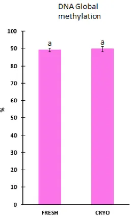
DNA methylation
The global DNA methylation between fresh and cryopreserved samples had no significant differences (p>0,05). The values obtained have a maximum of 89. 7 % of methylated DNA.
Figure 3.3 Global DNA methylation of European eel sperm. Fresh- Fresh sperm samples
and CRYO- Cryopreserved sperm samples. The results showed as mean ± 1.4 SEM and n of N=9, with the p>0.05. The letters on the top represent the significant difference
37
39
Receptor expression
The present study confirms the expression of five membrane and two nuclear progestin receptors through spermatogenesis in European eel testis. The nuclear and membrane progestin receptors are known to be highly expressed in fish testis (Hanna and Zhu, 2009) and it is confirmed in eels (Figure 3.1).
It has been showed that progestin induce early meiosis during spermatogenesis (Miura et al., 1991) and it regulates sperm maturation (Miura et al., 1991). The study settles these pattern for European eel spermatogenesis (Figure 3.1). In more detail the study suggests that in the European eel, the mPRalpha, mPRAL1 and mPRAL2 are involved in the regulation of sperm maturation, proposing an implication of these receptors on the final stage of spermatogenesis. Due to its expression profile (showed high expression in the spermatozoa stage, increasing gradually from the beginning to the end of spermatogenesis (Figure 3.1)), these receptor could be consider potential indicators of gametogenesis process. In the case of
mPRgamma and pgr2 an opposite profile was expressed (high expression in the
spermatogonia stage, showing a decrease, until spermatozoa stage.
Recently, it has been demonstrated that the concentrations on sperm plasma membranes of mPRalpha are positively correlated to sperm motility as well as sperm sensitivity to progestin, in flounder (Paralichthys lethostigma) (Tan et al., 2014). Adding these to the expression profiles of mPRalpha, reported and reminding that endocrine signalling controls are essential to physiological processes such as reproduction (Scholz and Mayer, 2008), mPRalpha is a potential biomarkers, for both spermatogenesis and sperm quality. Moreover, it was reported by Tubbs (2007) that mPRalpha is expressed in all testicular germ cell stages in Atlantic croaker (Micropogonias undulatus) and that mPRalpha revealed to be consistent with its purposed function as a maturation inducing steroid receptor in fish. Plus it is know that mPRalpha regulates motility in fish (Thomas et al., 2006).The presence of mPRalpha levels increases both sperm motility and fertilization success (Tan et
al., 2014). These facts suggest that the signalling pathway mediated by mPRalpha in sperm
is highly conserved in teleosts (Tubbs et al., 2010). These facts confirm its potential as a biomarker as well as mPRAL1 and mPRAL2, due to the similarities in its expressed profiles.
40
For the expression profile of nuclear pgrs1 the results were not very conclusive for its specific function on the European eel, being possible to say that its potential to become a biomarker is as low in the studied specie.
The industry would benefit with these biomarkers, because there is a need of reliable markers of sperm quality (Rurangwa et al., 2004). Naturally, the cost of the equipment would obstruct its access to these quality analyses, but collaboration between institutes and the laboratories could be seen as a solution. Plus, the research on these biomarkers can allow the design of specific markers for “sperm quality” and consequently increase the reproductive success, contributing to guarantee a high quality of the progeny.
These molecular biomarkers would provide knowledge about sperm expose to damages, such as cryopreservation or even toxicity, related with environmental contaminants, which are proven to interfere with hormonal regulation in vertebrates and consequently in reproduction (Scholz and Mayer, 2008).
Future research should be done to validate these progestin receptors in sperm samples. It would be important to make test on samples of good quality and of poor quality, since concentrations on sperm plasma membranes of mPRalpha are positively correlated to sperm motility (Tan et al., 2014) as mentioned.
Sperm quality analysis
Several protocols and investigation has been made in the comparison of fresh and cryopreserved sperm quality for eels (Pérez et al., 2000, 2003; Asturiano et al 2002; 2003; 2004; Muller et al., 2004; Szabó; Tanaka et al.,2002). The most successful protocols for artificial reproduction of Japanese eel had been published by Otha group (1996;1997) .The chosen protocol, for this study, was the one designed by Asturiano et al., (2003) for the European eel, based on the published studies made in the Japanese eel.
Fresh samples with the motility higher than 75% (as mentioned) were used to
proceed with analysis (high quality sperm is recommended for cryopreservation (Herráez et
41
recommend that early and late sperm should not be used for cryopreservation purposes (Billard and Zhang, 2001; Kopeika et al., 2007).
Even though, the cell survival of frozen-thawed sperm is affected by many factors such as: the male, freezing medium, cryoprotectant, thawing rates and the sperm quality (Chen et al., 1998 and Paredes and Bellas, 2009). It has been reported the possibility of cryopreserve eel sperm (Pérez et al., 2000, 2003; Asturiano et al 2002; 2003; 2004; Muller
et al., 2004; Szabó; Tanaka et al., 2002) and the present study reconfirmed that (~19% of
total motility, ~5% of progressive motility, ~100µm/s of VAP,~ 60µm/s of VSL and ~ 40µm/s of VCL) (Figure 3.2). It was reconfirm that spermatozoa mobility is affected by cryopreservation in all check parameters. However, lower motility values do not necessarily mean unsuccessful fertilization. In the salmonids case it was reported that post-thaw motility did not exceed 20-30% (total motility, 19% in the present study), yet this sperm was used for fertilization purposes, with success of 70-80% (800 eggs could be fertilized with 10µ1 of semen) (Scott and Baynes, 1980). The obtained results in the present study are toward to the observations of Tanaka et al. (2001) on the Japanese eel, that revealed a significant difference between frozen sperm samples and fresh sperm samples and still fertilization and hatching rates with the cryopreserved sperm were possible.
The preservation of ell sperm has been accomplish in both Japanese eel and European eel, but no optimal protocol has been developed so far, plus it is relatively low the post-thawing motility obtained in comparison with the fresh samples. These may indicate that new tools/different approaches are necessary, in order to understand better, the damaging points affecting post-thaw sperm quality, in order to improve the methods. Several parameters have been tested in the European eel (cell viability, motility parameters, cell morphology) but none has justified until know some damage occurring during freezing/thawing.
DNA methylation
It is known that the design of standard procedures/protocols for fish sperm cryopreservation remains, mostly, in the difficulty of guaranteeing homogeneous sperm quality post-thaw. Studies have reported the difficulty in to predict the quality of the freeze samples and different results are obtained, even when cryopreserved samples had the same apparent quality (Cabrita et al., 2008). In order to transfer cryopreservation methods to the
42
industry, and to standardize freezing protocols, it is very important to identify the parameters whose evaluation is required to guarantee post-thaw quality (Pérez-Cerezales et al., 2010). DNA is one of the cell components predisposed to suffer cryodamage; and several studies, in fish sperm quality, have pointed out the importance of maintaining DNA integrity during sperm cryopreservation (Labbé et al., 2001; Zilli et al., 2003; Cabrita et al., 2005; Pérez-Cerezales et al., 2009).
The mechanisms that lead to DNA damage sometimes are unclear and it is commonly believed that, especially DNA methylation, have an important role in it (Jenkins et al., 2014). Methylation damage in the DNA can be related to the use of cryoprotectants used for cryopreservation purposes as it has been describe for other fish species. The evaluation of DNA damage in fresh sperm should be seen as development of cryopreservation procedures (Pérez-Cerezales et al., 2010).
A quality marker is then important to ensure that the use of reproductive biotechnologies will not affect the sperm quality. LUMA method as a way to determine global methylation process, was assumed as a presumed quality marker for eel sperm, due to LUMA robustness, highly sensitivity, versatility and to the fact that it permits to accurate determine DNA methylation to a large number of samples, as reported by Karimi et al. (2006). LUMA proved that DNA methylation is unaffected by cryopreservation (Figure 3.3), suggesting that the protocol used seems to be secure in terms of DNA stability, to storage the sperm material of European eel.
On the other hand, it is important to emphasize that LUMA analyses the global DNA methylation and for this reason it could be more difficult to detected specific alterations. The nature of the test does not analyze the methylation on a specific gene. Therefore, in the present study DNA methylation determined by LUMA test could not prove to be a good marker of sperm quality due to this limitation. Several authors reported that DNA methylation patterns at specific loci and repetitive sequences are associated with human male infertility (Urdinguio et al., 2015), reinforcing the idea that DNA methylation is important to determine the quality of sperm.
43
45
In the present study it was concluded that, in the testis, the nuclear pgr2 and
mPRgamma1 and mPRgamma2 are involved on the induction of meiosis, and mPRalpha, mPRAL1 and mPRAL2 are involved in the process of final sperm maturation. It was also
settle, that the expression pattern of mPRalpha mPRAL1 and mPRAL2 are really good potential biomarkers for gametogenesis process and sperm quality.
The study also, reconfirmed that full sexual maturation and spermiation can be successfully induced in European eel. Plus, the used protocol for sperm cryopreservation damages the quality of the sperm in the tested parameters (Motility, VLS, VCL and VAP).
The present results for DNA methylation analysis, revealed that LUMA technique it cannot be considered a good biomarker for the quality of the sperm of European eel.
47
49
Aarestrup K, Økland F, Hansen MM, Righton D, Gargan P, Castonguay M, Bernatchez L, Howey P, Sparholt H, Pedersen MI, McKinley RS, 2009: Oceanic spawning migration of the European eel (Anguilla anguilla). Science 25, 1660.
Alemayehu L, 2011: Effect of Cryopreservation on Sperm Quality and Fertility, Artificial Insemination in Farm Animals. in: Manafi M, Effect of Cryopreservation on Sperm Quality and Fertility. Ethiopia : 14, 192-195.
Aranda A, Pascual A, 2001: Nuclear hormone receptors and gene expression.Physiological. Reviews 81, 1269–1300.
Asturiano JF, Jiménez MF, Pérez L, Balash S, Garzón DL, Peñarada DS, Vicent JS, Castr VMP, Jover M, 2006: Effects of hCG as spermiation inducer on European eel sperm quality. Theriogenology 66, 1012-1020.
Asturiano JF, Pérez L, Marco Jiménez F, Olivares L, Vicente JS, Jover M, 2003: Media and methods for the cryopreservation of European eel (Anguilla anguilla) sperm. Fish Physiology and Biochemistry 28, 501–502.
Asturiano JF, Pérez L, Tomás A, Zegrari S, Espinós FJ, Jover M, 2002: Hormonal induction of gonadal maturation and spawning in European eels females Anguilla anguilla): morphological changes and oocyte development. Boletin del Instituto Español de Oceanografía 18, 127-137.
Asturiano JF, Pérez L, Garzón DL, Jiménez M, Peñaranda DS, Vicente JS, Jover M, 2004: Physio-Chemical Characteristics of Seminal Plasma and Development of Media and Methods for the Cryopreservation of European eel Sperm. Fish Physiology and Biochemistr 30, 283-293.
Basavaraja N, Hegde SN, 2004: Cryopreservation of the endangered mahseer (Tor Khudree) spermatozoa: I. Effect of extender composition, cryoprotectants, dilution ratio, and storage period on post-thaw viability. Cryobiology 49, 149–156.
Benedikte HP, 2003: Induced sexual maturation of the European eel Anguilla anguilla and fertilisation of the eggs. Aquaculture 224, 323–338.
Bieniarz K, Epler P, 1977: Investigations on inducing sexual maturity in the male eel
Anguilla anguilla. Journal of Fish Biology 10, 555–559.
Billard R, Ginsburg SA, 1973: La spermiogenese et le spermatozoy d’Anguilla anguilla L. E’tude ultrastructurale. Annual Biology Animals Biochemistry Biophysics 13, 523-534.
Billard R, Zhang T, 2001: Techniques of genetic resource banking in fish, in: Watson PF, Holt WV, Cryobanking the genetic resource. London: 145–170.
Bissonnette JM, Knopp SJ, Maylie J, Thong T, 2009: Autonomic cardiovascular control in methyl-CpG-binding protein 2 (Mecp2) deficient mice. Autonomic Neuroscience 136, 82–89.
50
Blackmore PF, Neulen J, Lattanzio F, Beebe SJ, 1991: Cell surface-binding sites for progesterone mediated calcium-uptake in human sperm. Biology Chemistry 28, 18655–18659.
Bobe J and Labbe C, 2009: Egg and sperm quality in fish. General and Comparative Endocrinology 165, 48-535.
Boëtius I, Boëtius J, 1967: Studies in the European Eel, Anguilla anguilla. Experimental induction of the male sexual cycle, its relation to temperature and other factors. Meddelelser fra Danmarks Fiskeri-Og Havundersogelse 4, 339-405.
Boëtius I, Boëtius J, 1980: Experimental maturation of female silver eels, Anguilla anguilla. Estimates of fecundity and energy reserves for migration and spawing. Dana 1, 1-28. Cabrita E, Páramo SM, Gavaia PJ, Riesco MF, Valcarce DG, Sarasquete C, Herráez MP, Robles V, 2014: Factors enchancing fish sperm quality and emerging tools for sperm analysis. Aquaculture 432, 389-401.
Cabrita E, Robles V, Herráez MP, 2008: Marine and Freshwater Species. in: Cabrita E, Robles V, Herráez MP. Methods in Reproductive Aquaculture. 385–389.
Cabrita E, Sarasquete C, Martínez-Páramo S, Robles V, Beirão J, Pérez-Cerezales S, Herráe MP, 2010: Cryopreservation of fish sperm: applications and perspectives. Journal of Applied Ichthyology 26, 623-635.
Chen JD, Tsay T, Shie JM, Chern RH, 2012: Generation of transgenic tilapia by using cryopreserved electroporated sperm to mediate gene transfer. Fish Society of Taiwan 25, 265–272.
Demanno DA, Goetz FW, 1987: Steroid-induced final maturation in brook trout (Salvelinus
fontinalis) oocytes in vitro: the effects of forskolin and phosphodiesterase
inhibitors. Biology Reproduction 36, 1321–1332.
Dollerup J, Graver CM, 1985: Repeated induction of testicular maturation and spermiation, alternating with periods of feeding and growth in silver eels, Anguilla anguilla. Dana 4, 19-39.
Donnelly ET, Steele EK, McClure N, Lewis SEM, 2001: Assessment of DNA integrity and morphology of ejaculated spermatozoa from fertile and infertile men before and after cryopreservation. Human Reproduction 16, 1191-1199.
Dressing GE, Thomas P, 2007: Identification of membrane progestin receptors in human breast cancer cell lines and biopsies and their potential involvement in breast cancer. Steroids 72, 111–116.
Dufour S, Burzawa-Gerard E, Belle L, Sbaihi M, Vidal B 1983: Reproductive endocrinology of the European eel, Anguilla anguilla. in: Yamauchi Y, Eel Biology. Tokyo: 373– 383.
51
Evenson DP, Jost LK, Marshall D, Zinaman MJ, Clegg E, Purvis K, Angelis P, Claussen OP, 1999: Utility of the sperm chromatin structure assay as a diagnostic and prognostic tool in the human fertility clinic. Human Reproduction 14, 1039-1049.
Falkenstein E, Heck M, Gerdes D, Grube D, Christ M, Weigel M, Buddhikot M, Meizel S, Wehling M, 1999: Specific progesterone binding to a membrane protein and related nongenomic effects on Ca2+-fluxes in sperm. Endocrinology 140, 5999–6002. FAO (2014). World Review of Fisheries and Aquaculture for 2014 (Fisheries and
Aquaculture Technical Paper) Food and Agriculture Organization: Rome 15-16. FAO (2015). Statistical Pocketbook World Food and Agriculture for 2015. Food and
Agriculture Organization: Rome 14-22.
FEAP (2004-2015) European Aquaculture Production Report 2004-2015. Federation of European Aquaculture Producers. France.
Fernández JL, Muriel L, Rivero MT, Goyanes V, Vazquez R, Alvarez JG, 2003: The Sperm Chromatin Dispersion Test: A Simple Method for the Determination of Sperm DNA Fragmentation. Journal of Andrology 24, 56-66.
Feunteun E, 2002: Management and restoration of European eel population (Anguilla
anguilla): an impossible bargain. Ecological Engineering 18, 575-591.
Fontaine M, Bertrand L, Lopez L, Callamand L, 1964: Sur la maturation des organes génitaux de I' Anguille femelle (Anguilla anguilla) et l'émission spontané des œufs en aquarium. Comptes Rendus de l'Académie des Science 259, 2907-2910.
Gallego V, Carneiro PCF, Mazzeo I, Vílchez MC, Peñaranda DS, Soler C, Pérez L, Asturiano JF, 2013: Standardization of European eel (Anguilla anguilla) sperm motility evaluation by CASA software. Theriogenology 79, 1034-1040.
Gallego V, Mazzeo I, Vílchez MC, Peñaranda DS, Carneiro PCF, Pérez L, Asturiano JF, 2012: Study of the effects of thermal regime and alternative hormonal treatments on the reproductive performance of European eel males (Anguilla anguilla) during induced sexual maturation. Reproduction, Fertility and Development 354, 7-16. Garcia-Herrero S, Garrido N, Martinez-Conejero JA, Remohi J, Pellicer A, Meseguer M,
2011: Differencial transcriptomic profile in spermatozoa achieving pregnancy or not via ICSI. Reproductive Biomedicine Online 22, 25-36.
Garzón DL, Peñaranda DS, Pérez L, Jiménez FM, Espert X, Müller T, Jover M and Asturiano JF, 2008: Effects of pH, Sodium Bicarbonate, Cryoprotectants and Foetal Bovine Serum on the Cryopreservation of European Eel Sperm. Reproduction in Domestic Animals 43, 99-105.
Ginneken VGT and Maes G, 2005: The European eel (Anguilla anguilla, Linnaeus), its Lifecycle Evolution and Reproduction 15, 67-398.
52
Guerra SM, Valcarce DG, Cabrita E, Robles V, 2013: Analysis of transcripts in gilthead seabream sperm and zebrafish testicular cells: mRNA profile as a predictor gamete quality Aquaculture 406, 28-33.
Gwo JC, Ohta H, Okuzawa K, Wu HC, 1999: Cryopreservation of sperm from the endangered Formosan landlocked salmon (Oncorhynchus masou formosanus). Theriogenology 51, 569–582.
Hanna RN and Zhu Y, 2009: Expression of membrane progestin receptors in zebrafish (Danio rerio) oocytes, testis and pituitary. General and Comparative Endocrinology 1, 153-161.
Hanna RN, Daly SC, Pang Y, Anglade I, Kah, O, Thomas P, Zhu Y, 2010: Characterization and Expression of the Nuclear Progestin Receptor in Zebrafish Gonads and Brain. Biological. Reproduction. 82, 112-122.
Harper CV, Barratt CL, Publicover SJ, 2004: Stimulation of human spermatozoa with progesterone gradients to simulate approach to the oocyte. Induction of [Ca(2+)](i) oscillations and cyclical transitions in flagellar beating. Journal of Biological Chemistry 279, 46315–46325.
Herráez MP, Robles V, Cabrita E, 2008: Rainbow trout (Oncorhynchus mykiss) sperm cryopreservation.
Honaramooz A, Snedaker A , Boiani M, Schöler H, Dobrinski I, Schlatt S, 2002: Sperm from neonatal mammalian testes grafted in mice. Nature 15, 81-778.
Horváth A, Urbanyi B, 2000: The effect of cryoprotectants on the motility and fertilizing capacity of cryopreserved African catfish Clarias gariepinus sperm. Aquaculture
Research 31, 317– 324.
Huertas M, Scott AP, Hubbard PC, Canario AVM, Cerdá J, 2006: Sexually mature European eels (Anguilla anguilla) stimulate gonadal development of neigth-bouring males: possible involvement of chemical communication. General and Comparative Endocronology 147, 304-313.
Jaiswal BS, Tur-Kaspa I, Dor J, Mashiach S, Eisenbach M, 1999: Human sperm chemotaxis: is progesterone a chemoattractant?. Biology of Reproduction 60, 1314–1319.
Jenkins TG, Aston KI, Pflueger C, Cairns BR, Carrell DT, 2014: Age-Associated Sperm DNA Methylation Alterations: Possible Implications in Offspring Disease Susceptibility. Plos Genetics 10, 7-10
Jiméneza, D.L. Garzónb, D.S. Peñarandab, L. Pérezb, M.P. Viudes-de-Castroa, J.S. Vicentec, M. Joverb, J.F. Asturianob, 2006: Cryopreservation of European eel (Anguilla anguilla) spermatozoa: Effect of dilution ratio, foetal bovine serum supplementation, and cryoprotectants. Cryobiology 53, 51-57.
53
Johnson GD, Lalancette C, Linnemann AK, Leduc F, Boissonneault G, Krawetz SA, 2011: The sperm nucleus: chromatin, RNA and the nuclear matrix. Reproduction 141, 21-36.
Kagawa H, Kasuga Y, Adachi J, Nishi A, Hashimoto H, Imaizumi H, Kaji S, 2006: Effects of continuous administration of human chorionic gonodotropin, salmon pituitary extract, and gonodotropin-releasing hormone using osmotic pumps on induction of sexual maturation in male Japanese eel, Anguilla japonica. Aquaculture 296, 117-122.
Kamiya M and Utid S, 1968: Changes in activity of sodium-potassium-activated adenosinetriphosphatase in gills during adaptation of the Japanese eel to sea water. Comparative Biochemistry and Physiology 26, 675–685.
Karimi M, Johansson S, Stach D, Corcoran M, Grandér D, Schalling M, Bakalkin G, Lyko F, Larsson C, Ekström TJ, 2006: LUMA (LUminometric Methylation Assay)-a high throughput method to the analysis of genomic DNA methylation 312, 1989-1995. Karteris E, Zervou S, Pang Y, Dong J, Hillhouse EW, Randeva HS, Thomas P 2006:
Progesterone signaling in human myometrium through two novel membrane G protein coupled receptors: potential role in functional progesterone withdrawal at term. Mollecular Endocrinology 20, 1519–1534.
Khan IA, Lopez E, Leloup-Hâtey J, 2002: Induction of spermatogenesis and spermiation by a single injection of human chorionic gonadotropin in intact and hypophysectomized immature European eel (Anguilla anguilla). General and Comparative Endocrinology 68, 91-103.
Kime DE, Van Look KJW, McAllister BG, Huyskens G, Rurangwa E, Ollevier F, 2001: Computer-assisted sperm analysis (CASA) as a tool for monitoring sperm quality in fish. Comparative Biochemistry and Physiology 130, 425-433.
Kirkman-Brown JC, Barratt CL, Publicover SJ, 2004: Slow calcium oscillations in human spermatozoa. Biochemical Journal 378, 827–832.
Kopeika E, Kopeika J, Zhang T, 2007: Cryopreservation of fish sperm. Methods Mol Biol 368, 203–217.
Kottelat M, Freyhof J, 2007: Handbook of European freshwater fishes. in: Kottelat C, Freyhof J. Berlin: 646.
Kurokura H, Hirano R, Tomita M, Iwahashi M, 1984: Cryopreservation of carp sperm. Aquaculture 37, 267-273.
Labbe C, Martoriati A, Devaux A, Maisse G, 2001: Effect of sperm cryopreservation on sperm DNA stability and progeny development in rainbow trout. Molecular Reproduction and Developemnt 60, 397–404.
Lalancette C, Miller D, Li Y, Krawetz SA, 2008: Paternal contributions: New functional insights for spermatozoal RNA. Cell Biochemestry 104, 1570-1579.
54
Laverty G, Skadhauge E, 2012: Adaptation of teleosts to very high salinity. Comparative and Biochemistry Physiological 163, 1–6.
Leloup-Hâtey L, 1985: Testicular steroidogenesis during gonadotropin induced spermatogenesis in male European eel (Anguilla anguilla). in: Oudinet JP, Lopez E, Softs B, Holmes WN, Current Trends in Comparative Endocrinology. Hong Kong: 229-239.
Liang Y, Huang GY, Liu SS, Zhao JL, Yang YY, Chen XW, Tian F, Jiang Y, Ying G, 2015: Long-term exposure to environmentally relevant concentrations of progesterone and norgestrel affects sex differentiation in zebrafish. Aquatic Toxicology 160, 172–179. Liu D, Dillon JS, 2002: Dehydroepandrosterone activates endotherlial cell nitricoxide synthetase by a specific plasma membrane receptor coupled to Gα12,3. Biology Chemestry 277, 21379–21388.
Luconi M, Bonaccorsi L, Maggi M, Pecchioli P, Krausz C, Forti G, Baldi E, 1998: Identification and characterization of functional nongenomic progesterone receptors on human sperm membrane. Clinical Endocrinology Metabolism 83, 877–885. Magyary I, Urbányi B, Horváth L, 1996: Criopreservation of common carp (Cyprinus
carpio). Journal of Applied Ichthyology 12, 117–119.
Maller JL, 1998: Recurring themes in oocyte maturation. Biology Cell 90, 453–460
Matallanas, J. (2005): A world overview of species of interest to fisheries. Chapter: Anguilla
anguilla. FishBase 2203.
Mazzeo I, Peñaranda DS, Gallego V, Baloche S, Nourezadeh-Lillabade R, Tveiten H, Dufour S, Asturiano JF, Weltzien FA, Pérez L, 2014: Temperature modelates the progression of vitologenesis in the European eel. Aquaculture 434, 38-47.
Meske C, 1973: Experimentaly induced sexual maturity in artificially reared male eels (Anguilla anguilla). in: Schröder JH, Genetics and Mutagenesis of Fish. Berlin: 161-170.
Meyer C, Christ M, Wehling M, 1995: Characterization and solubilization of novel aldosterone binding proteins in porcine liver microsomes. Biochemesttry 229, 736– 740.
Migliaccio A, Piccolo D, Castoria G, Di Domenico M, Balancio A, Lombardi M, Gong W, Beato M, Auricchio F, 1998: Activation of the Src/p21ras /Erk pathway iby
progesterone receptor via cross-talk with estrogen receptor. EMBO 17, 101–111. Miura T and Miura CI, 2001: Molecular control mechanisms of fish spermatogenesis. Fish
Physiology and Biochemistry 1, 181-186.
Miura T, Yamauchi K, Nagahama Y, Takahashi H, 1991:Indoctuon of spermatogenesis in male Japanese eel (Anguilla japonica) by a single injection of human chorionic