ELISABETH DUPIN, CARLA REAL and NICOLE LeDOUARIN
Institut d’Embryologie Cellulaire et Moléculaire (CNRS FRE 2160) 49 bis Avenue de la Belle Gabrielle, 94136 Nogent-sur-Marne, France
Manuscript received on July 19, 2001; accepted for publication on July 26, 2001; presented byVivaldo Moura Neto
ABSTRACT
How the considerable diversity of neural crest (NC)-derived cell types arises in the vertebrate embryo has long been a key question in developmental biology. The pluripotency and plasticity of differentiation of the NC cell population has been fully documented and it is well-established that environmental cues play an important role in patterning the NC derivatives throughout the body. Over the past decade,in vivoandin vitrocellular approaches have unravelled the differentiation potentialities of single NC cells and led to the discovery of NC stem cells. Although it is clear that the final fate of individual cells is in agreement with their final position within the embryo, it has to be stressed that the NC cells that reach target sites are pluripotent and further restrictions occur only late in development. It is therefore a heterogenous collection of cells that is submitted to local environmental signals in the various NC-derived structures. Several factors were thus identified which favor the development of subsets of NC-derived cellsin vitro. Moreover, the strategy of gene targeting in mouse has led at identifying new molecules able to control one or several aspects of NC cell differentiationin vivo. Endothelin peptides (and endothelin receptors) are among those. The conjunction of recent data obtained in mouse and avian embryos and reviewed here contributes to a better understanding of the action of the endothelin signaling pathway in the emergence and stability of NC-derived cell phenotypes.
Key words: neural crest, avian embryo, cell differentiation, endothelin3, peripheral nervous system, melanocytes, Schwann cells, mesectoderm.
INTRODUCTION
The neural crest (NC) arises at the junction between the prospective epidermis and the prospective neural plate in vertebrates, regardless of whether the NC cells ultimately detach from the open neural folds (as in mammals and amphibians), the closed neural tube (as in birds), or from an ectodermal thickening at the neural plate-epidermis boundary (as in fish). Shortly afterward the morphologically identical NC
∗Invited paper
Correspondence to: Dr. Elisabeth Dupin
E-mail: [email protected] / Fax: 33+1 48734377
cells migrate extensively throughout the embryo and later give rise to a wide array of derivatives in a variety of sites.
TABLE I
Summary of the neural crest-derived cell types in the vertebrate embryo.
N e u r a l c r e s t d e r i v a t i v e s
Neuronal cells
Sensory ganglia of cranial nerves V, VII, IX, X
Spinal ganglia
Ganglion cells of the autonomic nervous system
Supportive cells of the nervous system
Glial cells of the peripheral ganglia
Schwann cells of peripheral nerves
Meninges of the anterior brain
Pigment cells– (except for pigmented retina)
Endocrine and paraendocrine cells
Adrenomedullary cells
Calcitonin-producing cells
Type I cells of the carotid body
Mesectodermal derivatives– (cephalic neural crest)
Visceral and facial skeleton
Cranial vault
Walls of large arteries derived from the aortic arches
Connective tissue of thymus and parathyroid glands
Dermis of neck and facial regions
cells of the sensory and autonomic ganglia of the peripheral nervous system (PNS), to the entire en-teric nervous system, and to various glandular and endocrine cell types. In addition, the cephalic NC from the level of the diencephalon yields mesenchy-mal cell types forming the so-called mesectoderm, including the facial and head dermis and connec-tive tissue, the smooth muscles of the wall of arte-rial trunks and most of the skull vault (i.e., parietal and frontal bones) (Couly et al. 1993). In addi-tion, the NC yields the pigment cells (except those of the pigmented retina). Melanocytes are generated along almost the entire axis of the embryo whereas other NC derivatives such as the chromaffin cells of the adrenal medulla and the enteric nerve cells are
em-bryo. Nevertheless, the trunk NC cells are unable to contribute to mesectodermal structures when grafted at cephalic level, therefore suggesting an early de-velopmental restriction of NC cells caudal to somite 5 as compared to the more rostral cephalic NC.
THE GENERATION OF NEURAL CREST CELLULAR DIVERSITY; THE NEURAL CREST STEM
CELL CONCEPT
A key question in understanding the ontogeny of NC derivatives is to elucidate how cellular diversity is generated from the apparently homogeneous pop-ulation of NC cells. Two different scenarios can be made to explain the diversity of cell phenotypes arising from such a pluripotent population of embry-onic cells. One possibility is that the NC consists of a heterogeneous assortment of committed unipotent progenitor cells, each programmed to give rise to a particular derivative. These progenitor cells could migrate in a directed fashion to their proper loca-tions and differentiate according to prescribed fates, or they could migrate randomly and those localized in inappropriate sites must either fail to differentiate or die. A second possibility is that the NC is a ho-mogeneous population of multipotent cells, capable of generating any crest derivative under appropriate conditions. Those multipotent cells might migrate randomly and differentiate according to instructive cues encountered along their migratory pathways or at their final sites.
With the establishment of clonal cultures of NC cells and with the complement in vivosingle cell tracing experiments, it was revealed that the reality lies somewhere in between these two extremes.
Using a permissive and constant culture medium, that theoretically permitted the differenti-ation of all NC-derived cell types, it has been pos-sible to study the potentialities of individual cells in vitro. In the 1970s, Cohen and co-workers were first able to establish cultures of trunk NC cells from the quail neural primordium. NC cells in a colony assay generated clones either entirely pigmented, partially pigmented or entirely without pigment ob-tained (Cohen and Konigsberg 1975). The
produc-tion of antibodies to neural antigens later allowed a better characterization of multipotent avian trunk NC cells, by identifying the existence of both cate-cholaminergic and sensory neurons in multipheno-typic colonies (Sieber-Blum 1989).
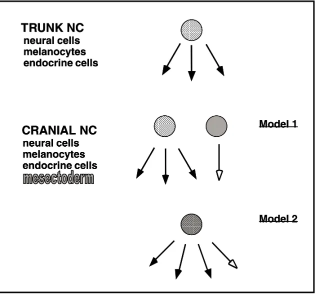
In vitroclonal analysis permitted us to address the controversial question of the origin of mesecto-dermal lineages from the cephalic NC. Two models could be proposed (Figure 1), a first one assum-ing an early segregation in the premigratory NC of mesectodermal precursors from the progenitors of other NC-derived (neural, melanocytic and en-docrine) lineages, and a second model where the NC includes highly multipotent cells capable to gener-ate both mesectoderm and other NC derivatives. By seeding migratory NC cells individually on a feeder layer of growth-inhibited 3T3 fibroblasts and ana-lyzing their progeny with lineage-specific markers, evidence was provided for extremely diverse devel-opmental and proliferative potentialities of NC cells taken from the mes-metencephalic level (Baroffio et al. 1988, 1991, Dupin et al. 1990).
Fig. 1 – Models for the origin of mesectodermal derivatives from the cephalic NC. Cranial and trunk NC share common derivatives (neural cells, melanocytes and endocrine cells) which derive from a multipotent NC stem cell in the trunk (striped circle). In addition to these trunk-like derivatives, the cephalic NC yields the mesectoderm. Two models can be proposed : Either the mesectoderm is generated from a specific precursor (grey circle) which is segregated early from trunk-like multipotent NC cells (striped circle) (Model 1) ; The other possibility is that common precursors (striped grey circle) are endowed with all cephalic NC potentialities, including the ability to yield mesectodermal derivatives (Model 2).
NC cells divide during migration, progressive re-striction takes place in the developmental potentials of the cells generated from an initial pool of multi-potent stem cells.
Pluripotency of NC cells was confirmed for mammalian cells by in vitro clonal analysis of mouse (Ito et al. 1993) and rat (Stemple and
Fig. 2 – Model for the segregation of cell lineages derived from the cephalic NC. Diagram illustrating the different progenitors from quail mesencephalic NC cells grown in clonal cultures. The presence of cartilage (C), neurons (N), glial cells (G), adrenergic cells (A) and melanocytes (M) was recorded in the colonies. Progenitors have been classified according to the number of these distinct phenotypes in their progeny. The results are consistent with the generation of unipotent progenitors from a ‘totipotent’ stem cell through several intermediate oligopotent precursors. Filiations between precursors are only hypothetical.
to give rise to multiple phenotypes and to self-renew (Stemple and Anderson 1992).
However, it is possible that crest cells isolated in vitromay behave differently than they do in the living embryo, due to the influence of signals coming from thein vivoenvironment and from cell-cell in-teractions. Therefore, an important complement to thein vitroexperiments is prospective cell lineage analysis in the intact embryo where an individual
that NC cells predetermined to the sensory neuron phenotype populate the dorsal root ganglion, thus confirming earlier evidence for the presence of com-mitted sensory neuronal precursors in the premigra-tory NC (Ziller et al. 1983).
Taken together, these results show that NC cells are heterogeneous in their developmental poten-tialities, including multipotent stem cell-like pro-genitors as well as already committed precursors. Therefore, appropriated signals from the environ-ment are required to promote the differentiation of the proper NC-derived cell types in the various derivatives. Knowing the factors that influ-ence the expression of developmental potentialities of NC cells locally and are responsible for final fate decisions is of great importance for understanding the molecular mechanisms underlying NC multipo-tency.
GROWTH FACTOR CONTROL OF NEURAL CREST CELL FATES
The evidence for the existence of multipotent pro-genitors in the NC raised the question of how these progenitors generate distinct phenotypes in the vari-ous NC derivatives. On one hand, cell fate could be assigned by lineage or other cell-autonomous mechanisms. On the other hand, cell fate could be influenced or determined by cell-extrinsic signals. Similar to the model proposed for the hematopoi-etic system, it seems likely that both types of mech-anisms operate in NC cell specification. In this model, multipotent stem cells generate progenitors committed to one or more sublineages, which then proliferate, survive and differentiate in response to specific environmental cues. The process of differ-entiation necessarily involves many changes in gene expression, which are brought about by regulatory transcription factors acting in the cell nucleus. One goal of studying NC differentiation is to understand the relationship between the action of environmental signals at the cell surface and the changes in tran-scriptional regulation that these signals bring about in target cells.
In general, it is thought that NC cells are
di-rected toward particular fates by signals localized along migratory pathways and at differentiation sites. Many such factors appear to instruct multi-potent NC cells to form specific derivatives. Others appear to act selectively, affecting the proliferation or survival of particular NC progenitors.
glial lineage commitment. The generation of glia in PNS ganglia, i.e., in a local environment containing strong instructive neurogenic factors (BMP2), is due to expression by the neuroblasts of Notch ligands, which in turn impairs neighboring cells to enter the neuronal pathway. The loss of neurogenic capity promoted by activation of Notch signaling is ac-quired rapidly and irreversibly, leading to glial cell differentiation (Morrison et al. 2000, Wakamatsu et al. 2000).
Another factor that plays a role in NC cell dif-ferentiation is retinoic acid (RA), a natural derivative of vitamin A. RA is known to inducein vitrothe dif-ferentiation of many cell lines of NC origin (Amos and Lotan 1990) and to cause NC embryopathies after deprivation or administration in excess during vertebrate embryonic development (Morriss-Kay 1993). In vitro experiments have shown that RA favors the development of neuronal lineages derived from the crest (Henion and Weston 1994, Dupin and Le Douarin 1995, Ito and Morita 1995, Rockwood and Maxwell 1996). In cephalic and trunk quail NC clonal cultures, it influences the differentiation of cells when they are in pluripotent state by specif-ically increasing the number of adrenergic cells in their progeny. RA also stimulates pigment synthesis in melanoblasts differentiatingin vitro(Dupin and Le Douarin 1995).
The mouse genetics proved to be very useful for the knowledge of the factors that influ-ence the migration, survival and differentiation of NC melanocytic precursors. Mouse mutants with alterations in coat color have permitted the study of pigment cell development. Two well-known mouse spotting mutants with clear defects in melanogenesis arewhite dominant-spotting(w) andSteel(sl). Het-erozygote mutants have a patchy coat color, whereas homozygotes are either completely white or die dur-ing embryogenesis, although the extent of pigmenta-tion deficit varies with different mutant alleles. The analysis of the responsible genes revealed a molec-ular relationship between the two mutant strains: the w locus encodes the receptor tyrosine kinase c-kit, whereas thesllocus encodes its cognate
lig-and, variously known as steel factor (SLF), stem cell factor or mast cell factor (for references, see Reith and Bernstein 1991, Williams et al. 1990, 1992, Galli et al. 1993). A number ofin vitrostudies of mouse and avian NC suggest that SLF promotes sur-vival and moderate proliferation of melanocytes or their precursors (Murphy et al. 1992, Lahav et al. 1994, Reid et al. 1995). Lahav et al. reported that SLF promotes the appearance of an early marker of melanoblasts/melanocytes in cultured quail NC cells (Lahav et al. 1994).
THE ROLE OF ENDOTHELIN 3 IN NEURAL CREST CELL DIFFERENTIATION
The endothelins are another determinant group of factors responsible for the differentiation of certain NC derivatives. The endothelins 1, 2 and 3 (EDN1, EDN2 and EDN3) are a family of 21 aa peptides that activate, in mammals, one or both of the two heptahelical, G-protein-coupled endothelin recep-tors type A and B (EDNRA and EDNRB). EDNRA exhibits different affinities for endothelin peptides and EDNRB accepts all three peptides equally. The EDN1/EDNRA signaling pathway thus acts on cra-nial NC derivatives. Target disruption ofEDN1and EDNRAgenes in mouse results in blood pressure el-evation and cranio-facial abnormalities. The devel-opment of thyroid, tongue and other facial structures is impaired (Kurihara et al. 1994, 1995). Many of these defects are related to abnormal development of NC-derived mesectoderm, which forms most of the connective tissue, cartilage and bones of the head and face. The breakthrough in deciphering the role of EDN3/EDNRB in NC development was the iden-tification of the genetic defects responsible for the Piebald (s) and Lethal spotting (ls) mutations in mouse. These mutations reside in the genes encod-ing EDNRB (Puffenberger et al. 1994) and its lig-and EDN3, respectively (Greenstein-Baynash et al. 1994).
phenotype of coat color spotting and aganglionic ter-minal gut (megacolon).
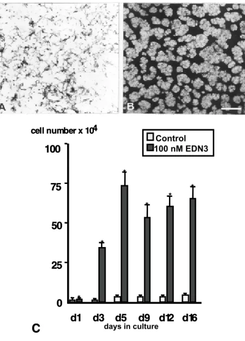
Both EDN3 and its receptor EDNRB are there-fore necessary for the development of NC-derived melanocytes and posterior enteric nerve cells. In vitrostudies have shown that EDN3 favors the de-velopment of melanocyte progenitors in mouse and avian NC cultures (Lahav et al. 1996, Reid et al. 1996) (Figure 3). The time at which melanocyte precursors appear probably corresponds to the stage before crest cells need the action of EDNRB/EDN3 receptor-ligand system. In mouse, requirement for functionalEDNRBcorresponds to a restricted pe-riod in melanoblast early migration, before entry into the epidermis (Shin et al. 1999). In avians, hu-man EDN3 peptide is a potent mitogen for NC cells well before the appearance of melanogenic differ-entiation (Lahav et al. 1996). These observations therefore suggest thatin vivoEDN3 is essential for increasing the early NC cell population to ensure a sufficient supply of melanoblasts needed for colo-nizing the entire skin. In addition, the analysis of the effects of EDN3 on single quail NC cells in cul-ture indicates that EDN3 promotes the survival and proliferation of uni- and bipotent melanocyte pre-cursors (Lahav et al. 1998).
In the avian embryo, NC cells initially express EDNRBbefore they leave the neural primordium, all along the neural axis (Nataf et al. 1996). At later stages,EDNRBis expressed along the medio-lateral pathway of NC cell migration in rat embryos (Gariepy et al. 1998). By contrast, EDNRB tran-scripts are not detected in avian melanocytes and melanoblasts. However, it was found that EDNRB2, a third EDNR identified in birds (Lecoin et al. 1998), is expressed by melanoblasts as soon as they begin to migrate from the migrating staging area near the dorsal neural tube, then when they progress along the medio-lateral pathway and later on during fur-ther differentiation of melanocytes. Expression of EDNRB2in the NC is thus restricted to the cells of the melanocytic lineage which develop in close contact to the epidermal source of the EDN3 ligand (Nataf et al. 1998).
In vitroprolonged exposure to EDN3 causes trunk NC cells to switch fromEDNRBtoEDNRB2 expression, which eventually results in the positive selection of melanogenic cells (Lahav et al. 1998). In vivo,EDNRB, which is expressed by virtually all premigratory and early migratory avian NC cells, is switched off by crest cells which migrate medio-laterally under the ectoderm and which start to ex-press EDNRB2 (Figure 3). It is therefore likely that, during avian skin development, EDN3 syn-thesized by the epidermis triggers, via activation of EDNRB2, the survival and proliferation of melano-blasts and eventually melanocytes.
PLASTICITY OF NEURAL CREST-DERIVED PHENOTYPES
Once they have differentiated, functional cells usu-ally maintain their cellular specificity in the multi-cellular organization of animals. In some pathologi-cal conditions, however, such as reparative regener-ation, tumorigenesis and carcinogenesis, conversion of cellular specificities can frequently be observed. Lens regeneration observed in thegenus Triturushas been considered to be the clearest and most repre-sentative example of cell type conversion, as newt lens can be regenerated from the dorsal iris by meta-plasia of its pigment cells. The whole neural retina of the newt can also be regenerated from the retinal pigment epithelium. The same results were obtained using the pigment epithelium of chick eyes and, un-der spreading culture conditions, it was observed the transdifferentiation of chick neural retina into lens and pigment cells (for references, Eguchi and Ko-dama 1993).
Fig. 3 – Effects of EDN3 on melanogenesis and cell division by quail NC cellsin vitro. (A, B) Effect of EDN3 on the pigmentation by trunk NC cells after 11 days in culture. In control medium (A), melanocytes remain dispersed whereas EDN3-treated cultures (B) show reproducible pattern of pigment cells and unpigmented pre-melanocytes. (C) Quantification of EDN3-induced changes of the total cell number at different time points. Cultures of quail trunk NC cells were analyzed during a culture period of 16 days. Significant differences are indicated by a star at the top of columns. (From Lahav et al. 1996).
glial-specific proteins (Dupin et al. 2000). There-fore, epidermal pigment cellsin vitrocan be reversed to the bipotent stage of glia-melanocytic NC progen-itor, which suggests that proliferative signals such as EDN3 may alter the stability of the pigment cell
phenotypein vivo.
melanocytes. The onset of melanogenesis by divid-ing Schwann cells occurs progressively, by sequen-tial induction ofEDNRB2, early melanocyte anti-gens and later on melanin synthesis (Dupin, Real, Glavieux, Vaigot and Le Douarin, in preparation).
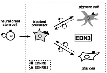
Fig. 4 – Summary of the action of EDN3 on NC precursors and NC-derived cells. EDN3 favor the survival and proliferation of bipotent glial-melanocytic progenitors derived from NC stem cells in the early NC. Activation by EDN3 of the melanogenic dif-ferentiation program involves the induction of EDNRB2 expres-sion by developing melanoblasts, whereas differentiating glial cells continue to express EDNRB. Whether the bipotent cells are able to co-express both types of receptors remains to be ascer-tained. At later stages, differentiated pigment cells in the epi-dermis and glial cells in peripheral nerves are able to revert to the immature bipotent stage of their common NC ancestor under prolongedin vitroexposure to EDN3.
Taken together, these results show that the se-lection and expansion of melanogenic precursors de-pend on the local action of EDN3 on bipotent glial-melanocytic NC cells derived from NC stem cells (Figure 4). The immature bipotent stage of this common ancestor can be recapitulatedin vitroby differentiated pigment and glial cells, thus provid-ing evidence for the plasticity of NC cell fate which is represented here by the reciprocal conversion be-tween pigment and glial phenotypes. This suggests that EDN3, due to its strong proliferative activity on NC precursors and NC-derived cells, could play a rolein vivoin several pathologies implicating both glia and melanocytes, such as neurofibromatosis and
tumors of mixed origin.
Abbreviations:EDN3, endothelin 3; EDNRB, en-dothelin receptor B; NC, neural crest; PNS, periph-eral nervous system; SLF, steel factor.
ACKNOWLEDGEMENTS
We wish to thank Michèle Scaglia for prepar-ing bibliography. This work was supported by the Centre National pour la Recherche Scientifique and Collège de France and by a grant from the As-sociation pour la Recherche contre le Cancer (N◦ 5578). C.R. is recipient of a doctoral fellowship from Fundação para Ciência e Tecnologia, Portugal (SFRH/BD/990/2000).
RESUMO
O modo como a diversidade dos tipos celulares derivados da crista neural (CN) surge, no embrião de vertebrado, tem sido uma pergunta chave na biologia do desenvolvimento.
A pluripotência e a plasticidade na diferenciação da
popu-lação de células da CN têm sido intensivamente
documen-tadas, ficando deste modo estabelecido que os factores
ambientais têm um papel importante na correta
diferen-ciação dos derivados da CN no organismo. Na década
passada, estudosin vivoein vitro, revelaram as
poten-cialidades de diferenciação de células de CN individuais
levando à descoberta de células ‘‘stem’’ (células tronco)
da CN. Embora seja claro que a diferenciação celular
fi-nal das células individuais está de acordo com a sua
lo-calização final no embrião, é necessário realçar que as
células da CN que atingem locais alvo são pluripotentes
e as restrições impostas nas suas potencialidades
ocor-rem somente numa fase mais tardia do desenvolvimento.
A CN é, deste modo, um conjunto heterogêneo de
célu-las que é submetida a sinais ambientais localizados nas
várias estruturas derivadas da CN. Foram assim
identifi-cados vários factores que favorecem a diferenciação de
subgrupos de células derivadas da CN,in vitro. Para
além destes factores foram também identificadas novas
moléculas capazes de controlar um ou vários aspectos da
diferenciação celular da CNin vivo, através da
estraté-gia molecular de ‘‘gene targeting’’ em ratinho. As
moléculas. O conjunto dos resultados obtidos
recente-mente em embriões de ratinho e de galinha e descritos no
presente artigo, contribuíram para uma melhor
compreen-são do modo de ação da via de sinalização das endotelinas
no controlo do aparecimento e da estabilidade dos
fenoti-pos celulares derivados da CN.
Palavras-chave: crista neural, embrião de ave, diferen-ciação celular, endotelina3, sistema nervoso periférico,
melanocitos, células de Schwann, mesectoderme.
REFERENCES
Amos B and Lotan R.1990. Retinoid-sensitive cells and cell lines. Methods Enzymol 190: 217-225.
Baroffio A, Dupin E and Le Douarin NM. 1988. Clone-forming ability and differentiation potential of migratory neural crest cells. Proc Natl Acad Sci USA 85: 5325-5329.
Baroffio A, Dupin E and Le Douarin NM. 1991. Common precursors for neural and mesectodermal derivatives in the cephalic neural crest. Development 112: 301-305.
Bronner-Fraser M and Fraser SE.1988. Cell lineage analysis reveals multipotency of some avian neural crest cells. Nature 335: 161-164.
Bronner-Fraser M and Fraser SE.1989. Develop-mental potential of avian trunk neural crest cellsin situ. Neuron 3: 755-766.
Cohen AM and Konigsberg IR.1975. A clonal ap-proach to the problem of neural crest determination. Dev Biol 46: 262-280.
Couly GF, Coltey PM and Le Douarin NM.1993. The triple origin of skull in higher vertebrates: a study in quail-chick chimeras. Development 117: 409-429.
Dupin E and Le Douarin NM.1995. Retinoic acid promotes the differentiation of adrenergic cells and melanocytes in quail neural crest cultures. Dev Biol 168: 529-548.
Dupin E, Baroffio A, Dulac C, Cameron-Curry P
and Le Douarin NM.1990. Schwann-cell differ-entiation in clonal cultures of the neural crest, as evidenced by the anti-Schwann cell myelin protein monoclonal antibody. Proc Natl Acad Sci USA 87: 1119-1123.
Dupin E, Glavieux C, Vaigot P and Le Douarin
NM.2000. Endothelin 3 induces the reversion of melanocytes to glia through a neural crest-derived glial-melanscytic progenitor. Proc Natl Acad Sci USA 97: 7882-7887.
Eguchi G and Kodama R.1993. Transdifferentiation. Curr Opin Cell Biol 5: 1023-1028.
Galli SJ, Zsebo KM and Geissler EN.1993. The kit ligand, Stem Cell Factor. Adv Immunol 55: 1-96.
Gariepy CE, Williams SC, Richardson JA, Hammer
RE and Yanagisawa M.1998. Transgenic expres-sion of the endothelin-B receptor prevents congeni-tal intestinal aganglionosis in a rat model of Hirsch-sprung disease. J Clin Invest 102: 1092-1101.
Greenstein-Baynash A, Hosoda K, Giaid A,
Richardson JA, Emoto N, Hammer RE and
Yanagisawa M. 1994. Interaction of endothelin-3 with endothelin-B receptor is essential for develop-ment of epidermal melanocytes and enteric neurons. Cell 79: 1277-1285.
Henion PD and Weston JA.1994. Retinoic acid se-lectively promotes the survival and proliferation of neurogenic precursors in cultured neural crest cell populations. Dev Biol 161: 243-250.
Ito K and Morita T.1995. Role of retinoic acid in mouse neural crest cell developmentin vitro. Dev Dyn 204: 211-218.
Ito K, Morita T and Sieber-Blum M.1993. In vitro Clonal Analysis of Mouse Neural Crest Develop-ment. Dev Biol 157: 517-525.
Kurihara Y, Kurihara H, Suzuki H, Kodama T,
Mae-mura K, Nagai R, Oda H, Kuwaki T, Cao WH,
Kamada N et al. 1994. Elevated blood pressure and craniofacial abnormalities in mice deficient in endothelin-1. Nature 368: 703-710.
Kurihara Y, Kurihara H, Oda H, Maemura K, Nagai
R, Ishikawa T and Yazaki Y. 1995. Aortic arch malformations and ventricular septal defect in mice deficient in endothelin-1. J Clin Invest 96: 293-300.
Lahav R, Lecoin L, Ziller C, Nataf V, Carnahan
JF, Martin FH and Le Douarin NM.1994. Effect of the Steel gene product on melanogenesis in avian neural crest cell cultures. Differentiation 58: 133-139.
1996. Endothelin 3 promotes neural crest cell prolif-eration and mediates a vast increase in melanocyte number in culture. Proc Natl Acad Sci USA 93: 3892-3897.
Lahav R, Dupin E, Lecoin L, Glavieux C, Champeval
D, Ziller C and Le Douarin NM.1998. Endothe-lin 3 selectively promotes survival and proliferation of neural crest-derived glial and melanocytic precur-sorsin vitro. Proc Natl Acad Sci USA 95: 14214-14219.
Lecoin L, Sakurai T, Ngo MT, Abe Y, Yanagisawa M
and Le Douarin NM.1998. Cloning and character-ization of a novel endothelin receptor subtype in the avian class. Proc Natl Acad Sci USA 95: 3024-3029.
Le Douarin N.1982. The neural crest. Cambridge: Cambridge University Press. 259p.
Le Douarin NM and Kalcheim C.1999. The Neu-ral Crest (second edition). New York: Cambridge University Press. 445p.
Morriss-Kay G.1993. Retinoic acid and craniofacial development molecules and morphogenesis. Bioes-says 15: 9-15.
Morrison SJ, Perez SE, Qiao Z, Verdi JM, Hicks C,
Weinmaster G and Anderson DJ.2000. Transient Notch activation initiates an irreversible switch from neurogenesis to gliogenesis by neural crest stem cells. Cell 101: 499-510.
Murphy M, Reid K, Williams DE, Lyman SD and
Bartlett PF.1992. Steel factor is required for main-tenance, but not differentiation, of melanocyte pre-cursors in the neural crest. Dev Biol 153: 396-401.
Nataf V, Lecoin L, Eichmann A and Le Douarin NM.
1996. Endothelin-B receptor is expressed by neural crest cells in the avian embryo. Proc Natl Acad Sci USA 93: 9645-9650.
Nataf V, Amemiya A, Yanagisawa M and Le Douarin
NM.1998. The expression pattern of endothelin 3 in the avian embryo. Mech Dev 73: 217-220.
Puffenberger EG, Hosoda K, Washington SS,
Nakao K, Dewit D, Yanagisawa M and
Chakravarti A. 1994. A missense mutation of the endothelin-B receptor gene in multigenic Hirschsprung’s disease. Cell 79: 1257-1266.
Reid K, Nishikawa SI, Bartlett PF and Murphy M.
1995. Steel factor directs melanocyte development
in vitrothrough selective regulation of the number of c-kit(+) progenitors. Dev Biol 169: 568-579.
Reid K, Turnley AM, Maxwell GD, Kurihara Y,
Kurihara H, Bartlett PF and Murphy M.1996. Multiple roles for endothelin in melanocyte devel-opment: regulation of progenitor number and stim-ulation of differentiation. Development 122: 3911-3919.
Reith AD and Bernstein A.1991. Molecular biology of theWandStellloci. In: Genome analysis. Vol. 3. Genes and Phenotypes. New York: Cold Spring Harbor Laboratory Press, p. 105-133.
Rockwood JM and Maxwell GD.1996. An analysis of the effects of retinoic acid and other retinoids on the development of adrenergic cells from the avian neural crest. Exp Cell Res 223: 250-258.
Shah NM and Anderson DJ.1997. Integration of mul-tiple instructive cues by neural crest stem cells reveals cell-intrinsic biases in relative growth factor respon-siveness. Proc Natl Acad Sci USA 94: 11369-11374.
Shah NM, Marchionni MA, Isaacs I, Stroobant P
and Anderson DJ.1994. Glial growth factor re-stricts mammalian neural crest stem cells to a glial fate. Cell 77: 349-360.
Shah NM, Groves AK and Anderson DJ.1996. Al-ternative neural crest cell fates are instructively pro-moted by TGF beta superfamily members. Cell 85: 331-343.
Shin MK, Levorse JM, Ingram RS and Tilghman
SM.1999. The temporal requirement for endothe-lin receptor-B signalendothe-ling during neural crest devel-opment. Nature 402: 496-501.
Sieber-Blum M.1989. Commitment of neural crest cells to the sensory neuron lineage. Science 243: 1608-1611.
Stemple DL and Anderson DJ.1992. Isolation of a stem cell for neurons and glia from the mammalian neural crest. Cell 71: 973-985.
Wakamatsu Y, Maynard TM and Weston JA.2000. Fate determination of neural crest cells by NOTCH-mediated lateral inhibition and asymmetrical cell division during gangliogenesis. Development 127: 2811-2821.
White PM, Morrison SJ, Orimoto K, Kubu CJ, Verdi
undergo cell-intrinsic developmental changes in sen-sitivity to instructive differentiation signals. Neuron 29: 57-71.
Williams DE, Eisenman J, Baird A, Rauch C,
Van Ness K, March CJ, Park LS, Martin
U, Mochizuki DY, Boswell HS, Burgess GS,
Cosman D and Lyman SD. 1990. Identification of a ligand for the c-kit proto-oncogene. Cell 63: 167-174.
Williams DE, de Vries P, Namen AE, Widmer MB
and Lyman SD.1992. The Steel factor. Dev Biol 151: 368-376.
Ziller C, Dupin E, Brazeau P, Paulin D and Le