In Vivo
Transplantation of Enteric Neural
Crest Cells into Mouse Gut; Engraftment,
Functional Integration and Long-Term Safety
Julie E. Cooper1, Conor J. McCann1, Dipa Natarajan1, Shanas Choudhury1,
Werend Boesmans2, Jean-Marie Delalande1,3, Pieter Vanden Berghe2, Alan J. Burns1,4*, Nikhil Thapar1,5*
1Stem Cells and Regenerative Medicine, UCL Institute of Child Health, 30 Guilford Street, London, United Kingdom,2Laboratory for Enteric NeuroScience, Translational Research Center for GastroIntestinal Disorders, Department of Clinical and Experimental Medicine, University of Leuven, Leuven, Belgium, 3Centre for Digestive Diseases, Blizard Building, Barts & The London School of Medicine & Dentistry, Queen Mary, University of London, London, United Kingdom,4Department of Clinical Genetics, Erasmus MC University Medical Centre, Rotterdam, The Netherlands,5Department of Gastroenterology, Great Ormond Street Hospital NHS Foundation Trust, Great Ormond Street, London, United Kingdom
*[email protected](NT);[email protected](AJB)
Abstract
Objectives
Enteric neuropathies are severe gastrointestinal disorders with unsatisfactory outcomes. We aimed to investigate the potential of enteric neural stem cell therapy approaches for such disorders by transplanting mouse enteric neural crest cells (ENCCs) into ganglionic and aganglionic mouse gutin vivoand analysing functional integration and long-term safety.
Design
Neurospheres generated from yellow fluorescent protein (YFP) expressing ENCCs
selected from postnatalWnt1-cre;R26R-YFP/YFPmurine gut were transplanted into gangli-onic hindgut of wild-type littermates or agangligangli-onic hindgut ofEdnrbtm1Ywamice (lacking functional endothelin receptor type-B). Intestines were then assessed for ENCC integration and differentiation using immunohistochemistry, cell function using calcium imaging, and long-term safety using PCR to detect off-target YFP expression.
Results
YFP+ ENCCs engrafted, proliferated and differentiated into enteric neurons and glia within recipient ganglionic gut. Transplanted cells and their projections spread along the nous myenteric plexus to form branching networks. Electrical point stimulation of endoge-nous nerve fibres resulted in calcium transients (F/F0 = 1.16±0.01;43 cells, n = 6) in YFP+ transplanted ENCCs (abolished with TTX). Long-term follow-up (24 months) showed trans-planted ENCCs did not give rise to tumours or spread to other organs (PCR negative in OPEN ACCESS
Citation:Cooper JE, McCann CJ, Natarajan D, Choudhury S, Boesmans W, Delalande J-M, et al. (2016)In VivoTransplantation of Enteric Neural Crest Cells into Mouse Gut; Engraftment, Functional Integration and Long-Term Safety. PLoS ONE 11(1): e0147989. doi:10.1371/journal.pone.0147989
Editor:Wenhui Hu, Temple University School of Medicine, UNITED STATES
Received:October 20, 2015
Accepted:January 11, 2016
Published:January 29, 2016
Copyright:© 2016 Cooper et al. This is an open access article distributed under the terms of the
Creative Commons Attribution License, which permits unrestricted use, distribution, and reproduction in any medium, provided the original author and source are credited.
Data Availability Statement:All relevant data are within the paper and its Supporting Information files.
extraintestinal sites). In aganglionic gut ENCCs similarly spread and differentiated to form neuronal and glial networks with projections closely associated with endogenous neural networks of the transition zone.
Conclusions
Transplanted ENCCs successfully engrafted into recipient ganglionic and aganglionic gut showing appropriate spread, localisation and, importantly, functional integration without any long-term safety issues. This study provides key support for the development and use of enteric neural stem cell therapies.
Introduction
Enteric neuropathies represent some of the most severe clinical gastrointestinal (GI) disorders and are characterised by failure of normal propulsive contractile activity leading to functional
obstruction to the flow of luminal contents.[1–3] Archetypal disorders include the congenital
disorder Hirschsprung disease and acquired oesophageal achalasia, which result from an abso-lute absence or loss of intrinsic enteric neurons in the distal and proximal parts of the GI tract respectively. More subtle neuronal defects underlie other disorders such as intestinal pseudo-obstruction and slow transit constipation, which show evidence of neuropathy on histology or
on contractile profiles from intestinal physiological studies.[4–7]
Enteric neuropathies are generally devastating conditions, which left untreated are life threatening. Current available treatments are largely limited to surgery to decompress the intestine or resect its most abnormal segments and the provision of specialised nutrition, often parenteral, to preserve growth and development. Outcomes of such interventions are often
unsatisfactory and associated with significant complications[8,9] highlighting the need for
alternative approaches.
Tremendous advances in regenerative medicine have brought the prospect of neural regen-eration to replace deficient or defective enteric neurons as a potential therapeutic strategy for
enteric neuropathies to the forefront.[8,10–12] Several cell types have been identified as
poten-tial sources of donor cells for a cell replacement therapy, such as skin-derived precursors and
central nervous system progenitors [13–16]. Arguably the most promising progress towards
this ultimate goal has been made using enteric neural crest cells (ENCCs) which comprise enteric neural stem cells, neurons and glia and are derived from the original population of migratory neural crest cells (NCC) that, during embryogenesis, colonise the gut to form the
enteric nervous system (ENS) [17–22].
The isolation of enteric neural stem cells was first described over a decade ago from murine
gut, including models of Hirschsprung disease.[23] Since then there has been steady progress
emanating from the isolation of such cells from human intestine including from the gut
mucosa of patients obtained by conventional endoscopy[24]. We and others have shown that
ENCCs contained within discrete cellular aggregates or neurospheres (including enteric neural
stem cells, neurons and glia) formed in culture can colonise recipient bowelin vitro.[23–32].
Most recently,in vivostudies have confirmed that ENCCs, predominantly from embryonic
mouse gut, are capable of migration, proliferation and differentiation and they retain some
functionality after transplantation into postnatal mouse gut.[16,33–36] Although these initial
studies have addressed aspects of feasibility of ENS stem cell transplants, a number of chal-lenges remain, including questions concerning long-term safety and the ability of transplanted
Centre at Great Ormond Street Hospital for Children NHS Foundation Trust and University College London. The funders had no role in study design, data collection and analysis, decision to publish or preparation of the manuscript.
cells to form neural networks, which connect to the endogenous enteric neural network. The
ability to form an integrated and functional ENS after transplantation,in vivo, will be critical in
the treatment of both aganglionic and euganglionic enteric neuropathies.
In this paper we confirm that postnatal mouse-derived ENCCs can colonise ganglionic
mouse gutin vivoand form neural networks, which functionally integrate with endogenous
ENS. We show that ENCCs similarly colonise aganglionic bowel. The transplanted cells do not appear to pose any long-term safety risk within the recipient animals.
Materials and Methods
Animals
Animals used for this study were maintained and the experiments performed were in accor-dance with the local approvals and the UK Animals (Scientific Procedures) Act 1986 under
licence from the Home Office (PPL70/7500).Wnt1-cre;R26R-YFP/YFPmice[37–39] in
which NCC express yellow fluorescent protein (YFP) were used to obtain YFP+ enteric
NCC (ENCCs).R26R-YFP/YFPlittermates (in which YFP is not expressed in the absence of
cre) were used as ganglionic recipient bowel into which YFP+ ENCCs were transplanted.
Ednrbtm1Ywamice lacking endothelin receptor type B (a model for Hirschsprung Disease) (obtained from Jackson Laboratory (JAX#003295)) were used as aganglionic recipient bowel for YFP+ ENCCs.
Isolation, cell sorting and culture of ENCCs
YFP+ ENCCs were obtained from early postnatal (P2-P4)Wnt1-cre;R26R-YFP/YFPmice.
Muscle strips of bowel (containing muscle layers and myenteric and submucosal plexuses) were dissociated enzymatically (Collagenase, Sigma, 1mg/ml) for 30 minutes at 37°C. Dissoci-ated cells were washed with neurosphere medium (NSM) (DMEM F12 supplemented with B27 (InvitrogenLife Technologies, UK), N2 (Life Technologies, UK), 20ng/ml EGF (Peprotech, UK), 20ng/ml FGF (Peprotech, UK), and Primocin antibiotic (Invivo Gen, UK)) and filtered
through a 100μm and 40μm mesh.
ENCCs were isolated from the total cell population using fluorescence activated cell sorting (FACS) for YFP. Dissociated cells were resuspended in NSM with 2% foetal calf serum before undergoing FACS using a MoFloXDP cell sorter (Beckman Coulter).
ENCC were washed with NSM before plating on fibronectin-coated wells of 6-well dishes in NSM. They were maintained in culture and generated neurospheres from around 1 week. Pri-mary neurospheres generated in cultures maintained for a maximum of 30 days were used for the transplants.
Transplantation of ENCCs into gut in vivo
YFP expressing ENCCs derived fromWnt1-cre;R26R-YFP/YFPmice were transplanted into
the ganglionic distal colon of‘wild-type’R26R-YFP/YFPlittermates (which did not express
YFP in the absence of cre) via laparotomy at weaning (postnatal day 21) (n = 66). The same
YFP expressing ENCCs were transplanted into Ednrbtm1Ywamutants (identified by their
pie-bald markings) as early as practically possible at postnatal day 9–13 (n = 5).
mouth pipette and inserted into a small pocket in the serosa of the bowel using the bevel of a syringe needle. The exteriorised bowel was returned to the abdomen and the laparotomy inci-sion was closed using a combination of absorbable sutures (Ethicon) and wound clips (World Precision Instruments), which were removed 7 days post-surgery.
Bromodeoxyuridine (BrdU) (Sigma, UK; 10mM (10μl/g weight of 10 mg/ml BrdU) in PBS)
was injected intraperitoneally at the time of surgery with additional application 24 hours post surgery.
TransplantedR26R-YFP/YFPmice were checked periodically following surgery and of those
transplanted 4 (of 66) were in obvious discomfort in the 12 hours following surgery and were euthanised according to protocol. The cause was determined to be perforation of the bowel. The rest were typically maintained for around 4 weeks post-transplantation before sacrifice and removal of bowel for analysis, although some animals were maintained up to 24 months to
obtain long-term safety data. Three of the 5 transplanted Ednrbtm1Ywamutants developed
abdominal swelling and loss of condition without obvious intestinal perforation 2–4 days after transplant and were euthanized. The other 2 animals were euthanized at day 5 post-transplant.
PCR
In order to ascertain whetherWnt1-cre;R26R-YFP/YFP-derived transplanted cells migrated to
locations other than the gut following transplantation intoR26R-YFP/YFPrecipients, PCR was
performed to identify the cre transgene. Brain, lungs, heart, liver, spleen, kidneys, adrenal
glands and gut mesentery were collected on sacrifice and frozen.Wnt1-cre;R26R-YFP/YFP
pos-itive control tissue was obtained fromWnt1-cre;R26RYFP/YFPgut tissue, transplanted gut, YFP+
neurospheres and aWnt1-cre;R26RYFP/YFPear biopsy. DNA was extracted using DNAReleasy
(Anachem) diluted 1μl in 4μl water and heated to 75°C for 5min followed by 96°C for 2 min.
PCR was performed using cre primers (ACCCTGATCCTGGCAATTTCGGC and 5’-GATGCAACGAGTGATGAGGTTCGC) and a cycle with a 60°C annealing temperature.
Immunohistochemistry
Cells and transplanted bowel were fixed and analysed using immunohistochemistry as
described previously[24,40]. The primary and secondary antibodies used in the study are listed
inTable 1andTable 2.
Briefly, for wholemount immunolabelling, gastrointestinal tracts were fixed in 4% PFA (1h),
washed (2x10min, PBS) and then blocked (1h–overnight, wholemount blocking solution (WBS)
(PBS, 2% Triton X-100, 10% sheep serum). Primary antibodies were applied for 48h at 4°C before washing (2x1h, PBS with 1% Triton X-100) and addition of secondary antibodies (24h, 4°C). Sec-ondary antibodies were washed and samples mounted on glass coverslips for imaging.
For labelling with BrdU antibody, samples were first labelled with other primary antibodies as described above. Samples were post fixed (4% PFA,10 min, RT) and washed 3x in PBS. This was followed by treatment with 2M HCl for 15minutes at RT. After washing (3x15min), Rat anti-BrdU (Oxford Biotech, UK, 1:20) was applied overnight at 4°C, followed by goat anti-rat 568 (1:500) secondary antibody. Post antibody washings and mounting were carried out as described above.
Images were acquired on a Zeiss LSM 710 confocal microscope (Zeiss, Cambridge, UK) and
were processed using ImageJ and Adobe Photoshop software[41,42].
Calcium imaging of transplanted mouse ENCCs
120.9 NaCl, 5.9 KCl, 1.2 MgCl2, 2.5 CaCl2, 11.5 glucose, 14.4 NaHCO3 and 1.2 NaH2PO4). After removing the mucosa, gut preparations were pinned tightly, serosal side up, in a Sylgard-lined chamber. Transplanted YFP+ cells were identified and imaged at this stage.
Tissues were then loaded with the fluorescent Ca2+indicator Fluo-4AM (Molecular Probes,
Invitrogen; 5mM) and Cremophor EL (Fluka Chemika, Buchs, Switzerland; 0.01%) in Krebs solution at room temperature for 20 minutes with continuous oxygenation.
After loading, tissues were washed (2x10 min, Krebs) prior to imaging. Live fluorescence imaging was performed on a Zeiss Examiner microscope equipped with a 20x (NA 1) water dipping lens, Poly V monochromator (TILL Photonics, Gräfelfing, Germany) and cooled CCD camera (Imago QE; TILL Photonics, Gräfelfing, Germany). The experimental chamber volume was maintained at 3ml via a gravity-fed perfusion system ensuring continuous perfusion (1ml/min) with 95% oxygen/5% carbon dioxide-gassed Krebs solution (at RT), excess solution was removed via a peristaltic suction pump. Fluo-4 was excited at 475nm, and its fluorescence
emission was collected at 525/50 nm. Images (640X512 pixels2) were acquired at 2 Hz.
Electrical train stimulation (2 s, 20 Hz of 300μs electrical pulses; Grass Instruments,
Quincy, Massachusetts, USA) was applied via a platinum electrode (diameter 25μm), inserted
at a distance of 200μm from the most peripheral YFP+ cell or fibre in the observed transplanted
region of interest. Care was taken during this insertion to ensure contact was restricted to endogenous (non YFP+) fibres. To confirm the neuronal origin of the signal in YFP+
trans-planted cells, experiments were conducted in the presence of tetrodotoxin (1μM, Sigma,
Bor-nem, Belgium). Local application of high K+was also used as a means of determining basic
functionality of transplanted cells within host tissues. Table 1. Primary antibodies for immunohistochemistry studies.
PRIMARY ANTIBODY CONCENTRATION COMPANY
Mouse anti-TuJ1 1:500 Covance
Mouse anti-GFAP 1:500 Dako
Rabbit anti-GFP 1:500 Invitrogen
Mouse anti-GFP 1:500 Invitrogen
Chicken anti-GFP 1:500 Abcam
Goat anti-Sox10 1:300 Santa Cruz
Rabbit anti-S100 1:400 Dako
Rabbit anti-nNOS 1:400 Invitrogen
Rabbit anti-VIP 1:400 AbD Serotec
Goat anti-ChAT 1:300 Millipore
Rat anti-BrdU 1:20 Oxford Biotech
Mouse anti-Synaptophysin 1:250 AbD Serotec
doi:10.1371/journal.pone.0147989.t001
Table 2. Secondary antibodies for immunohistochemistry studies.
SECONDARY ANTIBODY ALEXA FLUOR CONCENTRATION COMPANY
Goat anti-rabbit 488 1:500 Invitrogen
Goat anti-mouse 488 1:500 Invitrogen
Anti-chicken 488 1:500 Abcam
Anti-mouse 568 1:500 Invitrogen
Anti-rabbit 568 1:500 Invitrogen
Anti-mouse 647 1:500 Invitrogen
Anti-rabbit 647 1:500 Invitrogen
Images were collected using TILLVision software (TILL Photonics) and post acquisition analysis performed in IGOR PRO (Wavemetrics, Lake Oswego, Oregon, USA). Movement arti-facts were removed by registering the image stack to the first image. Regions of interest (ROI) were drawn over each cell, fluorescence intensity was normalised to basal fluorescence for each
ROI(F/F0), and peaks analysed [43–45].
Results
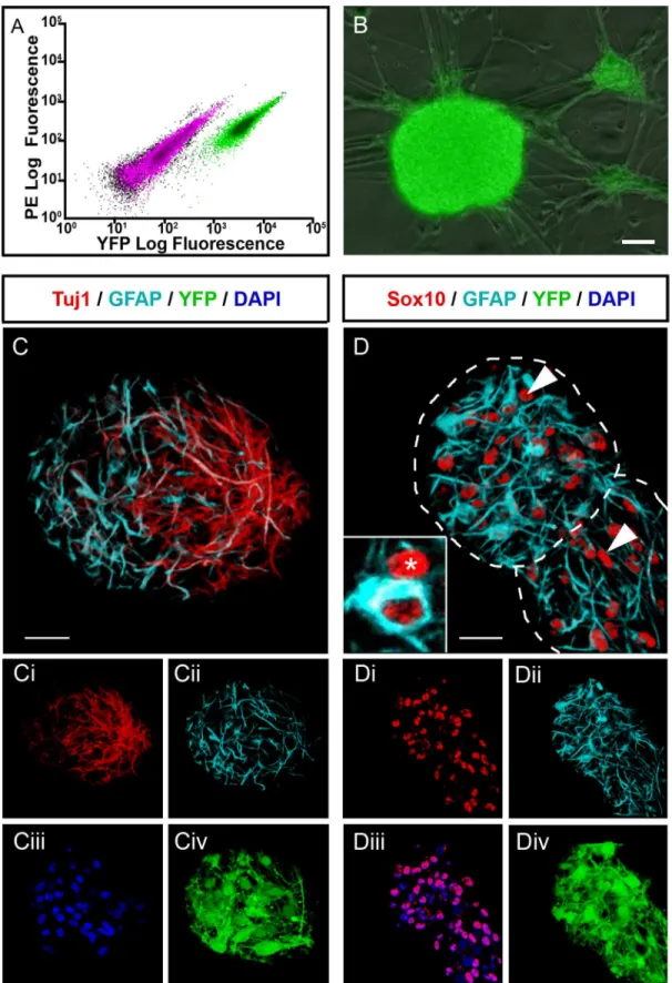
YFP+ ENCCs form neurospheres containing neurons, glia and
presumptive enteric neural stem cells
Intestinal muscle strips were taken fromWnt1-cre;R26R-YFP/YFPmice aged P2-4 in which
NCC and their derivatives express YFP. Following gut dissociation, YFP+ ENCCs were selected
by FACS where these cells accounted for 16.5±1.9% (n = 6) of the total gut cell population (Fig
1A). Selected YFP+ mouse ENCCs formed characteristic neurospheres of approximately 20μm
in diameter within 2 weeks in culture, which increased in size to approximately 120μm within
a month (Fig 1B). The neurospheres comprised cells expressing ENS markers such as the
pan-neuronal marker TuJ1 (31.7%±3.2% of cells within the neurosphere (n = 3)) and the glial marker GFAP (27.7%±5.9% (n = 3)), which maintained their YFP expression throughout
cul-ture (Fig 1C). They also contained cells that were immunopositive for Sox10 but negative for
GFAP (Fig 1D), the expression profile of presumptive enteric neural stem cells (Sox10+ cells
accounted for 67.7%±8.7% of cells within the neurosphere (n = 3), thus Sox10+;GFAP- enteric neural stem cells accounting for around 40%).
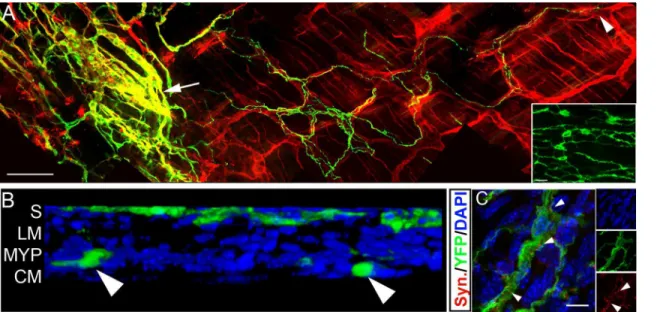
Transplanted ENCCs show appropriate colonisation, localisation and
formation of ENS-like networks in ganglionic host gut
YFP+ neurospheres were transplanted into postnatal wild-type mouse distal hindgut. YFP+ transplanted ENCCs were subsequently identified within the gut wall of 56/62 animals exam-ined (90.3%), up to 24 months post-transplantation. Within the gut, YFP+ ENCCs spread orally and anally from the site of transplantation and formed extensive branching networks
co-located with the endogenous ENS (Fig 2Aincluding inset andS1 Figpanel A). By 15 weeks
post-transplantation YFP+ networks, on average, covered an area of 4.3±3.1mm2and cell
bod-ies were observed at a distance of 1.4±0.4mm from the site of transplantation (n = 10) (S1 Fig
panel B and C). These parameters, as well as the proximal-distal spread of transplanted cells,
showed positive correlation to the number of days post-transplantation (S1 Figpanel B
(r = 0.68, n = 32, p<0.01); 1C (r = 0.54, n = 28, p<0.01); and 1D (r = 0.51, n = 28 p<0.01)
respectively). There was no significant difference between the mean oral (0.9±0.38mm) and
aboral (0.9±0.4mm) spread (t = 0.45, n = 6, p<0.5).
Cell bodies (Fig 2B) of, and projections (arrowhead,Fig 2A) from, transplanted YFP+TuJ1+
neuronal cells were located within the endogenous myenteric plexus and projected within it for several millimetres. Punctate labelling around transplanted cells with the synaptic vesicle
pro-tein synaptophysin (arrowheads,Fig 2C) suggested synapse formation within the host gut.
Transplanted YFP+ ENCCs show functional integration within host gut
In order to test the functional integration of transplanted YFP+ ENCCs within the host gut
mus-culature, we examined [Ca2+]iupon electrical stimulation of the endogenous enteric neural
net-work (Fig 3A) or used application of high K+as a means of neuronal activation. Electrical point
stimulation of endogenous enteric nerve fibres resulted in calcium transients (F/F0 = 1.16±0.01;
S1 Movie). These calcium transients were abolished in the presence of 1μM TTX, confirming
their neuronal identity (Fig 3D and 3E; 43 cells, n = 6) (S1 Movie). Local application of high K+
also resulted in similar widespread calcium transients throughout YFP+ transplanted cell net-works and contributed to large contractions of the musculature (data not shown).
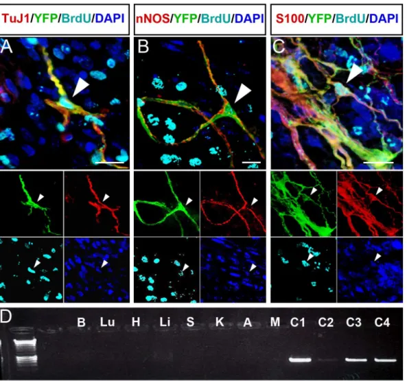
Transplanted YFP+ ENCCs generate a range of ENS cell types in vivo
The ENS cell-types of transplanted YFP+ ENCCs were analysed by immunofluorescence 4-weeks after transplantation. 53±14% of transplanted cells expressed the neuronal marker
TuJ1 (n = 13) (Fig 4AandS2 Figpanel A andS3 Figpanel A). nNOS expressing cells accounted
for the majority of neurons (50±5% of transplanted cells;Fig 4BandS2 Figpanel B andS3 Fig
panel A). Transplanted cells expressed other neuronal markers, such as ChAT, Calbindin and
VIP (S2 Figpanels C, D and E respectively) but these cell types were less numerous (data not
shown). Transplanted cells also expressed the glial cell markers S100 and GFAP (Fig 4CandS2
Figpanel F and G). S100+ cells comprised 64±22% of transplanted cells (S3 Figpanel A; n = 3).
YFP+ ENCCs proliferate post-transplant giving rise to enteric neurons
and glia
The proliferative capability of transplanted YFP+ cells was assessed by BrdU incorporation. BrdU pulses were administered at transplantation and 24h later. BrdU incorporation was
iden-tified within YFP+ cells and quaniden-tified in different cell types (S3 Figpanel B). 26±19% of
(outlined by dotted lines) also contain cells (asterisk and arrowheads) that express Sox10 (red;D, DiandDiii,arrowheads) but are negative for GFAP (cyan; D, Dii) i.e. presumptive ENSSCs. Inset inDshows a cross section through a Sox10+/GFAP- cell (red only; asterisk; presumptive ENSSC) adjacent to a Sox10+/GFAP+ cell (red and cyan; presumptive glial cell). Scale bar inB-D= 20μm.
doi:10.1371/journal.pone.0147989.g001
Fig 2. ENCCs from transplanted murine neurospheres show appropriate colonisation, localisation and formation of ENS-like networks in recipient wild-type gut. A.Wholemount gut preparation showing YFP+ transplanted cells (green) projecting along endogenous TuJ1+ (red) ENS nerve fibres. Arrow indicates cell bodies at the presumptive site of transplantation and arrowhead indicates distal extent of projections of transplanted cells. YFP+ transplanted cells also expressing the neuronal marker TuJ1 are seen as yellow. Inset shows a high power image taken from boxed region inS1 Figpanel A revealing interconnections and network formation between YFP+ cell bodies.B.Confocal 3D reconstruction showing YFP+ transplanted cells (green) located at the site of transplantation on the serosal surface (S) and within the myenteric plexus (MYP; arrowheads) between the inner circular (CM) and outer longitudinal (LM) muscle layers.C.YFP+ transplanted cells (green) co-locate with the synaptic marker synaptophysin (red; arrowheads). Scale bar inA= 100μm;C=
25μm. Inset in C shows individual channels.
TuJ1+ cells (Fig 4AandS3 Figpanels B and C; n = 13), 36±19% of nNOS+ cells (Fig 4BandS3
Figpanel B, n = 3) and 32±17% of S100+ cells (Fig 4CandS3 Figpanel B; n = 3) showed BrdU
incorporation. This suggests that varying proportions of transplanted cells expressing neuronal and glia markers are derived from cells that proliferate during the 48hrs following
transplantation.
Transplanted YFP+ ENCCs do not show uncontrolled proliferation, form
tumours or spread to other organs
Long-term studies of mice that were the recipients of YFP+ ENCC transplants were con-ducted to assess safety of these transplants. Although initial BrdU exposure (48 hours after transplantation) resulted in BrdU incorporation in transplanted cells, exposure 48hrs prior to culling at 4 weeks post-transplantation did not show BrdU incorporation (data not shown) suggesting there was no uncontrolled proliferation of transplanted cells. Macro-scopic and PCR examination of transplanted animals and tissues aged 19–25 months (including brain, lungs, heart, liver, spleen, kidneys, adrenal glands and gut mesentery)
failed to identify any YFP fluorescence orcretransgene within organs other than the gut
and positive controls (Fig 4D).
Fig 3. Transplanted YFP+ cells show functional integration within host gut. A.Schematic of experimental protocol demonstrating electrical point stimulation of host enteric nerve fiber at a site distant from YFP+ transplanted cells.B,C.Representative images of YFP+ cells before (B) and after (C) Fluo4-AM loading. Arrows indicate transplanted neurons (TP cell) from which Ca2+responses are plotted in(D). D.Representative traces showing Ca2+ responses recorded as F/F0 from TP cell in control conditions (solid lines) and after addition of TTX (dotted lines).E.Summary data demonstrating abolition of Ca2+responses in the presence of TTX (43 cells, n = 6). Also seeS1 Movie.
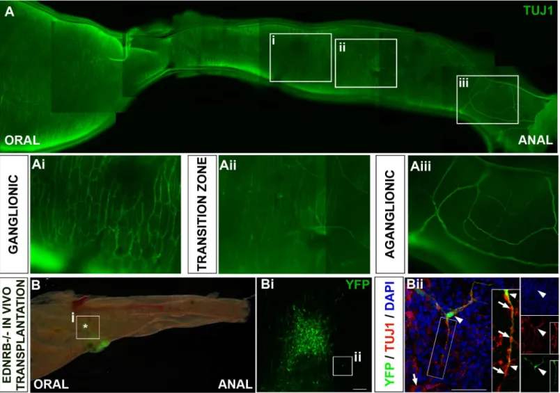
YFP+ ENCCs transplanted into aganglionic Ednrb
tm1Ywagut form
branching neuronal networks
Ednrbtm1Ywamice are characterised by a variable length of aganglionosis in the distal colon and
mutants within our colony survive for around two weeks. Tuj1 immunohistochemistry showed
the proximal colon appeared normally ganglionated (Fig 5A and 5Ai) progressing distally to
the hypoganglionated region of transition zone (Fig 5A and 5Aii) and to the distal-most bowel,
which, although showing innervation by extrinsic fibres, lacks an intrinsic ENS (Fig 5A and
5Aiii).
YFP+ neurospheres were transplanted into the most distal portion of hindgut accessible
from the peritoneal cavity of Ednrbtm1Ywamice (n = 5). Transplanted cells survived and could
be identified by their YFP fluorescence in 4 out of 5 guts after 3–5 days (Fig 5B). Transplanted
Fig 4. Transplanted YFP+ mouse ENCCs proliferate and generate enteric neurons and glia but do not spread beyond transplanted gut. A-C. Z-projections in which transplanted YFP+ cells co-express ENS markers (yellow; arrowheads) including the pan-neuronal marker TuJ1 (A), the inhibitory neuronal marker nNOS (B), and the glial marker S100 (C). Transplanted cells co-expressing ENS markers also demonstrate BrdU incorporation (cyan; arrowheads;A-Cand insets). DAPI labels nuclei in blue (A-Cand insets). Insets show individual channels.D.Wnt1-cre;R26RYFP/YFPexpressing transplanted cells (YFP+) were identified withinR26RYFP/YFPrecipient mice by the presence of the cre transgene on PCR. Representative cre-PCR to identify cre expressing cells within the major organs from a transplanted mouse. Brain (B), lungs (Lu), heart (H), liver (Li), spleen (S), kidneys (K), adrenal glands (A) and gut mesentery (M) were all negative for cre. Control tissues:Wnt1-cre;R26RYFP/YFP
gut tissue (C1), transplanted gut (C2), YFP+ neurospheres (C3) and Wnt1-cre;R26RYFP/YFPear biopsy (C4) are cre+. Scale bar inA-C= 20μm.
cells included both Tuj1+ neurons (Fig 5Bii) and S100+ glia (data not shown) and had spread
out to form interconnected neural networks (Fig 5Bi). Furthermore projections from
trans-planted neuronal cells associated closely with the endogenous neurons of the transition zone (Fig 5Biiand inset in 5Bii).
Discussion
Our studies confirm the feasibility of thein vivotransplantation of post-natally sourced
ENCCs into both ganglionic and aganglionic post-natal intestine. Not only are transplanted cells able to engraft successfully into significant segments of intestine they are able to show crit-ical functional integration with the endogenous neural networks. Importantly, our work sug-gests that ENCC transplantation is safe in the long-term.
Fig 5. YFP+ ENCC colonise aganglionic Ednrbtm1Ywagut in vivo. A.Neurons in an Ednrbtm1Ywacolon immunolabelled with TuJ1 progressing from
proximal ganglionated gut (Ai), through the partially ganglionated transition zone (Aii) to the distal aganglionic gut (Aiii).B.Ednrbtm1Ywagut 3 days after transplantation with YFP+ ENCCs (green) which could be identified in distal colon. Boxed area is area enlarged in Bi and asterisk denoted presumptive site of transplantation in this region.Bi. Interconnections are visible between transplanted cells that have spread from the site of transplantation.Bii.Z-projections in which endogenous neurons express the neuronal marker Tuj1 (red; arrow) and YFP+ transplanted cells (green) also coexpress Tuj1 (yellow; arrowhead). Projections from YFP+;Tuj1+ transplanted cells closely associate with projections from endogenous Tuj1+ neurons present in the transition zone (insets in Bii;arrowheads and arrows respectively). Scale bar inBi= 100μm;Bii= 50μm. DAPi is shown in blue.
Only one other study to date has robustly tested engraftment and functional integrity of
ENCCs followingin vivotransplantation [32]. Our study, however, extended this work
signifi-cantly across a number of domains from assessment of engraftment, post-transplant cell behaviour and functional integration with endogenous neurons, through to long-term studies
of viability and safety. We were also able to assessin vivotransplantation into aganglionic
intestine.
Our exclusive use of postnatal intestine in thein vivosetting as both donor and recipient of
ENCCs served to best recapitulate the clinical setting of autologous transplantation. We have previously shown that ENCCs can be harvested from human post-natal gut, even utilising min-imally invasive techniques such as endoscopy. Such cells successfully colonised recipient agan-glionic gut either embryonic gut maintained on chorioallantoic membrane or human gut
maintainedin vitro[24]. Although human ENCCs remain to be tested in thein vivosetting our
murine studies confirm that the therapeutic strategy of autologous transplantation of ENCCs into aganglionic and euganglionic intestine is a viable option.
Previous studies have suggested that recipient intestine with established endogenous enteric
neurons is poorly receptive to transplanted cells [10]. Our studies and those of others [16,33,
35,46] confirm this not to be the case opening up the considerable potential for ENCC therapy
for a range of enteric neuropathies not characterised by aganglionosis such as slow transit con-stipation, intestinal pseudo-obstruction or those occurring following injury.
In our studies transplanted ENCCs showed significant proliferation in recipient intestine following transplantation. BrdU assays confirmed that although highly proliferative following delivery (giving rise to both neurons and glia in approximately equal proportions), trans-planted cells did not continue to proliferate and seem to achieve a steady state within a few
weeks. This mimics the behaviour of the endogenous ENS after insult or injury [47] implying
that transplanted cells may be responding to endogenous signalling mechanisms tasked with maintaining the structural and functional integrity of the ENS, perhaps at the site of injury sec-ondary to implantation of the neurospheres. Nonetheless, this capacity to expand until
pre-sumably astatus quois achieved holds huge promise given it suggests that sufficient
‘therapeutic’cells may be generated following transplant to colonise and integrate within the recipient ENS.
Of course therapeutic success is reliant on appropriate differentiation and functional
inte-gration. In our studies, and as recently reported [32], we utilised YFP+ ENCCs derived from
postnatalWnt1-cre;R26R-YFP/YFP mice to generate YFP+ neurospheres, comprised of mature
neurons and glia as well as enteric neural stem cells but devoid of non-ENS cells (e.g. smooth muscle and fibroblast-like cells). Upon transplantation, the YFP+ ENCCs were capable of gen-erating both neurons and glia including a range of neuronal subtypes. Importantly nNOS+ cells were the predominant neuronal subtype evident in transplanted ENCCs. This may be crit-ical for therapy of enteric neuropathies, many of which are characterised by loss of ENS or more specifically of inhibitory nNOS neurons with tonic contraction and a failure of adequate
relaxation of the affected intestine[48,49]. Our further studies will aim to determine whether
the nNOS component of transplanted cells is capable of restoring the inhibitory response in models of nNOS deficiency
Transplanted ENCCs localised to the myenteric plexus and with their projections followed established networks for considerable distances, which, accompanied by expression of the syn-aptic marker synaptophysin, suggested integration with the host neuromusculature. In work by Hotta et al the authors made intracellular recordings of individual transplanted ENCCs con-firming them to be functionally active neurons and suggesting their functional integration
within recipient gut [35]. Using the different approach of intracellular calcium imaging we
widespread calcium transients throughout multiple cells within YFP+ transplanted neural net-works. Such firing of multiple transplanted cells suggests integration of circuitry, which would be required to impart functional improvements in models of neuropathy. This technique has
been used widely to demonstrate ENS functionality [44,45]. These studies do suggest
func-tional integration of the transplanted networks with the endogenous neuromusculature but do not confirm that this translates to changes in colonic motility as measured by contractile func-tion or indeed transit of luminal contents. The use of wild-type ganglionic intestine precludes this assessment given it is unlikely that one is able to see a supra-physiological change in motil-ity. This will need to be done in the context of models of enteric neuropathy or aganglionosis.
To date studies demonstrating the colonisation of aganglionic gut by transplanted ENCCs
have been almost completely limited toin vitroexperiments [24,28] given the very poor
sur-vival of the mouse models of Hirschsprung disease. We have been able to progress these studies intoin vivotransplantation with data that supports the ability of ENCCs to rebuild neural net-works. Although, the survival of recipient EDNRB null animals was limited we were able to
deliver the cellsin vivoand show that they were able to engraft successfully. Accepting a short
duration for assessment transplanted ENCCs survived, spread out and formed close associa-tions with endogenous neurons, providing promise that in a therapeutic setting transplanted ENCCs may be able to make the connections with the endogenous ENS required to make func-tional circuitry. Although, in keeping with the experience of others, our experiments continue to be hampered by the poor survival of mouse models of Hirschsprung Disease (including Ednrb and monoisoformic Ret51), novel strategies may facilitate future studies in such animals. Stamp et al recently report a surgical model in a rat model of Hirschsprung disease whereby formation of an intestinal stoma enabled good post-natal survival and well-being theoretically
facilitating assessment of transplants[50]. Alternatives include the use of less affected models of
enteric neuropathy, such as the nNOS null mutant, which is compatible with survival, but has
detectable neurological deficits [51], or other models of aganglionosis such as chemical ablation
of the ENS using treatments such as benzalkonium chloride (BAC) [47,52].
A strength of our studies is the robust assessment of long-term safety up to 24 months post transplantation. We could consistently visualise transplanted YFP+ cells within hindguts of recipient mice in these end stage experiments, however we never observed YFP-derived tumours in any organ. Additionally PCR analysis demonstrated that transplanted cells were restricted to the distal colon with no evidence of spread or seeding to sites away from the target organ. This containment of transplanted cells taken together with their restricted proliferative capacity to the period immediately following transplantation provides critical safety data for the application of any future cellular therapy. Although we did not assess transplanted cells for genetic alterations our studies strongly support the long-term safety and suggest a minimal risk of malignant transformation of ENCC transplants or metastatic spread.
In conclusion, our findings demonstrate that within the context ofin vivotransplantation
postnatal ENCCs are able to engraft successfully and safely within recipient mouse bowel. These observations significantly support and advance the development of cell replacement strategies for a range of enteric neuropathies but this needs to be verified by further studies
detailingin vivotransplantations into more robust models of these devastating disorders.
Supporting Information
S1 Fig. Spread of YFP+ mouse ENCC following transplantation intoin vivogut. A.YFP + transplanted cells migrate from the presumptive site of transplantation (asterisk) to form
branching networks. Arrowheads indicate oral- and anal-most cells.B.Quantification of the
plotted over time (days post-transplant), (R = 0.54; n = 28; p<0.01).C.Quantification of the
maximal migration of transplanted YFP+ cells from the presumptive site of transplantation
plotted as a function of time (days post-transplant) (R = 0.68; n = 32; p<0.01).D.
Quantifica-tion of the maximal proximal-distal spread of transplanted YFP+ cells plotted as a funcQuantifica-tion of
time (days post-transplant) (R = 0.51; n = 28; p<0.01). Scale bar inA= 250μm.
(TIF)
S2 Fig. Transplanted YFP+ ENCC generate neurons and glia, including different neuronal subtypes. A-G.3D reconstructions (low and high magnification) of z-stacks taken from whole-mount gut preparations in which YFP+ transplanted cells (green) are immunohistochemically labelled with a range of ENS markers (red; co-expression yellow and arrowheads).
Trans-planted cells express the pan neuronal marker TuJ1 (A), inhibitory neuronal markers nNOS
and VIP (B, E), excitatory neuronal markers ChAT and Calbindin (C, D), and the glial markers
S100 and GFAP (F, G). DAPI labels nuclei in blue. Scale bar inA-Glow magnification = 50μm;
high magnification = 10μm. Insets show individual channels.
(TIF)
S3 Fig. Proliferation and safety data for transplanted YFP+ mouse ENCC. A.Percentage of TuJ1+, nNOS+ and S100+ cells in the total population of transplanted cells (n = 13, 3, 3
respec-tively).B.Percentage of TuJ1+, nNOS+ and S100+ transplanted cells showing BrdU
incorpo-ration (n = 13, 3, 3 respectively).C.Percentage of TuJ1+ transplanted cells showing BrdU
incorporation with high inter-sample variability, but low within-sample variability. (TIF)
S1 Movie. Ca2+responses of transplanted YFP+ mouse neurons following electrical stimu-lation of endogenous ENS.Representative videos of Ca2+responses following point stimula-tion of the endogenous ENS. Left panel shows activastimula-tion of transplanted YFP+ cells in control
conditions. In the presence of 1μM TTX, evoked Ca2+responses are abolished (middle panel)
and are restored after washout (right panel). Equivalent transplanted cells (Fig 2B), Ca2+
response traces (Fig 2D) and cumulative data plots (Fig 2E) are presented in the main
manu-script. (AVI)
Author Contributions
Conceived and designed the experiments: NT JC CM DN AJB JMD. Performed the experi-ments: JC CM DN WB SC. Analyzed the data: JC CM NT. Contributed reagents/materials/ analysis tools: JC CM DN NT AJB PVB WB. Wrote the paper: JC NT DN CM AJB JMD.
References
1. Amiel J, Sproat-Emison E, Garcia-Barcelo M, Lantieri F, Burzynski G, Borrego S, et al. Hirschsprung disease, associated syndromes and genetics: a review. J Med Genet. 2008; 45(1):1–14. Epub 2007/
10/30. doi:10.1136/jmg.2007.053959PMID:17965226.
2. Chuenkova MV, Pereiraperrin M. Neurodegeneration and neuroregeneration in Chagas disease. Adv Parasitol. 2011; 76:195–233. Epub 2011/09/03. doi:10.1016/B978-0-12-385895-5.00009–8PMID:
21884893.
3. McKeown SJ, Stamp L, Hao MM, Young HM. Hirschsprung disease: a developmental disorder of the enteric nervous system. Wiley Interdiscip Rev Dev Biol. 2013; 2(1):113–29. Epub 2013/06/27. doi:10.
1002/wdev.57PMID:23799632.
4. Facer P, Knowles CH, Thomas PK, Tam PK, Williams NS, Anand P. Decreased tyrosine kinase C expression may reflect developmental abnormalities in Hirschsprung's disease and idiopathic slow-transit constipation. Br J Surg. 2001; 88(4):545–52. Epub 2001/04/12. doi:10.1046/j.1365-2168.2001.
5. Geramizadeh B, Hayati K, Rahsaz M, Hosseini SV. Assessing the interstitial cells of Cajal, cells of enteric nervous system and neurotransmitters in slow transit constipation, using immunohistochemistry for CD117, PGP9.5 and serotonin. Hepatogastroenterology. 2009; 56(96):1670–4. Epub 2010/03/11.
PMID:20214215.
6. Giorgio V, Borrelli O, Smith VV, Rampling D, Koglmeier J, Shah N, et al. High-resolution colonic manometry accurately predicts colonic neuromuscular pathological phenotype in pediatric slow transit constipation. Neurogastroenterol Motil. 2013; 25(1):70–8 e8-9. Epub 2012/10/04. doi:10.1111/nmo.
12016PMID:23030503.
7. Wedel T, Spiegler J, Soellner S, Roblick UJ, Schiedeck TH, Bruch HP, et al. Enteric nerves and intersti-tial cells of Cajal are altered in patients with slow-transit constipation and megacolon. Gastroenterology. 2002; 123(5):1459–67. Epub 2002/10/31. PMID:12404220.
8. Burns AJ, Thapar N. Neural stem cell therapies for enteric nervous system disorders. Nat Rev Gastro-enterol Hepatol. 2014; 11(5):317–28. Epub 2013/12/11. doi:10.1038/nrgastro.2013.226PMID:
24322895.
9. De Giorgio R, Camilleri M. Human enteric neuropathies: morphology and molecular pathology. Neuro-gastroenterol Motil. 2004; 16(5):515–31. Epub 2004/10/27. doi:10.1111/j.1365-2982.2004.00538.x
PMID:15500508.
10. Hotta R, Anderson RB, Kobayashi K, Newgreen DF, Young HM. Effects of tissue age, presence of neu-rones and endothelin-3 on the ability of enteric neurone precursors to colonize recipient gut: implica-tions for cell-based therapies. Neurogastroenterol Motil. 2010; 22(3):331–e86. Epub 2009/09/25. doi:
10.1111/j.1365-2982.2009.01411.xPMID:19775251.
11. Hotta R, Natarajan D, Burns AJ, Thapar N. Stem cells for GI motility disorders. Curr Opin Pharmacol. 2011; 11(6):617–23. Epub 2011/11/08. doi:10.1016/j.coph.2011.09.004PMID:22056114.
12. Hotta R, Thapar N. Advances in enteric neurobiology: how close are we to clinical use? J Pediatr Gas-troenterol Nutr. 2011; 53 Suppl 2:S43–5. Epub 2012/01/12. PMID:22235473.
13. Wagner JP, Sullins VF, Dunn JC. Skin-derived precursors generate enteric-type neurons in aganglionic jejunum. J Pediatr Surg. 2014; 49(12):1809–14. Epub 2014/12/10. doi:10.1016/j.jpedsurg.2014.09.
023PMID:25487489; PubMed Central PMCID: PMC4261145.
14. Wagner JP, Sullins VF, Dunn JC. Transplanted skin-derived precursor stem cells generate enteric gan-glion-like structures in vivo. J Pediatr Surg. 2014; 49(8):1319–24; discussion 24–5. Epub 2014/08/06.
doi:10.1016/j.jpedsurg.2014.01.061PMID:25092099; PubMed Central PMCID: PMC4122864. 15. Kwok CK, Tam PK, Ngan ES. Potential use of skin-derived precursors (SKPs) in establishing a
cell-based treatment model for Hirschsprung's disease. J Pediatr Surg. 2013; 48(3):619–28. Epub 2013/03/
14. doi:10.1016/j.jpedsurg.2012.08.026PMID:23480922.
16. Findlay Q, Yap KK, Bergner AJ, Young HM, Stamp LA. Enteric neural progenitors are more efficient than brain-derived progenitors at generating neurons in the colon. Am J Physiol Gastrointest Liver Phy-siol. 2014; 307(7):G741–8. Epub 2014/08/16. doi:10.1152/ajpgi.00225.2014PMID:25125684.
17. Le Douarin N, Kalcheim C. The Neural Crest. 2nd ed. Cambridge, United Kingdom: Cambridge Uni-versity Press; 1999. 445 p.
18. Burns AJ, Thapar N. Advances in ontogeny of the enteric nervous system. Neurogastroenterol Motil. 2006; 18(10):876–87. Epub 2006/09/12. doi:10.1111/j.1365-2982.2006.00806.xPMID:16961690.
19. Furness JB. Novel gut afferents: Intrinsic afferent neurons and intestinofugal neurons. Auton Neurosci. 2006; 125(1–2):81–5. Epub 2006/02/16. doi:10.1016/j.autneu.2006.01.007PMID:16476573.
20. Burns AJ. Migration of neural crest-derived enteric nervous system precursor cells to and within the gastrointestinal tract. Int J Dev Biol. 2005; 49(2–3):143–50. Epub 2005/05/21. doi:10.1387/ijdb.
041935abPMID:15906227.
21. Kapur RP, Yost C, Palmiter RD. A transgenic model for studying development of the enteric nervous system in normal and aganglionic mice. Development. 1992; 116(1):167–75. Epub 1992/09/01. PMID:
1483385.
22. Sasselli V, Pachnis V, Burns AJ. The enteric nervous system. Dev Biol. 2012; 366(1):64–73. doi:10.
1016/j.ydbio.2012.01.012PMID:22290331.
23. Bondurand N, Natarajan D, Thapar N, Atkins C, Pachnis V. Neuron and glia generating progenitors of the mammalian enteric nervous system isolated from foetal and postnatal gut cultures. Development. 2003; 130(25):6387–400. Epub 2003/11/19. doi:10.1242/dev.00857PMID:14623827.
25. Almond S, Lindley RM, Kenny SE, Connell MG, Edgar DH. Characterisation and transplantation of enteric nervous system progenitor cells. Gut. 2007; 56(4):489–96. Epub 2006/09/16. doi:10.1136/gut.
2006.094565PMID:16973717; PubMed Central PMCID: PMC1856871.
26. Belkind-Gerson J, Carreon-Rodriguez A, Benedict LA, Steiger C, Pieretti A, Nagy N, et al. Nestin-expressing cells in the gut give rise to enteric neurons and glial cells. Neurogastroenterol Motil. 2013; 25(1):61–9 e7. Epub 2012/09/25. doi:10.1111/nmo.12015PMID:22998406; PubMed Central PMCID:
PMC3531577.
27. Hagl C, Schafer KH, Hellwig I, Barrenschee M, Harde J, Holtmann M, et al. Expression and function of the Transforming Growth Factor-b system in the human and rat enteric nervous system. Neurogas-troenterol Motil. 2013; 25(7):601–e464. Epub 2013/03/29. doi:10.1111/nmo.12119PMID:23534441.
28. Lindley RM, Hawcutt DB, Connell MG, Almond SL, Vannucchi MG, Faussone-Pellegrini MS, et al. Human and mouse enteric nervous system neurosphere transplants regulate the function of aganglio-nic embryoaganglio-nic distal colon. Gastroenterology. 2008; 135(1):205–16 e6. Epub 2008/06/03. doi:10.1053/
j.gastro.2008.03.035PMID:18515088.
29. Lindley RM, Hawcutt DB, Connell MG, Edgar DH, Kenny SE. Properties of secondary and tertiary human enteric nervous system neurospheres. J Pediatr Surg. 2009; 44(6):1249–55; discussion 55–6.
Epub 2009/06/16. doi:10.1016/j.jpedsurg.2009.02.048PMID:19524749.
30. Metzger M, Bareiss PM, Danker T, Wagner S, Hennenlotter J, Guenther E, et al. Expansion and differ-entiation of neural progenitors derived from the human adult enteric nervous system. Gastroenterology. 2009; 137(6):2063–73 e4. Epub 2009/06/25. doi:10.1053/j.gastro.2009.06.038PMID:19549531.
31. Rauch U, Hansgen A, Hagl C, Holland-Cunz S, Schafer KH. Isolation and cultivation of neuronal pre-cursor cells from the developing human enteric nervous system as a tool for cell therapy in dysganglio-nosis. Int J Colorectal Dis. 2006; 21(6):554–9. Epub 2005/11/04. doi:10.1007/s00384-005-0051-z
PMID:16267668.
32. Binder E, Natarajan D, Cooper J, Kronfli R, Cananzi M, Delalande JM, et al. Enteric neurospheres are not specific to neural crest cultures: implications for neural stem cell therapies. PLoS One. 2015; 10(3): e0119467. doi:10.1371/journal.pone.0119467PMID:25799576.
33. Dettmann HM, Zhang Y, Wronna N, Kraushaar U, Guenther E, Mohr R, et al. Isolation, expansion and transplantation of postnatal murine progenitor cells of the enteric nervous system. PLoS One. 2014; 9 (5):e97792. Epub 2014/05/30. doi:10.1371/journal.pone.0097792PMID:24871092; PubMed Central PMCID: PMC4037209.
34. Hetz S, Acikgoez A, Voss U, Nieber K, Holland H, Hegewald C, et al. In vivo transplantation of neuro-sphere-like bodies derived from the human postnatal and adult enteric nervous system: a pilot study. PLoS One. 2014; 9(4):e93605. Epub 2014/04/05. doi:10.1371/journal.pone.0093605PMID: 24699866; PubMed Central PMCID: PMC3974735.
35. Hotta R, Stamp LA, Foong JP, McConnell SN, Bergner AJ, Anderson RB, et al. Transplanted progeni-tors generate functional enteric neurons in the postnatal colon. J Clin Invest. 2013; 123(3):1182–91.
Epub 2013/03/05. doi:10.1172/JCI65963PMID:23454768; PubMed Central PMCID: PMC3582137. 36. Nishikawa R, Hotta R, Shimojima N, Shibata S, Nagoshi N, Nakamura M, et al. Migration and
differenti-ation of transplanted enteric neural crest-derived cells in murine model of Hirschsprung's disease. Cyto-technology. 2014. Epub 2014/09/19. doi:10.1007/s10616-014-9754-8PMID:25230796.
37. Danielian PS, Muccino D, Rowitch DH, Michael SK, McMahon AP. Modification of gene activity in mouse embryos in utero by a tamoxifen-inducible form of Cre recombinase. Curr Biol. 1998; 8 (24):1323–6. Epub 1998/12/09. PMID:9843687.
38. Druckenbrod NR, Epstein ML. The pattern of neural crest advance in the cecum and colon. Dev Biol. 2005; 287(1):125–33. Epub 2005/10/04. doi:10.1016/j.ydbio.2005.08.040PMID:16197939.
39. Srinivas S, Watanabe T, Lin CS, William CM, Tanabe Y, Jessell TM, et al. Cre reporter strains produced by targeted insertion of EYFP and ECFP into the ROSA26 locus. BMC Dev Biol. 2001; 1:4. Epub 2001/ 04/12. PMID:11299042; PubMed Central PMCID: PMC31338.
40. Bondurand N, Natarajan D, Barlow A, Thapar N, Pachnis V. Maintenance of mammalian enteric ner-vous system progenitors by SOX10 and endothelin 3 signalling. Development. 2006; 133(10):2075–86.
doi:10.1242/dev.02375PMID:16624853.
41. Schneider CA, Rasband WS, Eliceiri KW. NIH Image to ImageJ: 25 years of image analysis. Nature methods. 2012; 9(7):671–5. Epub 2012/08/30. PMID:22930834.
42. Schindelin J, Arganda-Carreras I, Frise E, Kaynig V, Longair M, Pietzsch T, et al. Fiji: an open-source platform for biological-image analysis. Nature methods. 2012; 9(7):676–82. doi:10.1038/nmeth.2019
PMID:22743772; PubMed Central PMCID: PMC3855844.
44. Hao MM, Boesmans W, Van den Abbeel V, Jennings EA, Bornstein JC, Young HM, et al. Early emer-gence of neural activity in the developing mouse enteric nervous system. The Journal of neuroscience: the official journal of the Society for Neuroscience. 2011; 31(43):15352–61. Epub 2011/10/28. doi:10.
1523/JNEUROSCI.3053-11.2011PMID:22031881.
45. Boesmans W, Martens MA, Weltens N, Hao MM, Tack J, Cirillo C, et al. Imaging neuron-glia interac-tions in the enteric nervous system. Frontiers in cellular neuroscience. 2013; 7:183. Epub 2013/10/25. doi:10.3389/fncel.2013.00183PMID:24155689; PubMed Central PMCID: PMC3801083.
46. Goto K, Kawahara I, Inada H, Misawa H, Kuniyasu H, Nabekura J, et al. Activation of 5-HT receptors facilitates neurogenesis from transplanted neural stem cells in the anastomotic ileum. J Physiol Sci. 2015. Epub 2015/09/04. doi:10.1007/s12576-015-0396-1PMID:26335766.
47. Laranjeira C, Sandgren K, Kessaris N, Richardson W, Potocnik A, Vanden Berghe P, et al. Glial cells in the mouse enteric nervous system can undergo neurogenesis in response to injury. J Clin Invest. 2011; 121(9):3412–24. Epub 2011/08/26. doi:10.1172/JCI58200PMID:21865647; PubMed Central PMCID:
PMC3163972.
48. Ghoshal UC, Daschakraborty SB, Singh R. Pathogenesis of achalasia cardia. World journal of gastro-enterology: WJG. 2012; 18(24):3050–7. doi:10.3748/wjg.v18.i24.3050PMID:22791940; PubMed
Central PMCID: PMC3386318.
49. Takahashi T. Pathophysiological significance of neuronal nitric oxide synthase in the gastrointestinal tract. J Gastroenterol. 2003; 38(5):421–30. doi:10.1007/s00535-003-1094-yPMID:12768383.
50. Stamp LA, Obermayr F, Pontell L, Young HM, Xie D, Croaker DH, et al. Surgical Intervention to Rescue Hirschsprung Disease in a Rat Model. J Neurogastroenterol Motil. 2015; 21(4):552–9. Epub 2015/10/
02. doi:10.5056/jnm15079PMID:26424040.
51. Dickson EJ, Heredia DJ, McCann CJ, Hennig GW, Smith TK. The mechanisms underlying the genera-tion of the colonic migrating motor complex in both wild-type and nNOS knockout mice. Am J Physiol Gastrointest Liver Physiol. 2010; 298(2):G222–32. Epub 2009/12/05. doi:10.1152/ajpgi.00399.2009
PMID:19959818; PubMed Central PMCID: PMC2822500.