ISSN 0001-3765 www.scielo.br/aabc
Production of transgenic goat (
Capra hircus
) with human Granulocyte
Colony Stimulating Factor (hG-CSF) gene in Brazil
VICENTE J.F. FREITAS1, IRINA A. SEROVA2,5, LYUDMILA E. ANDREEVA3, GUENNADI A. DVORYANCHIKOV4, EDILSON S. LOPES-Jr.1, DÁRCIO I.A. TEIXEIRA1, LUCIENE P.B. DIAS5, SUELY R.G. AVELAR1, RAYLENE R. MOURA1, LUCIANA M. MELO1,
ALEXSANDRA F. PEREIRA1, JOÃO B. CAJAZEIRAS1, MARIA L.L. ANDRADE1, KARLLIELY C. ALMEIDA1, FRANCISCO C. SOUSA1, ANTONIO C.C. CARVALHO5
and OLEG L. SEROV2
1Universidade Estadual do Ceará, Laboratório de Fisiologia e Controle da Reprodução
Av. Paranjana 1700, 60740-000 Fortaleza, CE, Brasil
2Institute of Cytology and Genetics, Russian Academy of Sciences, Lavrentev av. 10, 630090 Novosibirsk, Russia 3Institute of Molecular Genetics, Russian Academy of Sciences, Kurchatov sq. 2, 123182 Moscow, Russia
4School of Medicine, University of Miami, Miami, Florida 33124, USA
5Universidade Federal do Rio de Janeiro, Instituto de Biofísica Carlos Chagas Filho, CCS Bloco G,
Ilha do Fundão, 21949-900 Rio de Janeiro, RJ, Brasil
Manuscript received on December 1, 2006; accepted for publication on September 25, 2007; contributed byANTONIOC.C. CARVALHO*
ABSTRACT
In order to produce transgenic goats with hG-CSF, a total of 24 adult Saanen and 48 adult undefined breed goats were used as donors and recipients, respectively. Donors were estrus-synchronized with vaginal sponges and superovulated by a treatment with 200 mg FSH given twice daily in decreasing doses over 3 days starting 48 h before sponge removal. Ovulation was induced by injecting 100µg GnRH 36 h after sponge removal. The recipients also received an estrus
synchronization treatment. Donors were mated with fertile Saanen bucks and, approximately 72 h after sponge removal, zygotes were recovered surgically by flushing oviducts. The recovered zygotes were briefly centrifuged to a reliable visualization of the pronuclei. The DNA construct containing hG-CSF gene flanked by goat and bovineαs1-casein
sequences was injected into pronuclei of 129 zygotes. The microinjected embryos (3-6 per female) were transferred to 27 recipients. Ten recipients became pregnant and 12 kids were born. One transgenic male founder was identified in the group of kids. This is the first report of a birth of a transgenic goat in Latin America.
Key words:transgenesis, goat, hG-CSF, DNA microinjection, embryo.
INTRODUCTION
Production of valuable proteins of pharmaceutical inter-est in the milk of transgenic farm animals has become an attractive alternative to microbial and animal cell biore-actors. The generation of transgenic large ruminants (cattle) is, however, very expensive because of the long gestation period, small litter size and high maintenance
*Member Academia Brasileira de Ciências Correspondence to: Vicente José de F. Freitas E-mail: [email protected]
cited for use in treatment of other human health prob-lems, such as myocardial infarction (Oh et al. 2006) and cerebral ischaemia (Lu and Xiao 2006). In addition, Ko et al. (2000) have developed a transgenic female goat harboring goat-casein promoter/hG-CSF fusion gene by microinjection into fertilized one-cell goat zygotes.
Following a feasibility study in which transgenic mice that secrete high levels of hG-CSF into their milk were produced (Dvoryanchikov et al. 2005) at the In-stitute of Biophysics Carlos Chagas Filho (Universidade Federal do Rio de Janeiro, Brazil), we initiated a project to produce hG-CSF-transgenic goats in Brazil. Thus, the aim of this study was to examine the overall efficiency of production of goats that are transgenic for the hG-CSF. This study resulted in the production of a transgenic goat containing a goat s1-casein/human G-CSF fusion gene, the first transgenic goat produced in Latin America.
MATERIALS AND METHODS
CONSTRUCTION OFG-CSF EXPRESSIONVECTOR
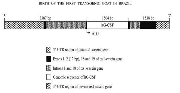
The hG-CSF gene was fused to both goat and bovine DNA sequences of s1-casein gene (CSN1S1). The DNA construct was inserted into a plasmid vector, named pGCm3 (Fig. 1). This expression vector was designed based on pGCm1 and pGCm2 previously described and used for production of transgenic mouse (Dvoryanchi-kov et al. 2005). The pGCm3 DNA insert (6429 bp) has a 5’flanking fragment (3387 bp) originated from goat CSN1S1 gene (Ramunno et al. 2004). This fragment in-cludes a promoter region, the first exon, the first intron and the 12 bp from second exon. In pGCm3 construct, there is the full-length hG-CSF gene (1504 bp) followed
0.25 mM EDTA, at pH 7.4.
EXPERIMENTALANIMALS ANDETHICS
A total of 23 adult Saanen goats (1-5 years old) were used as embryo donors in this study, while 48 undefined breed goats (2-5 years old) served as recipients for the micro-injected embryos. The study was carried out between mid-January and mid-February 2006. Studies were con-ducted in conformance with guidelines of animal care. This project was approved by the Animal Ethics Com-mittee of the State University of Ceará (CEUA/UECE) as well as the Biosecurity National Technical Committee (CTNBio).
ESTRUSSYNCHRONIZATION ANDSUPEROVULATION
The timing of estrus was synchronized in donors and recipients with intravaginal sponges (Progespon, Syntex, Buenos Aires, Argentina) containing 60 mg medroxypro-gesterone acetate for 10 days and an injection of 75µg
Fig. 1 – Construction pGCm3 with hG-CSF under control of 5‘-flanking sequence of goatαs1-casein
gene used to microinjection into goat zygotes.
EMBRYORECOVERY
Donors and recipients were deprived of food and wa-ter for 24 h prior embryo recovery and transfer. Em-bryos were surgically recovered 72 h following sponge removal. A low dose (0.1 mg/kg) of xylazine hydro-chloride (Rompun, Bayer, São Paulo, Brazil) was in-tramuscularly injected as a preanesthetic agent. After a peridural injection (7 mg/kg) of lidocaine (Anestésico L, Eurofarma, São Paulo, Brazil) a mid-ventral incision was made and the reproductive tract was exteriorized. Ovaries were observed for fresh ovulation sites to serve as an estimate of the number of embryos expected. The oviduct was then flushed retrogradely with 10-15 ml of sterile phosphate-buffered saline. The flushing medium was collected into sterile Petri dishes and examined under a stereomicroscope (Nikon SMZ-800, Kawasaki, Japan) for the presence of ova/embryos.
EMBRYOMANIPULATION ANDMICROINJECTION
The microinjection was carried out using only fertilized one-cell embryos. In order to visualize pronuclei all zygotes were briefly centrifuged at 13,400 rpm for 4-6 min. After centrifugation, if the pronuclei were not visible, they were placed in droplets of M-2 medium sup-plemented with 10% FCS and covered with mineral oil, and briefly cultured at 37◦C with 5% CO
2. The zygotes
with visible pronuclei were placed in droplets of M-16
medium supplemented with 10% FCS prior to microin-jection, which was performed under inverted microscope with DIC optics (Nikon TE2000, Kawasaki, Japan) and a pair of micromanipulators (Narishige, Tokio, Japan). The DNA fragment isolated from a plasmid containing the pGCm3 gene was injected into the one of two pronu-clei in volume of 1-2 pl. Noticeable swelling of the nuclei was the criterion for successful microinjection. Microinjected embryos were cultured for 1-2 h at 37◦C
with 5% CO2to evaluate post injection survival. The
sur-viving zygotes were maintained in culture until embryo transfer. Non-injected zygotes with invisible pronuclei were further cultured to confirm fertilization by cleavage.
EMBRYOTRANSFER ANDPREGNANCYDIAGNOSIS
total volume of 25µL. The amplification was conducted
under following conditions: denaturing for 3 minutes at 95◦C, then 35 cycles (30s at 95◦C, 30s at 55◦C and 30s
at 72◦C) and the final stage for 5min at 72◦C. The PCR products were analyzed by electrophoresis in a 1.5 or 3% agarose gel and visualized by ethidium bromide staining.
STATISTICALANALYSIS
Data were expressed as mean±SEM. Differences be-tween means were based ont-test. Probability of<0.05
was considered to be significant statistically.
RESULTS AND DISCUSSION
The response to superovulation and the results of em-bryo recovery and evaluations are presented in Table II. All donors showed estrus and were responsive to the superovulation treatment (≥5 ovulations/female). The superovulation response and the embryo recovery rate from donors found in the present study were superior when compared to those reported in previous studies (Gootwine et al. 1997, Lee et al. 2000, Freitas et al. 2003).
Altogether 379 oocytes/embryos were recovered, of which 75.5% were fertilized and most were at the one-cell stage. These results can be explained by the ef-fect of GnRH injection on the synchronization of ovu-lation. This way, the donors were hand mated at the good time in order to guarantee a high fertilization rate. In addition, the use of GnRH following sponge removal influenced the stage of development of embryos recov-ered, a key parameter for the success of a transgenic founder generation program (Baldassarre et al. 2004).
imal unusable for further embryo recoveries. A further refinement of the pronuclear microinjection technology has been reported in usingin vitroproduced zygotes from oocytes recovered by laparoscopic ovum pick-up (Wang et al. 2002).
In our experiment, as mentioned above, the micro-injectable one-cell embryos were a significant propor-tion of the recovered oocytes/embryos (Fig. 2A,B). However, previous results had showed that the zygote cytoplasm of Korean native strain (Lee et al. 2000) and BELE (Baldassarre et al. 2004) goats contains numer-ous lipid and opaque inclusions. The same was observed in this study with Saanen goats. This feature makes dif-ficult the pronuclei visualization before microinjection of recombinant DNA. In order to improve the pronu-clei visualization, zygotes were briefly centrifuged as de-scribed earlier (Lee et al. 1997). The procedure allowed us to visualize the pronuclei in some zygotes (Fig. 2C,D) so that total number of goat zygotes with visible pronu-clei was 55% (77 out of 129). In the remaining zygotes we were unable to observe a detailed morphology of the pronuclei and the microinjections were done in an area where the pronuclei were most probably located. In these cases, it was impossible to control the efficiency of injec-tion by observing such as an important change in pronu-cleus morphology as its swelling (Fig. 2D).
Number of treated donors 23 Interval sponge removal to onset of estrus (h) 27.0±2.2
Number of ovulations 20.4±7.3
Embryo recovery rate (%) 79.5
Fertilized oocytes (%) 75.5
TABLE III
Estimation of some factors on pregnancy rate of recipient goats after surgically transfer of the microinjected zygotes.
Parameter Recipients
Pregnant Non Pregnant Time of zygote culture before transfer (hours) 6.1±0.6 6.2±0.3
Number of transferred zygotes 4.8±0.4 4.8±0.4
Number of ovulations in recipients at time of transfer 3.1±0.4a 1.9±0.3b
a,b = P <0.01.
were located near the zygote center emerging from the lipid granules, which were shifted to the zygote poles by centrifuging (Fig. 2C). This stage apparently proceeds shortly the moment of the fusion of male and female pronuclei, and therefore corresponds to the late stages of zygote development. After 96 h of culture, non microin-jected goat embryos developed normally until morula stage (Fig. 2E).
The hormonal treatment used for estrus synchro-nization was effective to induce estrus in 87.5% of goat recipients. The estrus began at 26.0±1.6 h after sponge
removal and the ovulation rate was 1.3±0.2. These
results were similar to other studies performed in goats (Freitas et al. 1997).
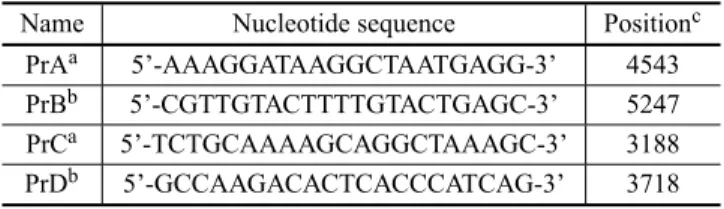
Twenty-seven recipients received the microinjected embryos. The pregnancy rate in this report (37.0 %) was comparable to previous reports (Gootwine et al. 1997, Lee et al. 2000). In our study, it was observed that the number of ovulations influenced the number of preg-nant goats (Table III). It was probably due to an ele-vated plasma progesterone concentration, provided by a high number of corpora lutea, during early pregnancy that could improve embryonic survival and growth (Pope et al. 1995). Altogether 10 recipients were preg-nant and produced 12 offspring. PCR analysis (Fig. 3) finally identified one transgenic goat in this study.
produc-Fig. 3 – Identification of transgenic goat using PCR analysis with PrA/PrB (A) and PrC/PrD (B) primers. Control amplifications with specific primers designed to 5‘-flanking sequence of the goatαs-casein gene (C). C−= negative control, genomic DNA of wild-type goat; 3 to 16 = DNA
sample of experimental goats (4 = “Carlos” positive for the pGCm3 transgene); C+ = a positive control (mixture of non-transgenic goat genomic DNA and the recombinant pGCm3 DNA); W = no template; M= a 100 bp ladder.
tion of human pharmaceuticals or other valuable pro-teins. Our results show that obtaining a reasonable rate of kidding following transfer of microinjected embryos is possible by using thein vivoembryo production and transfer to recipients with a high number of ovulations. In the present study one transgenic kid was produced (Fig. 2F). This transgenic goat represents 8.3% of the kids born and 0.7% of the embryos microinjected and transferred. At our knowledge, this is the first report of birth of transgenic goat in Latin America.
ACKNOWLEDGMENTS
This work was supported by grants from Ministério de Ciência e Tecnologia/Rede Nordeste de Biotecnologia/ Banco do Nordeste (MCT/Renorbio/BNB), Conselho Nacional de Desenvolvimento Científico e Tecnológico/ Programa de Apoio ao Desenvolvimento Científico e Tecnológico (CNPq/PADCT) and by the Program of the Russian Academy of Sciences “Dynamics of plant, ani-mal and human gene polls” (Russia). V.J.F. Freitas and A.C.C. Carvalho are senior investigators of CNPq (Brazil).
RESUMO
A fim de produzir caprinos transgênicos para o hG-CSF, uti-lizou-se 24 cabras Saanen adultas e 48 cabras sem raça defi-nida adultas como doadoras e receptoras, respectivamente. As doadoras tiveram o estro sincronizado por esponjas vaginais e foram superovuladas com 200 mg de FSH em doses decres-centes, duas vezes ao dia e iniciando 48 h antes da retirada da esponja. A ovulação foi induzida pela injeção de 100 µg de GnRH às 36 h após a retirada da esponja. As receptoras tam-bém receberam um tratamento de sincronização do estro. As doadoras foram cobertas por bodes Saanen férteis e, aproxi-madamente 72 h após a retirada da esponja, os zigotos foram colhidos cirurgicamente por lavagem dos ovidutos. Os zigotos colhidos foram rapidamente centrifugados para uma melhor visualização dos pró-núcleos. A construção de DNA, con-tendo o gene do hG-CSF flanqueado pelos genes caprino e bovino daαs1-caseína, foi injetada em 129 embriões. Os
Reprod Sci 82-83: 255–266.
BALDASSARREHET AL. 2003. Production of transgenic goats by pronuclear microinjection of in vitro produced zygotes derived from oocytes recovered by laparoscopy. Theriogenology 59: 831–839.
BALDASSARRE H, WANG B, GAUTHIER M, NEVEU N, LAZARISAANDKARATZASCN. 2004. Effect of GnRH injection timing in the production of pronuclear-stage zy-gotes used for DNA microinjection. Zygote 12: 257–261.
DVORYANCHIKOVGA, SEROVAIA, ANDREEVALE, DIAS
LPB, AZEVEDOSANDSEROVOL. 2005. Secretion of biologically active human granulocyte colony-stimulating factor (G-CSF) in milk of transgenic mice. Genetika 41: 1310–1318.
EBERTKMANDSCHINDLERJES. 1993. Transgenic farm animals: progress report. Theriogenology 39: 121–135. EBERT KM, SELGRATH JP, DITULLIO P, DENMAN J,
SMITHTE, MEMONMA, SCHINDLERJE, MONASTER
-SKYGM, VITALEJAANDGORDONK. 1991. Trans-genic production of a variant of human tissue-type plas-minogen activator in goat milk: generation of transgenic goats and analysis of expression. Biotechnology 9: 835– 838.
FREITASVJF, BARILGANDSAUMANDE J. 1997. Estrus synchronization in dairy goats: use of fluorogestone ac-etate vaginal sponges or norgestomet ear implants. Anim Reprod Sci 46: 237–244.
FREITASVJFET AL. 2003. Birth of normal kids after mi-croinjection of pronuclear embryos in a transgenic goat (Capra hircus) production program in Brazil. Gen Mol Res 2: 200–205.
LEECSET AL. 2000. Embryo recovery and transfer for the production of transgenic goats from Korean native strain,
Capra hircus aegragrus. Small Rum Res 37: 57-63.
LUCZANDXIAOBG. 2006. G-CSF and neuroprotection: a therapeutic perspective in cerebral ischaemia. Biochem Soc Trans 34: 1327–1333.
MORSTYNGANDBURGESSAW. 1988. Hemopoietic growth factors: A review. Cancer Res 48: 5624–5637.
OHJ, KIMDHANDKANGH. 2006. Granulocyte colony-stimulating factor and acute myocardial infarction. JAMA 296: 1968–1969.
POPEWF, CARDENASH, WILEYTMANDMCCLUREKE. 1995. Dose-response relationships of exogenous proges-terone shortly after ovulation on estrous cycle length, blas-tocyst development and fertility in sheep. Anim Reprod Sci 38: 109–117.
RAMUNNO L, CONSENZA G, RANDO A, ILLARIO R, GALLO D, DIBERARDINO D ANDMASINA P. 2004. The goat s1-casein gene: gene structure and promoter analysis. Gene 334: 105–111.
SAMBROOKJ, FRITSCHEFANDMANIATIST. 1989. Molec-ular Cloning: A Laboratory Manual. Cold Spring Harbor, New York: Cold Spring Harbor Lab. 1989.
SOUZALMET AL. 1986. Recombinant human granulocyte colony-stimulating factor: Effects on normal and leuke-mic myeloid cells. Science 232: 61–65.
VIRETF, GONCALVESA, TARPINC, CHABANNONCAND
VIENSP. 2006. G-CSF in oncology Bull Cancer 93: 463– 471.