UNIVERSIDADE DA BEIRA INTERIOR
Ciências da Saúde
Bicarbonate Transporters in Male Fertility:
Identification and Functionality in Testicular Cells
Raquel Alexandra Lages Bernardino
Master Degree Thesis in Biomedical Sciences
Ciências Biomédicas
(2
ndcycle of studies)
Supervisor: Prof. Pedro Fontes Oliveira, PhD
Co-Supervisor: Prof. Sílvia Socorro, PhD
UNIVERSIDADE DA BEIRA INTERIOR
Ciências da Saúde
Transportadores de Bicarbonato na Fertilidade
Masculina: Identificação e Funcionalidade nas
Células Testiculares
Raquel Alexandra Lages Bernardino
Dissertação para a obtenção de Grau de Mestre em
Ciências Biomédicas
(2º Ciclo de Estudos)
Orientador: Prof. Doutor Pedro Fontes Oliveira
Co-orientador: Prof. Doutora Sílvia Socorro
O conteúdo do presente trabalho é da exclusiva responsabilidade do autor:
ii
Agradecimentos
A realização desta tese de mestrado contou com importantes apoios e incentivos, sem os quais esta etapa da minha formação académica não se teria tornado uma realidade. Desejo expressar os meus sinceros agradecimentos a todos aqueles que direta ou indiretamente me ajudaram a cumprir os meus objetivos.
Ao meu orientador, Professor Doutor Pedro Fontes Oliveira, pela disponibilidade demonstrada, conhecimento científico, acompanhamento e conselhos dados durante todo este percurso, assim como pelas críticas, sugestões e correções que contribuíram para o melhoramento deste trabalho. Sem a sua orientação este trabalho não seria possível.
À minha co-orientadora, Professora Doutora Sílvia Socorro, pela competência científica, correções e sugestões que contribuíram para a elaboração desta tese.
Ao Doutor Marco Alves por todo o apoio, criticas, correções, sugestões e os valiosos conselhos que foram essenciais para melhorar a minha tese.
Aos meus colegas de laboratório: Ricardo, Luís, Sara, Cátia, Margarida, Inês, Tânia, Aline, Tito, Nelson, Gonçalo, Mário e em especial à Ana Martins por toda a ajuda, disponibilidade e acompanhamento no laboratório.
Às amigas que fiz durante este percurso, em especial: Sónia Miguel, Ana Costa e Catarina Silva, por toda amizade e compreensão demonstrados durante estes anos. Sem o vosso apoio não seria possível ultrapassar os momentos menos bons.
À minha família, em especial aos meus pais e à minha irmã por acreditarem sempre que eu era capaz, pelo apoio incondicional, o incentivo e paciência demonstrados durante todo este percurso.
iii
Resumo
A formação de espermatozóides competentes é um processo complexo dependente do ambiente criado ao longo do trato reprodutor masculino. A regulação do conteúdo iónico dos fluidos luminais é essencial para a maturação dos espermatozóides. O bicarbonato é essencial não só para a homeostase iónica, como também tem um papel fundamental na manutenção do pH dos diversos fluidos ao longo do trato reprodutor masculino.
A diabetes mellitus (DM) representa uma das maiores ameaças à saúde na sociedade moderna, e afeta cada vez mais homens em idade reprodutiva. A DM é uma doença metabólica caracterizada por hiperglicémia, resultante de defeitos na secreção e/ou ação da insulina. Esta doença pode ser dividida, maioritariamente, em dois subtipos, tipo 1 e tipo 2 (T2DM). A T2DM é conhecida como a diabetes não-insulino-dependente, e inclui indivíduos com resistência à insulina, em que geralmente a secreção de insulina é insuficiente. Este tipo de diabetes pode ser prevenido se for detetado precocemente, no estado de pré-diabetes, que usualmente antecede o aparecimento desta doença.
Tem sido descrito que a DM afecta a regulação do pH intracelular (pHi) em células de mamíferos, principalmente devido à alteração significativa da atividade de alguns transportadores iónicos, particularmente de alguns mecanismos de transporte dependentes de bicarbonato. Pouco se sabe, no entanto, sobre os efeitos desta patologia nos mecanismos de transporte de membrana envolvidos na regulação do pH em células do trato reprodutor masculino, e ainda menos se sabe sobre os efeitos das diferentes fases envolvidas na progressão desta patologia, particularmente do pré-diabetes.
O primeiro objetivo deste trabalho foi analisar possíveis alterações nos níveis dos transportadores de bicarbonato mais relevantes da família Slc4 (trocador aniónico 2 -AE2; trocador Cl-/HCO
3- dependente de Na+ - NDCBE; Na+/HCO3- co-transportador eletrogénico 1 -
NBCe1; Na+/HCO
3- co-transportador eletroneutro 1 - NBCn1) nos testículos e epidídimos de um
modelo animal de pré-diabetes. Foram avaliados os níveis de expressão de proteína e mRNA por western blot e real-time PCR, respetivamente. Assim, foi possível confirmar a presença de todos estes transportadores de bicarbonato da família Slc4 em testículo e epidídimo. A nível testicular, embora não tenham sido detetadas alterações na expressão de proteína, os níveis de mRNA de NBCe1, NBCn1 e NDCBE encontraram-se significativamente aumentados em animais pré-diabéticos. Por outro lado, a nível epididimal, a condição pré-diabética causou um aumento significativo nos níveis proteicos de AE2 e uma diminuição significativa nos níveis de proteína de NDCBE. Estas variações poderão traduzir-se em alterações no fluxo transepitelial de HCO3- no epidídimo in vivo, que pode comprometer a sobrevivência dos
iv
espermatozoides durante o seu armazenamento e maturação. Deste modo, os nossos resultados podem correlacionar-se com resultados descritos anteriormente, que demonstraram um aumento significativo na anormalidade morfológica em espermatozoides de ratos pré-diabéticos.
Por outro lado, vários estudos apresentam uma associação direta entre homens com altos níveis de 17β-estradiol (E2) e o aumento do risco de diabetes e, para além disso, o E2 é
responsável pela modulação da expressão de transportadores iónicos específicos no trato reprodutor masculino. Assim, o segundo objetivo do nosso trabalho foi determinar o efeito desta hormona esteróide sexual na expressão e funcionalidade dos transportadores de bicarbonato selecionados da família Slc4, em culturas de células de Sertoli (SCs). Os quatro transportadores estudados foram identificados e quantificados nas SCs (usando RT-PCR e real time PCR, respetivamente). Nas células tratadas com E2 (100 nM) foi observado um aumento
significativo nos níveis de expressão de mRNA de AE2, NBCn1 e NBCe1. Posteriormente, foi também avaliado o efeito do E2 (100 nM) no transporte transcelular em SCs cultivadas em
suportes semipermeáveis, usando a técnica de Voltage-Clamp. As SCs tratadas com E2
apresentaram alterações significativas na variação da corrente de curto-circuito (∆Isc)
induzida por Adenosine-5-triphosphate (ATP), bem como na recuperação dessa corrente de curto-circuito (Isc) após estimulação com ATP. Estas alterações poderão ser promovidas pelo
aumento da expressão de AE2 observados em células tratadas com E2, visto que foi descrito
que nestas células o Isc envolvea secreção do Cl- através da membrana apical por canais de Cl
-ativados pelo ATP, e a sua absorção através da membrana basolateral por mecanismos dependentes de HCO3−. Um aumento dos níveis de AE2 certamente será responsável por
promover a variação de Isc após a estimulação com ATP.
Assim, os nossos resultados mostram uma relação do pré-diabetes, assim como do aumento dos níveis de E2, com a expressão/função dos transportadores de bicarbonato em
epidídimo e SCs de rato, fornecendo novas evidências sobre os mecanismos pelos quais esta fase precoce da DM e algumas das suas características podem afetar a função reprodutivas masculina.
Palavras- Chave:
Transportadores de bicarbonato, fertilidade masculina, transportadores membranares,
v
Resumo Alargado
O estabelecimento da fertilidade masculina envolve processos complexos que requerem a interação entre diferentes tecidos do trato reprodutor masculino. Nos mamíferos, os testículos são os elementos centrais do sistema reprodutor masculino, estando envolvidos na síntese de hormonas esteróides e na produção de gâmetas masculinos, os espermatozóides. A formação de espermatozóides competentes é um processo complexo dependente dos ambientes estabelecidos ao longo do trato reprodutor masculino. A regulação das propriedades iónicas do conteúdo dos diversos fluidos luminais é essencial para a maturação dos espermatozóides, bem como a regulação do seu pH. No interior dos túbulos seminíferos a espermatogénese é suportada pelas células de Sertoli (SCs), que promovem o suporte físico e nutricional das espermatogónias, espermatócitos, espermatídios e finalmente os espermatozóides, que são libertados no lúmen dos túbulos seminíferos. As SCs regulam entre outras coisas, a passagem de iões, água e metabolitos energéticos para o lúmen tubular. Depois de completa a espermatogénese, os espermatozóides são transportados até ao epidídimo, que com o seu microambiente luminal ajuda a transformar os espermatozóides imaturos e sem mobilidade em células competentes e capazes de fertilizar.
A normal ocorrência dos processos celulares necessita que o pH intracelular (pHi) e extracelular (pHo) sejam mantidos dentro de limites estreitos. O ajustamento do pH dos fluidos corporais é de extrema importância para uma função normal de todas as células e tecidos. Sabe-se que os transportadores de bicarbonato são de elevada importância para a regulação do pHi e pHo dos fluidos de muitos tecidos. Na realidade, na maioria dos tecidos, o bicarbonato é essencial não apenas para a manutenção do pH, mas tem também um papel fundamental na homeostase iónica e osmolaridade, e os tecidos reprodutivos masculinos não serão a exceção.
A diabetes mellitus (DM) representa uma das maiores ameaças à saúde na sociedade moderna. A sua incidência tem vindo a aumentar drasticamente e afeta cada vez mais pessoas jovens, ainda em idade reprodutiva. A DM é uma doença metabólica caracterizada por hiperglicémia, resultante de defeitos na secreção e/ou ação da insulina. Esta doença pode ser dividida em dois tipos, tipo 1 (T1DM) e tipo 2 (T2DM), ambos relacionados com várias complicações sistémicas. A T1DM geralmente desenvolve-se em idade jovem, e é causada pela destruição autoimune das células β do pâncreas. Requer uma terapia de reposição diária de insulina. A T2DM é conhecida como a diabetes não-insulino-dependente, e ocorre quando a produção de insulina pelas células β não é suficiente para manter os níveis fisiológicos no sangue. Este tipo de diabetes pode ser prevenido se for detetado precocemente, no estado de pré-diabetes, que usualmente antecede o aparecimento desta doença. A transição de um
vi
estado de pré-diabetes para T2DM ocorre quando a capacidade secretora das células β não é capaz de compensar a resistência à insulina.
Tem sido descrito que a DM altera a regulação do pHi em células de mamíferos, principalmente, devido à alteração significativa da actividade de alguns transportadores iónicos, nomeadamente de alguns mecanismos de transporte dependentes de bicarbonato. No entanto, pouco se sabe sobre os efeitos desta patologia nos mecanismos de transporte de membrana envolvidos na regulação do pH nas células do trato reprodutor masculino, e ainda menos se sabe sobre os efeitos das diferentes fases envolvidas na progressão desta patologia, particularmente o pré-diabetes.
O primeiro objetivo deste trabalho foi analisar possíveis alterações nos níveis dos transportadores de bicarbonato mais relevantes da família Slc4 (trocador aniónico 2 -AE2; trocador Cl-/HCO
3- dependente de Na+ - NDCBE; Na+/HCO3- co-transportador eletrogénico 1 -
NBCe1; Na+/HCO
3- co-transportador eletroneutro 1 - NBCn1) nos testículos e epidídimos de um
modelo animal de pré-diabetes. Foram avaliados os níveis de expressão de proteína e mRNA por western blot e real-time PCR, respetivamente. Assim, confirmamos a presença de todos estes transportadores de bicarbonato da família Slc4 em testículo e epidídimo. A nível testicular, embora não tenham sido detetadas alterações na expressão de proteína, os níveis de mRNA de NBCe1, NBCn1 e NDCBE encontravam-se significativamente aumentados em animais pré-diabéticos. Por outro lado, a nível epididimal, o pré-diabetes causa um aumento significativo nos níveis proteicos de AE2 e uma diminuição significativa nos níveis de proteína de NDCBE. Estas variações poderão traduzir-se em alterações no fluxo transepitelial de HCO3
-no epidídimo in vivo, que podem comprometer a sobrevivência dos espermatozóides durante o seu armazenamento e maturação. Deste modo, os nossos resultados podem correlacionar-se com resultados descritos anteriormente, que demonstraram um aumento significativo na anormalidade morfológica em espermatozoides de ratos pré-diabéticos.
Por outro lado, vários estudos apresentam uma associação entre homens com elevados níveis de 17β-estradiol (E2) e o aumento do risco de diabetes e, para além disso, o E2
é responsável pela modulação da expressão de transportadores iónicos específicos no trato reprodutor masculino. Assim, o segundo objetivo do nosso trabalho foi determinar o efeito desta hormona esteróide sexual na expressão e funcionalidade dos transportadores de bicarbonato selecionados da família Slc4, em culturas de SCs de rato. Os quatro transportadores estudados foram identificados e quantificados nas SCs (usando RT-PCR e real time PCR, respetivamente). Nas células tratadas com E2 (100 nM) foi observado um aumento
significativo nos níveis de expressão de mRNA de AE2, NBCn1 e NBCe1. Posteriormente, também foi avaliado o efeito do E2 (100 nM) no transporte transcelular em SCs cultivadas em
suportes semipermeáveis, usando a técnica de Voltagem-Controlada. As SCs tratadas com E2
apresentaram alterações significativas na variação da corrente de curto-circuito induzidas por ATP (∆Isc), bem como na recuperação da corrente de curto-circuito (Isc) depois da estimulação
vii
com ATP. Estas alterações podem ser promovidas pelo aumento reportado nos níveis de AE2 observados em células tratadas com E2, visto que nestas células o Isc envolvea secreção do Cl
-através da membrana apical, -através de canais de Cl- ativados por ATP, e a sua absorção
através da membrana basolateral por mecanismos dependentes de HCO3−. Um aumento dos
níveis de AE2 será certamente responsável por promover a variação de Isc após a estimulação
com ATP.
Assim, os nossos resultados mostram uma relação do pré-diabetes, assim como do aumento dos níveis de E2, com a expressão/função dos transportadores de bicarbonato em
epidídimo e SCs de rato, fornecendo novas evidências sobre os mecanismos pelos quais esta fase precoce da DM e algumas das suas características podem afetar a função reprodutivas masculina.
viii
Abstract
The formation of competent spermatozoa is a complex event that depends on the establishment of adequate environments throughout the male reproductive tract. The maintenance of a proper ionic content in the luminal milieus is crucial for spermatozoa maturation. Bicarbonate is not only essential to ionic homeostasis, as HCO3- concentration
plays an essential role in the pH maintenance along the male reproductive tract.
Diabetes mellitus (DM) is one the most prominent public health threats in modern societies and its incidence is drastically increasing in men with reproductive age. This metabolic disease is characterized by hyperglycaemia that can result from defects in insulin secretion and/or insulin action. There are two types of DM, type-1 DM and type-2 DM (T2DM). T2DM is referred to as non-insulin-dependent diabetes, and encompasses individuals who have insulin resistance and usually have a relative insufficient insulin secretion. This type of diabetes can be prevented if detected early, in a status called pre-diabetes, which usually precedes the appearance of the disease.
It has been reported that DM alters pHi regulation in mammalian cells mainly by markedly altering the activity in some ion transporters, particularly some bicarbonate-dependent mechanisms. Little is known on the effects of this pathology on the membrane transport mechanisms involved in pH regulation on male reproductive tract cells and even fewer on the effects of the different stages involved in the progression of this pathology, particularly during pre-diabetes.
The first objective of this work was to analyse possible alterations on the levels of the most relevant bicarbonate transporters of the Slc4 family (anion exchanger 2 -AE2; Na+-driven
Cl-/HCO
3- exchanger - NDCBE; electrogenic Na+/HCO3- co-transporter 1- NBCe1; electroneutral
Na+/HCO
3- co-transporter 1 - NBCn1) in testis and epididymis of a pre-diabetic animal model.
Protein and mRNA expression levels were evaluated by western blot and real-time PCR, respectively. We were able to confirm the presence of all the bicarbonate transporters of the Slc4 family studied both in testis and epididymis. At testicular level, although no alterations were detected in protein expression, the mRNA levels of NBCe1, NBCn1 and NDCBE were significantly increased in diabetic animals. On the other hand, at epididymal level, pre-diabetes caused a significant increase on AE2 protein levels and a significant decrease of NDCBE protein levels. Hence, these alterations might translate into changes of the HCO3−
transepithelial epididymal fluxes in vivo, which might represent a threat for sperm survival during storage in the epididymis. Our results might correlate with previous results that reported a significant increase in abnormal sperm morphology in pre-diabetic rats.
ix
Furthermore, as several studies support an association of men with higher 17β-estradiol (E2) levels and the increased risk of diabetes and, moreover, E2 is responsible for the
modulation of the expression of specific ion transporters in the male reproductive tract, the second objective of our work was to determine the effect of this sex steroid hormone on the expression and functionality of selected bicarbonate transporters of the Slc4 family in cultured Sertoli cells (SCs). All the selected four transporters were identified and quantified in SCs (using RT-PCR and real time PCR, respectively). In cells treated with E2 (100 nM) a
significant increase in mRNA expression levels of AE2, NBCn1 and NBCe1 was observed. Subsequently, we also evaluated the effect of E2 (100 nM) on transcellular transport in SCs,
grown in semi-permeable supports, using the Voltage-Clamp technique. E2-treated SCs
presented a significant alteration on the shift of the short-circuit current (∆Isc) induced by
ATP, as well as on short-circuit current (Isc) recovery after stimulation. These alterations may
be promoted by the increase of AE2 mRNA levels observed in E2-treated cells, as in these cells the Isc involves the secretion of Cl- through the apical membrane by an ATP-activated Cl
-conductance and its absorption via HCO3−-dependent mechanisms through the basolateral
membrane. An increase on AE2 levels will surely be responsible for a prompter effect of this transporter on Isc variation following ATP activation.
Thus, our results show a relation of the pre-diabetes, as well as increased E2 levels,
with the expression/function of bicarbonate transporters in rat epididymis and SCs,providing new evidence on the mechanisms by which this prodromal stage of DM and its associated features can affect male reproductive function.
Keywords:
Bicarbonate transporters, male fertility, membrane transporters, diabetes mellitus, estrogens.
x
Table of Contents
Agradecimentos ... ii Resumo ... iii Resumo Alargado ... v Abstract... viii Table of Contents ... xList of figures ... xii
List of tables ... xiii
Abbreviations ...xiv
I. Introduction ... 1
1. General aspects ... 2
2. Spermatogenesis and hormonal regulation ... 4
2.1 Sertoli cells ... 8
3. The mammalian epididymis ... 9
4. Diabetes mellitus and male fertility ... 12
5. pH Regulation ... 14
6. Bicarbonate transporters in the male reproductive tract ... 17
6.1 Slc4 family ... 18
6.1.1 Na+-independent bicarbonate transporters ... 19
6.1.2 Na+-dependent bicarbonate transporters ... 20
6.2 Slc26 family ... 24
7. Diabetes, estrogens and pH regulation ... 26
II. Aim of project ... 29
III. Material and methods ... 31
1. Chemicals ... 32
2. In vitro studies ... 32
2.1 Primary cultures of rat Sertoli cells ... 32
2.2 Hormonal treatment of rat Sertoli cells ... 33
xi
3.1 Establishment of the pre-diabetes animal model ... 33
4. RNA extraction ... 34
5. RT-PCR ... 34
6. Real time – PCR ... 34
7. Total protein extraction ... 35
8. Western blot ... 36
10. Voltage Clamp ... 36
11. Statistical Analysis ... 37
IV. Results ... 38
1.1 Pre-diabetic rodent model characterization ... 39
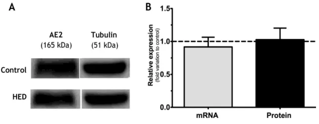
1.2 HED increases protein levels of AE2 in the epididymis ... 39
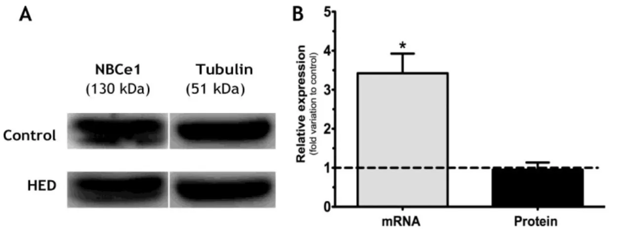
1.3 HED increases mRNA levels of NBCe1 in the testis ... 41
1.4 HED increases mRNA levels of NBCn1 in the testis ... 42
1.5 HED decreases protein levels of NDCBE in the epididymis ... 43
2. 17-Estradiol effects in bicarbonate transporters of Sertoli cells ... 44
2.1 Relative expression of bicarbonate transporters in cultured Sertoli cells ... 44
2.2 Elevated concentration of E2 alter mRNA expression of AE2, NBCn1 and NBCe1 in rat Sertoli cells ... 45
2.3 Alterations in transcellular transport in E2-treated rat Sertoli cells ... 46
V. Discussion ... 49
1. Bicarbonate transporters in testis and epididymis of HED-treated animals ... 51
2. Bicarbonate transporters in Sertoli cells: regulation by 17-Estradiol ... 53
VI. Conclusion ... 57
VII. References ... 59
xii
List of figures
Figure 1: Schematic representation of the mammalian testes and epididimys.. ... 2 Figure 2: Schematic illustration of seminiferous tubule, spermatogenesis and cells in
interstitial tissue outside the tubule.. ... 5
Figure 3: Hormonal regulation of male reproductive tract.. ... 6 Figure 4: Schematic and histological representation of the male reproductive tract and
excurrent ducts... 10
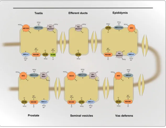
Figure 5: Schematic representation of the synthesis of bicarbonate. ... 14 Figure 6: Schematic representation of the distribution of transporters bicarbonate in male
reproductive system.. ... 18
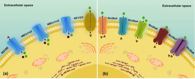
Figure 7: Bicarbonate transporters... 26 Figure 8: Effect of High Energy Diet (HED) on membrane transporter anion exchanger 2 (AE2)
mRNA and protein levels in rat testis. ... 40
Figure 9: Effect of High Energy Diet (HED) on membrane transporter anion exchanger 2 (AE2)
in mRNA and protein levels in rat epididymis.. ... 40
Figure 10: Effect of High Energy Diet (HED) on eletrogenic Na+/HCO3- cotransporters (NBCe1)
in mRNA and protein levels in rat testis.. ... 41
Figure 11: Effect of High Energy Diet (HED) on eletrogenic Na+/HCO3- cotransporters (NBCe1)
in mRNA and protein levels in rat epididymis.. ... 41
Figure 12: Effect of High Energy Diet (HED) on eletroneutral Na+/HCO3- cotransporters
(NBCn1) in mRNA and protein levels in rat testis. ... 42
Figure 13: Effect of High Energy Diet (HED) on eletroneutral Na+/HCO3- cotransporters
(NBCn1) in mRNA and protein levels in rat epididymis. ... 43
Figure 14: Effect of High Energy Diet (HED) on Na+-driven Cl-/HCO3- exchanger (NDCBE) in
mRNA and protein levels in rat testis. ... 43
Figure 15: Effect of High Energy Diet (HED) on Na+-driven Cl-/HCO3- exchanger (NDCBE) in
mRNA and protein levels in rat epididymis. ... 44
Figure 16: Identification and quantification of the mRNA levels of bicarbonate transporters.
... 45
Figure 17: Effect of 17β-estradiol (E2) 100 nM on bibarbonate transporters relative expression
of mRNA in rat Sertoli cells. ... 46
Figure 18: Short-circuit current (Isc) response to the addition of ATP to the apical bathing
solution.. ... 48
Figure 19: Diagram of a simplified cell with the bicarbonate membrane transport systems
considered.. ...511
Figure 20: Diagram of a simplified polarized Sertoli cell with the chloride channel and
xiii
List of tables
Table 1: Genes, sequence nucleotide and respectively conditions for PCR amplification of
xiv
Abbreviations
AE – Anion exchanger
AUCg - Area under the curve ATP – Adenosine-5-triphosphate BTB – Blood testes barrier CA – Carbonic anhydrase
CFTR – Cystic fibrosis transmembrane CLD – Congenital chloride diarrhea CSF – Cerebrospinal fluid
DM – Diabetes mellitus
DMEM: Ham’s F12 - Dulbecco’s Modified Eagle Medium Ham’s Nutrient Mixture F12 DRA – Down – regulated in adenoma
E2 - 17β-Estradiol
EDTA – Ethylene diamine tetra acetic acid EtOH- Etanol
ERα – Estrogen receptor α ERβ - Estrogen receptor β ErKO – ER knockout mouse EST – Expressed sequence tags FBS – Fetal bovine serum
FSH – Follicle – stimulating hormone GPER30 – G-Protein coupled receptor 30
xv
GnRH – Gonadotropin releasing hormoneHBSS - Hank’s Balanced Salts Solution HED – High energy diet
HPT – Hipothalamic-pituitary testis Isc – Short -circuit current
ITS supplement - Insulin-Transferrin-Sodium Selenite supplement LH – Luteinizing hormone
M-MLV RT - Moloney Murine Leukemia Virus Reverse Transcriptase mRNA - Messenger Ribonucleic Acid
NBCe – Electrogenic Na+/HCO
3- cotransporters
NBCn – Electroneutral Na+/HCO
3- cotransporters
NCBe - Electrogenic Na+ - coupled HCO
3- Transporters
NCBn - Electroneutral Na+ - coupled HCO
3- Transporters
NCBE - Na+ - coupled HCO
3- exchanger
NCBTs - Na+ - coupled HCO
3- Transporters
NDCBE - Na+ - driven Cl-/HCO
3- exchanger
NHE3 – Na+/H+ exchanger 3
P450arom – Aromatase enzyme cytochrome P450 PAT-1 – Putative anion transporter 1
PBS – Phosphate Buffered Saline PCR – Polymerase Chain Reaction PDS- Pendred syndrome
pHi - intracellular pH pHo – extracellular pH
xvi
qPCR -Real-time PCRRIPA - Radio-Immunoprecipitation Assay RNAt - total RNA
RTF – Rete testis fluid
sAC – Soluble adenylyl cyclase Slc4 – Solute carrier 4
Slc26 – Solute carrier 26 SCs- Sertoli cells
STF- Seminiferous tubular fluid T1DM – Type 1 Diabetes Mellitus T2DM – Type 2 Diabetes Mellitus TBS- Tris-buffered saline solution WHO- World Health Organization
1
2
1. General aspects
The establishment of male fertility is a complex process that requires concerted interactions between different tissues of the male reproductive tract and accessory glands, and between the different cell types that compose these organs. The male reproductive tract is composed of highly heterogeneous tissues, including testis, efferent ducts, epididymis and vas deferens (Figure 1) (R. Jones & Murdoch, 1996; Orgebin-Crist & Davies, 2003; Pastor-Soler, Piétrement, & Breton, 2005). The mammalian testis is a complex organ, divided in compartments, testicular lobules and each lobule encloses coiled seminiferous tubules (Shubhada, Glinz, & Lamb, 1993; W.H. Walker & Cheng, 2005), which contain the Sertoli cells (SCs) and the germinal cells in different development stages. These tubules are avascular and no nerves penetrate through their walls (B. Setchell, 1986). Besides, the lobules formed by seminiferous tubules, are separated by extensions of the tunica albuginea, that open on both ends into the rete testis (Figure 1) (Pastor-Soler et al., 2005; Saladin, 2003).
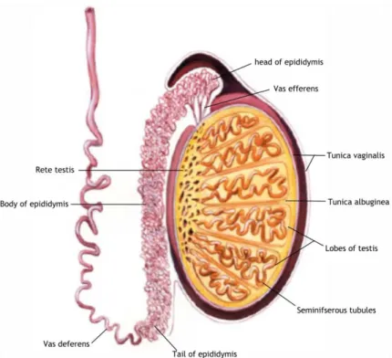
Figure 1: Schematic representation of the mammalian testes and epididymis. The testis is
encapsulated by two layers: the tunica vaginalis that is the most outer tunic; and tunica albuginea that divides the testicles into lobules, filled by seminiferous tubules. The seminiferous tubules end in the rete testis, which converge to vas efferens that is connected to the epididymis. The epididymis is attached to the vas deferens and can be divided into three sections: head, body and tail. Adapted from Pastor-Soler et al. (2005).
The interstitial space of the testis, that comprises all the spaces between the seminiferous tubules, contains all the blood and lymphatic vessels, which are essential for the
3
movement of hormones and nutrients into, and out of the testis (O’donnell, Robertson, Jones, & Simpson, 2001). In this interstitium we can also find the nerves, the Leydig cells which are the primary sites of steroidogenesis in the testis, and a significant population of macrophages (B. Setchell, 1986).
The testes perform two main functions: synthesis of steroid hormones and production of spermatozoa in a process called spermatogenesis (S. Carreau, Genissel, Bilinska, & Levallet, 1999; L. Rato, Socorro, Cavaco, & Oliveira, 2010; Saez, 1994). Indeed, a chain of complex local interactions involving the various testicular cells types, such as germ, Sertoli, peritubular and Leydig cells are responsible for the control of spermatogenesis (Shubhada et al., 1993; W.H. Walker & Cheng, 2005). Within the seminiferous tubules, the SCs reside on the basement membrane, under which are the lymphatic endothelium and the peritubular myoid cells (Dym & Fawcett, 1970). The SC plays a central role in the development of functional testis and, subsequently, in the manifestation of a male phenotype (Mruk & Cheng, 2004; R. M. Sharpe, McKinnell, Kivlin, & Fisher, 2003). Without the physical and metabolic support of the SCs, germ cell differentiation, meiosis and transformation into spermatozoa would not occur (R. Sharpe, 1994; R. M. Sharpe et al., 2003). It is well known that normal testicular development and maintenance of spermatogenesis are controlled by gonadotrophins and testosterone whose effects are modulated by locally produced factors. Moreover, estrogens are also pivotal to this process (S. Carreau et al., 1999; Saez, 1994).
Adjacent SCs form tight junctions with each other to form a basal and adluminal compartment. In these compartments, meiotic and post-meiotic steps of spermatogenesis proceed and occurs the formation of a fluid-filled lumen (Dym & Fawcett, 1970). As a result, the developing germ cells present in the adluminal compartment become effectively protected from direct access to plasma components and thus become dependent on the secretion of factors by the SC (L. Rato et al., 2012; R. Sharpe, 1994; R. M. Sharpe et al., 2003). The structural basis of the tubular barrier has been well characterized and has been reported to mainly reside in the specialized junctions between pairs of SCs (Dym, 1973; Dym & Fawcett, 1970). SCs also control the composition of the seminiferous tubular fluid (STF) and the physicochemical milieu where spermatogenesis occurs (L. Rato et al., 2010). These cells regulate, among other things, the passage of ions and the selective flow of water, steroids and carbohydrates into the tubular lumen (Abraham, 1991). After completion of spermatogenesis within the seminiferous tubules, spermatozoa are carried to the rete testis and from there across the efferent ductules to the epididymis. This movement is carried out by cilliary movement in the efferent ductules, for muscle contraction, and for fluid flow (Saladin, 2003). The efferent ductules concentrate the dilute testicular fluid and spermatozoa by reabsorbing approximately 90% of the fluid secreted by the testes (Clulow, Jones, Hansen, & Man, 1998; Newcombe, Clulow, Man, & Jones, 2000).
4
The functional aspects of the testes are very complex, its normal function depends on the vascular system delivering oxygen, nutrients and hormones into testicular interstitial fluid and removing waste and secretory products (Bergh & Damber, 1993). All these processes are dependent on numerous factors, which act in cascade, and any anatomical, physiological, hormonal or electrolytic abnormality can change reproductive parameters.
2. Spermatogenesis and hormonal regulation
Spermatogenesis is a process controlled by a network of endocrine and other regulatory factors (L. Rato et al., 2012; Verhoeven, Willems, Denolet, Swinnen, & De Gendt, 2010; W.H. Walker, 2011) in which immature germ cells undergo division, meiosis and differentiation to give rise to mature cells (
M.G. Alves et al.,2013a;
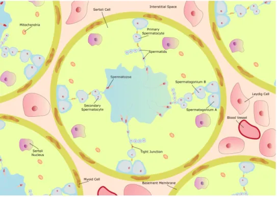
L. Rato et al., 2010). This process occurs in seminiferous tubules, the functional unities of the testis, through close association of germ cells with the epithelial somatic cells, the SCs (O’donnell et al., 2001; L. Rato et al., 2010; Shubhada et al., 1993; W.H. Walker & Cheng, 2005). The spermatogenesis can be divided into four different phases (Saladin, 2003) that include mitosis, meiosis, spermiogenesis and spermiation (Cheng, Wong, Yan, & Mruk, 2010). Spermatogonia are the immature germ cells in the testis, and include type A and type B spermatogonia, the last of which are committed to differentiation. The spermatogonia stay in basal compartment, in touch with the SCs. The spermatogonia migrate between SCs to the adluminal compartment, where undergo numerous mitotic cycles (L. D. Russell, Ettlin, Hikim, & Clegg, 1993; L. Rato et al., 2012; Saladin, 2003). After the last mitosis of type B spermatogonia, primary spermatocytes are formed (L. D. Russell et al., 1993). These cells replicate their DNA and thus initiating meiosis (O’donnell et al., 2001). They undergo the first meiotic division to yield secondary spermatocyte, that in turn undergo second meiotic division and to yield the haploid round spermatid (O’donnell et al., 2001). Spermiogenesis is the final stage of spermatogenesis, in which the maturation of spermatids into spermatozoa occurs. (L. D. Russell et al., 1993). Briefly, spermatids undergo morphological changes such as the establishment of the flagellum, the formation of the acrosome and the elongation of the nucleus (Figure 2) (M.G. Alves et al., 2013b). A great part of the cytoplasm is also eliminated and the chromatin is gradually condensed together with the changes of histones by transition proteins and then by protamines (Zini & Agarwal, 2011). When germ cell development is complete, the spermatozoa are released from the SCs into the tubule lumen (spermiation), and proceed through the rete testis, until they enter the epididymis via the efferent ducts. During passage through the epididymis, the spermatozoa undergo a series of biochemical changes to become motile and capable of fertilization (R. Jones & Murdoch, 1996; O’donnell et al., 2001; Orgebin-Crist & Davies, 2003).5
Figure 2: Schematic illustration of seminiferous tubule, spermatogenesis and cells in interstitial tissue outside the tubule. The Sertoli cells (SCs) reside on a basement membrane, under which are thelymphatic endothelium and the peritubular myoid cells. Two adjacent SCs abut a tight junction which limits intercellular transport and represents the blood testes barrier (BTB). Outside the BTB is the basal compartment, where spermatogonial renewal occurs, and inside the BTB is the adluminal compartment, where meiosis, spermiogenesis and spermiation occurs. Spermatogenesis is the process by which immature spermatogonium within the testis, divide and differentiate. Spermatogonium type A that divides mitotically in spermatogonium type B. After two meiosis, primary and secondary spermatocyte are formed. Then, takes place the spermiogenesis that produces spermatids, and the mature elongated spermatid that is subsequently released to the seminiferous epithelium (spermiation). Adapted from Alves et al.(2013b).
The endocrine glands of the male reproductive system includes the hypothalamus, the pituitary and the testes forming the hypothalamic-pituitary testis axis (HPT). Within this axis, neurons of the hypothalamus produce gonadotropin releasing hormone (GnRH). Pulsatile GnRH signals stimulate gonadotroph cells in the anterior pituitary to secret follicle-stimulating hormone (FSH) and luteinizing hormone (LH) that then act on the testis to regulate the spermatogenic potential (W.H. Walker & Cheng, 2005). The testicles are involved in the production of the sex steroid hormones that exert a negative feedback on the hypothalamus and the pituitary to control the secretion of the gonodotropins, LH and FSH (L. Rato eta al.,2012; O’donnell et al., 2001). GnRH enters the hypothalamic-pituitary portal system and binds to receptors on the plasma membranes of pituitary cells, resulting in the synthesis and release of LH and FSH (Saladin, 2003). LH binds to receptors on the surface of Leydig cells in the testis and stimulates the production of testosterone, a steroid hormone that diffuses into the seminiferous tubules. Within the seminiferous tubules, only SC possesses receptors for testosterone and FSH and thus the major targets of the ultimate hormonal signals that
6
regulate spermatogenesis (Figure 3) (M. G. Alves et al., 2013
c
; Hoesl, Saad, Pöppel, & Altwein, 2005; Mruk & Cheng, 2004; W.H. Walker & Cheng, 2005). SCs also produce glycoprotein hormones, inhibin, activin, and follistatin, which regulate the secretion of FSH. Testosterone, 17-estradiol (E2), inhibin, activin, and follistatin are major testicularhormones that regulate the release of the gonadotropins LH and FSH. Generally, testosterone, E2 and inhibin reduce the secretion of LH and FSH, whereas activin stimulates
the secretion of FSH and follistatin inhibits FSH secretion (Saladin, 2003).
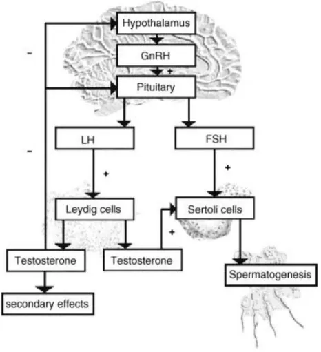
Figure 3: Hormonal regulation of male reproductive tract. The hypothalamus synthesizes the
gonadotropin releasing hormone (GnRH), which will stimulate the pituitary to produce the luteinizing hormone (LH) and follicle-stimulating hormone (FSH). LH and FSH bind membrane receptors on Leydig and Sertoli cells (SCs), respectively, and stimulate the testosterone production and spermatogenesis. The release of GnRH by the hypothalamus and LH by the pituitary is inhibited by increasing levels of testosterone. This androgen is responsible for male secondary sexual characteristics, and acts on SCs to stimulate spermatogenesis. Legend: + stimulating, - inhibition. Adapted from Hoesl et al. (2005).
Androgens are considered the sex male hormones, particularly testosterone, whereas estrogens are considered to be the sex female hormone, namely E2. Nevertheless, androgens
7
differences, but result from quantitative divergence in hormone concentrations and differential expressions of steroid hormone receptors (S. Carreau & Hess, 2010
; P. Oliveira et
al., 2011a
). Estrogens are produced from testosterone by aromatase enzyme cytochrome P450 (P450arom), encoded by the CYP19 gene (Boon, Chow, & Simpson, 2010; E. R. Simpson et al., 1994). This enzyme is involved in the irreversible conversion of androgens into estrogens and is present in the endoplasmic reticulum of many tissues (E. R. Simpson et al., 1994). In the mammalian testis it's well known that aromatase is mainly localized in Leydig cells (S. Carreau et al., 1999). In rodents, the source of testicular estrogens has been a considerable subject of interest. It has been reported that SCs are the major source of estrogens in immature animals but in adult rats only Leydig cells synthesize this hormone. This can be justified by low quantities of the aromatase transcripts in the adult SCs (S. Carreau et al., 2003). Moreover it has been reported the existence of P450arom transcripts in the epithelial cells of the rat epididymis (Wiszniewska, 2002). Expression of aromatase is stimulated by FSH and the maximum stimulatory effect of FSH in aromatase gene expression occurs in 20-days old rat SCs and seems to parallel SCs differentiation (S. Carreau, De Vienne, & Galeraud-Denis, 2008).The role of E2 in the development and physiology of male reproductive tract of
mammals is still a matter of debate, even though there is a growing body of evidence suggesting that E2 are playing a role via their specific receptors, estrogen receptor α (ERα)
estrogen receptor β (ERβ) and G-protein coupled receptor-30 (GPER30) ( M.G. Alves., 2013a; Fisher et al., 1997; R. Hess et al., 1997; Prossnitz et al., 2008). ERs are distributed all along the genital tract (S. Carreau et al., 2002; O’donnell et al., 2001; Scobie et al., 2002; Sirianni et al., 2008). Testicular estrogen should interact with ERs which in turn mediate the transcription of tissue specific genes (S Carreau et al., 2011). The biological significance of ER subtypes is unclear but may provide an explanation for the selective actions of estrogens in different target tissues.
The ERα are expressed in various cellular types of the male reproductive tract (Cavaco, Laurentino, Barros, Sousa, & Socorro, 2009; Fisher et al., 1997; R. A. Hess, Bunick, & Bahr, 1995), such as SCs (Lucas et al., 2008), Leydig cells, efferent ductules (Fisher et al., 1997; R. Hess et al., 1997),testis (Pelletier, Labrie, & Labrie, 2000), spermatocytes (Pentikäinen, Erkkilä, Suomalainen, Parvinen, & Dunkel, 2000), spermatids (Durkee, Mueller, & Zinaman, 1998) and spermatozoa (Durkee et al., 1998). The presence of ERβ has been described in reproductive tract tissues and was visualized in SCs (Pelletier et al., 2000), Leydig cells (Pelletier et al., 2000), spermatocytes (Saunders, Fisher, Sharpe, & Millar, 1998), spermatids (S Carreau et al., 2011), spermatogonia (Saunders et al., 1998), spermatozoa (Pentikäinen et al., 2000) and prostate (Weihua, Warner, & Gustafsson, 2002). GPER30 is able to mediate E2 action, has been identified in a variety of human and rodent estrogen target
8
including the testes and spermatogonia, highlight for a role of this receptor in controlling mouse spermatogonia cell proliferation in response to E2 (Sirianni et al., 2008).
Animal models with ER knockout (ERKO) presented compromised spermatogenesis, steroidogenesis and male fertility (S. Carreau & Hess, 2010; Joseph, Shur, & Hess, 2011). Hess and collaborators (1997) described that αERKO animals have reduced fertility because of abnormal fluid reabsorption in the efferent ductules, which leads to germ cell destruction, and diluted sperm into epididymis, rather than concentrated, resulting in infertility, this model also revealed several abnormalities in the epididymis (R. A. Hess, 2000). Spermatogenesis, steroidogenesis and fertility are not affected in βERKO animals (Hewitt, Harrell, & Korach, 2005). Otto and collaborators (2009) described that GPER30 deficient mice are fertile.
Spermatogenesis in rodents is therefore at least partly under the E2 control,
particularly stem germ cell number and spermatid/sperm formation (S. Carreau & Hess, 2010; Li, Papadopoulos, Vidic, Dym, & Culty, 1997; Shetty, Krishnamurthy, Krishnamurthy, Bhatnagar, & Moudgal, 1997). E2 are involved not only in some regulating steps of
spermatogenesis but also through, for instance, the cadherin synthesis that mediates Sertoli-germ cell interactions (R. M. Sharpe, 1998). Furthermore, E2 has an important role for sperm
motility (S. Carreau & Hess, 2010), as reported in aromatase-deficient men, in which motility and number of spermatozoa are reduced (Carani, Fabbi, Zirilli, & Sgarbi, 2002). Some in vivo and in vitro studies revealed that E2 can act as germ cell survival factor and that this effect is
dose-dependent (Pentikäinen et al., 2000). For example, E2 prevents apoptosis of germ cells
within human seminiferous tubules in vitro even in the absence of gonadotropins (Pentikäinen et al., 2000). However, proapoptotic effects of E2 on spermatogenesis have also been
observed (Mishra & Shaha, 2005
;
S. Laurentino et al., 2011; V. Simões et al., 2012).A better knowledge about the role of E2 and its receptors in the regulation of the
homeostasis and functions of male reproductive tract will be important for a deeper understanding of the control of male fertility.
2.1 Sertoli cells
Sertoli cells, the somatic constituents of the seminiferous epithelium, extend from the base to the apex of the epithelium, directly interacting with the developing germ cells (Mruk & Cheng, 2004). SCs are the first cells to recognizably differentiate in the indifferent foetal gonad, an event which enables seminiferous cord formation (Mackay, 2000). There appear to be fundamental differences between species as to SCs proliferative capacity. In rodents all proliferation occurs in foetal and neonatal life. In contrast, in humans, these periods are separated by a decade or more and in lower primates by periods of months (R. M. Sharpe et al., 2003). SCs are irregularly shaped, columnar cells that extend from the basal to
9
the adluminal compartment and occupy a volume of approximately 17–19% in the seminiferous epithelium of adult rats (L. D. Russell, Ren, Hikim, Schulze, & Hikim, 2005). They have an enormous surface area, which allows them to sustain a vast number of developing germ cells. This attribute, in itself, is crucial not only to spermatogenesis, but also to germ cell movement (Mruk & Cheng, 2004). SCs have several functions, including: (1) providing nourishment for the developing sperm cells; (2) destroying defective sperm cells; (3) secreting fluid that helps in the transport of sperm into the epididymis; (4) releasing of the hormone inhibin that helps regulate sperm production (Sikka & Wang, 2008
; M.G. Alves.,
2012
). Hence, these somatic cells are known as the “nurse cells” for their role in providing structural and nutritional support for the developing germ cells, and creating an immunologically protected space for the germ cells (Griswold, 1998; Mäkelä et al., 2011; Meroni, Riera, Pellizzari, & Cigorraga, 2002).Adjacent SCs form tight junctions with each other such that nothing larger than 1kDa can pass from the outside to the inside of the tubule (W. H. Walker, 2010
; M.G. Alves.,2013a
), regulating the movement of products, such as nutrients and wastes, both into and out of the seminiferous epithelium (Madara, 1998). The tight junctions between all SCs form the blood– testis barrier (BTB) that divides the seminiferous epithelium into basal and apical compartments. In the apical compartment occur spermiogenesis and spermiation, thus there are located the post-meiotic germ cells (B. Setchell, Scott, Voglmayr, & Waites, 1969), whereas outside of the barrier, are located germ cells at the beginning of meiosis (Mruk & Cheng, 2004). Once the germ cells move beyond BTB, they lose the access to serum constituents. These cells become high dependent upon SCs to supply nutrients and growth factors (Mruk & Cheng, 2004; W. H. Walker, 2010). The BTB controls the access of plasma substances to the seminiferous epithelium, maintaining different levels of substances between rete testis fluid and lymph or plasma. (B. Setchell et al., 1969).Thus, the differentiation of SCs and the formation of a competent BTB are essential to the establishment of normal spermatogenesis (Sikka & Wang, 2008).3. The mammalian epididymis
The epididymis consists of a highly coiled single tubule of approximately a few meters long in the mammals. These tubular structure are adherent to the testis by the epididymo-testicular connective tissue and distally by both, the caudal connective tissue and the epididymal fat pad (T. T. Turner, 2008). The 3 main segments of the epididymis include the caput (head), corpus (body), and cauda (tail), although additional segments are recognized by the microenvironments (Serre & Robaire, 1998). Epididymal lumen diameter gradually increases from the caput to the cauda, with the large diameter tubules of the cauda reflecting the cauda’s storage function (Foley, 2001). Human epididymis is approximately 43%
10
caput, 27% corpus, and 29% cauda (Figure 4) (Joseph et al., 2011; Robaire, Hinton, & Orgebin-Crist, 2006).
The epididymal epithelium consists of a pseudostratified columnar epithelium. The lining cells have a large brush border and are designed for secretive and resorptive functions, and many cells have prominent stereocilia. Five cell types have been described in epididymal epithelium: principal, narrow, clear, basal, and halo cells (Robaire & Hermo, 1988), all of which differing in their relative abundance depending on the epididymal region and species studies. The epididymal epithelial cells show cell-cell tight junctions composed of a number of cell adhesion molecules (Cyr et al., 2007), which impose a blood-epididymal barrier similar in effect to the BTB (Howards, Jessee, & Johnson, 1976), that is, the blood-epididymal barrier provides a specialized, immune-privileged microenvironment in which sperm remain isolated from other body compartments (Hinton & Keeper, 1985).
Figure 4: Schematic and histological representation of the male reproductive tract and excurrent ducts. Schematic (left) showing relative orientation of the efferent ducts and the proximal (IS and
caput) and distal segments (corpus and cauda) of the epididymis. The cauda connects to the ejaculatory duct or vas deferens. Sagittal section (right) of the efferent ducts and epididymis depicting the convoluted nature of the duct as well as its complex and changing epithelium. eff duct, efferent ductules; IS and init segment, initial segment. Bar =1.5 mm. Adapted from Joseph et al. (2011).
Epididymal function is dependent on circulating sex steroid hormones and testicular factors in the luminal fluid. The luminal microenvironment of the epididymis is important for sperm maturation. Indeed, the establishment of the epididymal-blood barrier allows the epididymis to regulate and modify the luminal fluid contents, which is crucial in sperm maturation. Both epididymal secretion and reabsorption of luminal fluids establish and modify
11
the epididymal microenvironment (Foley, 2001; Serre & Robaire, 1998). The epididymis performs several crucial functions for male fertility, namely in the transport, concentration and storage of spermatozoa. However, the main function is to provide a luminal environment that transforms spermatozoa info fully mature cells (Robaire et al., 2006).
Sperm are moved through the epididymis in part by hydrostatic pressures originated from fluids secreted in the seminiferous tubules (B. Setchell, 1974), and by peristaltic-like contractions of the tubules (Hinton & Setchell, 1978). The contractions of the tunica albuginea of the testis also potentially play a role in the generation of positive fluid pressure in the head of the epididymis (Banks et al., 2006). Peristaltic contractions of the peritubular myoid cells surrounding the epididymis originate positive hydrostatic pressure, which causes fluid movement of the caput to distal duct (Foley, 2001). Net transport rates are estimated to be rapid in the efferent ducts and proximal epididymis, where fluid is nonviscous and water is being rapidly absorbed from the lumen. However, the transport rates decrease in the more distal tubule where the lumen content becomes more viscous (T. Turner, Gleavy, & Harris, 1990). In the epididymal lumen fluid, the concentration of ions, small organic molecules, and specific proteins secreted by the epithelium is likely important for sperm maturation or for the regulation of downstream activities of the epididymal epithelium. (Foley, 2001).
The increase in sperm concentrations between the efferent ducts and cauda epididymis is caused by fluid reabsorption subsequent to electrolyte transport (Wong, Gong, Leung, & Cheuk, 2001). Ion transporters like the cystic fibrosis transmembrane (CTFR) and the sodium membrane transporters cause osmotic shifts that draw water movements from the epididymal lumen, through aquaporin channels (Da Silva, Piétrement, Brown, & Breton, 2006). This reabsorption of water results in a gradual increase in intraluminal sperm concentrations and, ultimately, to a dense sperm pack filling the lumen of the cauda epididymis (Foley, 2001). Approximately 55%–65% of the total epididymal sperm in humans are stored in the cauda epididymis (Amann, 1981). Electrolytes and small organic molecules change in characteristic patterns along the epididymis (T. Turner, 2002). The exposure of sperm to this ever-changing microenvironment is necessary for its full maturation (Robaire et al., 2006). In fact, the epididymal microenvironment continuously changes as the sperm move from the proximal to the distal epididymis. At any point along the duct, the luminal environment is the result of net secretory and absorptive processes of the epithelium, which continuously changes along this duct (Robaire et al., 2006). These changes include net of H2O,
Na+, Cl- and HCO
3- reabsorption, K+ secretion and luminal acidification (T. Turner, 2002).
Electrolyte and water transport in the epididymis is an important function of the epididymis because it affects the concentration of all major constituents in the epididymis. Furthermore, fluid transport has an immediate effect on sperm because spermatozoa are bathed in a milieu created by the epithelium. (Wong et al., 2001). In general, the epididymal secretions function
12
to protect, stabilize, or modify the sperm surface, with the end product being spermatozoa that are viable, motile, and capable to fertilize an egg (Robaire et al., 2006).
4. Diabetes mellitus and male fertility
Diabetes Mellitus (DM) is one of the most prominent public health threats in modern societies and its prevalence is drastically increasing. The World Health Organization (WHO) reported that in 2000 there were 177 million people with DM worldwide, but this number is likely to increase. In fact, the WHO estimates that there will be 300 million people living with the disease in 2025 (WHO, 2002).
DM is a chronic, metabolic disease characterized by hyperglycaemia that can result from defects in insulin secretion and/or insulin action (Association, 2012
; M.G. Alves et
al.,2013d
). It causes several systemic complications and co-morbidities such as renal failure or hypertension (Kumar, Nugent, Kalakunja, & Pirtle, 2003; M.G. Alves et al., 2013d
), besides a severe alteration in carbohydrate, lipid and protein metabolism (Association, 2012). The DM is a pathological process that affects the whole body system. Skeletal muscle, fat, and liver are considered as the insulin-sensitive tissues. Alterations of the functional status of these tissues may result in insulin resistance of the body (Ai et al., 2005). The vast majority of the diagnosed DM cases are classified as Type 1 Diabetes Mellitus (T1DM) or Type 2 Diabetes Mellitus (T2DM). T1DM is responsible for only 5-10% of those with DM and generally develops at young age with the great majority of the patients being diagnosed before the age of 30 (Agbaje et al., 2007). It is caused by autoimmune destruction of pancreatic -cells, requiring daily insulin replacement therapy, in addition to diet and physical activity. In this form of diabetes, the rate of -cell destruction is quite variable, being rapid in some individuals (mainly infants and children) and slow in others (mainly adults) (Association, 2012; Berdanier, 2001). Untreated T1DM is characterized by hyperglycaemia, hypoinsulinemia, ketonuria, and hyperlipidaemia, resulting from a general metabolic failure (Emilien, Maloteaux, & Ponchon, 1999). On the other hand, T2DM, which is responsible for 90-95% of the diagnosed DM patients, is referred to as non-insulin-dependent diabetes or adult onset diabetes, and encompasses individuals who have insulin resistance and usually have relative insufficient insulin secretion. Most patients with T2DM are obese, and obesity itself causes some degree of insulin resistance (Association, 2012).The complexity of DM diagnostic, especially in obese patients, led the investigators to establish an intermediate state, often called as pre-diabetic state. This intermediate state is characterized by resistance to insulin-mediated glucose disposal and compensatory hyperinsulinemia (M. Alves et al., 2013
b
; Reed et al., 2000). Pre-diabetes associates with the metabolic syndrome, representing a group of abnormalities, including overweight (visceral abdominal fat distribution), dyslipidaemia, hypertension, and impaired glucose metabolism,13
with insulin resistance as the postulated underlying pathogenic mechanism (Kasturi, Tannir, & Brannigan, 2008). Pre-diabetic patients have significant metabolic disorders that increase the risk for T2DM development (Engelgau, Narayan, & Herman, 2000) and associated complications. In addition to mild glycaemia, pre-diabetic individuals also present impaired glucose tolerance and insulin secretion as well as relative insulin insensitivity (M.G. Alves et al., 2013
b
; Bock et al., 2006; Engelgau et al., 2000). The transition from pre-diabetes to T2DM occurs when the secretory capacity of the pancreatic -cell is no longer able to compensate for the insulin resistance. However, hyperglycaemia in patients with T2DM is not associated with absolute hypoinsulinemia. Usually, day-long circulating insulin concentrations in patients with T2DM are comparable in absolute terms to the values in non-diabetics patients (Reed et al., 2000).Infertility is a major health problem in both, developed and developing world, with up to one in six couples requiring specialist investigation or treatments in order to conceive (Bener, Al-Ansari, Zirie, & Al-Hamaq, 2009). It is defined as the state in which a couple wanting a child cannot conceive after 1 year of unprotected intercourse. Male factor infertility accounts for up to half of all cases of infertility and affects one man in 20, in the general population (Tremellen, 2008). Male infertility can occur either as an isolated disorder or within the framework of a known complex disorder or syndrome. The number of causes of male infertility is broad, including gene mutations, aneuploidies, infectious diseases, ejaculatory duct occlusion, varicocele, radiation, chemotherapy, erectile dysfunction, anatomopathologic abnormalities, aging and drugs. However, nearly 50% of infertile men are classified as idiopathic (Ollero et al., 2001).
Relating the incidence of DM with fertility rates of modern societies, it shows that they are closely linked and increasing incidence of DM is often related with increasing infertility cases (
M.G. Alves et al., 2013d;
Lutz, 2006). This is partly due to the increasing incidence of DM in man with reproductive age. Sexual disorders, such as erectile dysfunction (Sexton & Jarow, 1997) or retrograde ejaculation (Fedele, 2005), are known to occur in diabetic individuals. It is also well known that DM alters the HPT axis, which is responsible for some of the problems related to DM, such as impotence (Ballester et al., 2004). Diabetes are also reported to significantly decreased seminiferous tubule diameter and increased testicular blood vessel numbers (Anderson & Thliveris, 1986). The endocrine control of spermatogenesis, is in fact severely altered in DM (Ballester et al., 2004), and sperm quality of diabetic men can be compromised. This may be a direct consequence of the unique characteristics of glucose metabolism that testicular cells present (M.G. Alves et al., 2013b
). In testes, glucose metabolism is also a pivotal event. Moreover, spermatogenesis maintenance in vivo depends upon glucose metabolism (Zysk, Bushway, Whistler, & Carlton, 1975), although there are low levels of this sugar in tubular fluid (Robinson & Fritz, 1981). SC is responsible for the conversion of glucose, a non metabolized substrate by developing germ cells, in lactate which14
is the preferential substrate for those cells (M.G. Alves et al., 2013
b
). Indeed, impairment of glucose metabolism is often related with increased fatty acid metabolism. Some studies reported that DM caused an increased endogenous oxygen uptake and reduced lactate production by testicular cells (Sharaf, Kheir El Din, Hamdy, & Hafeiz, 1978). However, the molecular mechanisms of testicular glucose metabolism in diabetic conditions are far from being disclosed.5. pH Regulation
Intracellular processes functioning occur only over a narrow pH range, and adjustment of body fluids pH is a process of paramount importance for the normal functions of the cells and tissues. Under a variety of physiological conditions, pH may change transiently, producing an acute alteration in the cell function as a component of a signal transduction process (J. M. Jones, Lorton, & Bavister, 1995). Given the large number of cellular processes that are sensitive to [H+], the study of pH, has become an emerging area of crucial interest for
understanding the regulation of cellular function. Changes in intracellular pH (pHi) affect the ionization state of all weak acids and weak bases, a bewildering array of cellular molecules that includes all peptides and proteins, and thus may potentially affect a wide array of biological processes (Boron, 2004).
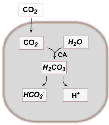
CO2 is the major end product of carbohydrate and lipid metabolism, and the
CO2/HCO3- buffer pair constitutes one of the most important pH buffers in nearly all body
compartments. Carbonic anhydrase (CA), an enzyme that catalyses the reversible hydration of CO2, is present in most body and subcellular compartments and serves to accelerate the
normally slow equilibration between CO2 and HCO3- (Figure 5) (S. L. Alper, Chernova, &
Stewart, 2002; Boron, 2004).
Figure 5: Schematic representation of the bicarbonate synthesis. The bicarbonate buffering system is
an important buffer system in acid-base homeostasis. Carbon dioxide (CO2) cross the plasma membrane
15
that catalyses this reversible hydration of CO2. H2CO3 can dissociate into H+ and HCO3-, to maintain the
ionic balance.
Cells possess in their plasmatic membrane a wide range of ion transporters that participate in pH regulation. HCO3- is the mobile physiological pH buffer that protects cells
from fast and local changes in pHi (Casey, Grinstein, & Orlowski, 2009). The membrane proteins which mediate the transport of HCO3- play an important role in maintaining both
intracellular and extracellular pH (pHo) within narrow limits (Boron, 2004). Acid-base
transporters are divided as to functionality into two groups: (1) acid-extruders (Na+-H+
exchangers, Na+-driven HCO
3-/Cl- transporters, Na+/HCO3- co-transporters, and V-ATPases)
which move H+ out of the cell and/or move bases such as HCO
3- and are utilized to increase
pHi when acidosis occurs, and (2) acid-loaders (Na+-independent HCO3-/Cl- transporters and
Na+/HCO
3- co-transporters), responsible for moving acids into the cell and/or bases out of the
cell and utilized to decrease pHi when alkalosis occurs (Boron, 2004). The ability to function
either as an acid loader or as an acid extruder also depends on the ionic gradients established through the membrane (P. Oliveira, Sousa, Barros, Moura, & Rebelo da Costa, 2009).
As said, almost all bicarbonate transporters are important for the regulation of pHi,
but some specific HCO3- transporters also play key roles in the regulation of cellular volume,
as well as on the transport of acid/base equivalents across epithelia. For instance, electrogenic Na+/HCO
3- co-transporters (NBCe) play key roles in bicarbonate reabsorption by
the renal proximal tubule (Boron, Chen, & Parker, 2009; Maunsbach et al., 2000) and bicarbonate secretion by the pancreatic duct (Abuladze et al., 1998); electroneutral Na+/HCO
3- co-transporters (NBCn) regulate pHi in vascular smooth muscle (I. Choi, Aalkjaer,
Boulpaep, & Boron, 2000; Damkier, Nielsen, & Praetorius, 2006) and in axons in the brain (M. D. Parker, Musa-Aziz, et al., 2008; C. Z. Wang, Yano, Nagashima, & Seino, 2000).
As it happens in other tissues, the control of HCO3- concentration is essential to
maintain pH along the male reproductive tract. In mouse, the luminal HCO3- concentration
reaches its highest value in the lumen seminiferous tubules, the lowest in the caput epididymis, and then slightly rises in the cauda epididymis and vas deferens (Levine & Marsh, 1971). In the acidic fluid of the cauda epididymis, the matured spermatozoa are rendered in quiescence with little motility and no ability to fulfill fertilization (M. Chang, 1951). Unlike this epididymal fluid which is acidic and contains very low concentration of HCO3- (Levine &
Marsh, 1971), the semen from mammals is rich in HCO3- and has an alkaline pH, which varies
in a broad range from 7.2 to 8.4 in human (Owen & Katz, 2005).
The control of the STF pH is crucial for male fertility and regulation of pHi of SCs, the somatic component of the BTB, should also play a major role in this process (Mruk & Cheng, 2004; Tuck, Setchell, Waites, & Young, 1970). pHi is kept mainly through the net balance
between production and elimination of protons and by intracellular buffers (Roos & Boron, 1981).The composition of the STF is influenced by net movements of water, K+ secretion, Na+,