BJRS
RADIATION SCIENCES
06-02-A (2018) 01-12Acepted: 2018-06-25
Implementation of internal monitoring programs for
workers occupationally exposed by
131I in nuclear medicine
services in Brazil
S. M. Oliveira; A. L. A. Dantas; B. M. Dantas
aInstituto de Radioproteção e Dosimetria - IRD, CEP - 22783-127, Rio de Janeiro - RIO DE JANEIRO, Brazil
ABSTRACT
In nuclear medicine services (NMS), workers routinely handle radionuclides for diagnostic and therapy. This practice represents a risk of incorporation by these radionuclides. The International Atomic Energy Agency (IAEA) recommends the implementation of an internal monitoring program on workers potentially exposed to annual effective doses higher than 1 mSv, as for example, those who handle 131I for therapy. Currently, in Brazil, there are not enough available laboratories qualified to provide internal monitoring services to attend all possible demand of internal monitoring if such regulation were applied by the Brazilian Nuclear Regulatory Board (CNEN). Therefore, the objective of this work is to provide simple and inexpensive methods for in vivo routine thyroid monitoring of 131I using equipment available in the clinics itself. Thus, diagnostic devices available in four public hospitals located in the city of Rio de Janeiro were calibrated for use in occupational internal monitoring. The devices evaluated in this work to measure 131I in workers´ thyroid presented enough sensitivity to estimate intakes that would deliver committed effective doses below 1 mSv per year.
1. INTRODUCTION
In Nuclear Medicine Services (NMS), a wide variety of radionuclides in the form of unsealed sources are handled routinely for diagnostic and therapeutic purposes. Such professional activity represents a significant risk of external exposure of occupationally exposed workers, as well as the possibility of intakes via inhalation and ingestion and the subsequent internal exposure.
In spite of a consensus that external exposure is predominant, significant intakes may occur simultaneously in nuclear medicine practice. However, depending on the exposures scenario, and based on international criteria of evaluation, the permanent risk of intakes of radionuclides requires the implementation of a routine monitoring plan, aiming to control and limit internal doses [1].
According to national and international regulations, the Radiation Safety Officer (RSO) of the facility is responsible for managing the Radiation Protection Programme (RPP) and, based on monitoring results, to implement the necessary measures to keep exposure levels as low as possible [2]. The RPP may include an evaluation of internal occupational exposures to be carried out through specific methodologies which allow identification and quantification of intakes as well as the estimation of the committed effective doses of the workers [3]. In Brazil, there are approximately 90 nuclear medicine clinics authorized to handle 131I for therapy purposes [4],
resulting in a significant number of workers routinely exposed to a risk of intake. It should also be pointed out that 131I presents the higher risk of internal exposure among the radionuclides used in
nuclear medicine. Although the national regulations require the implementation of internal monitoring programs of this group of workers, in Brazil there are not enough qualified laboratories available to offer internal dosimetry services and attend the possible demand of internal monitoring. Consequently, it would represent an impeditive high cost to the medical institutions if the Brazilian Nuclear Regulatory Board would apply the requirements of internal monitoring [5].
The objective of this work is to develop feasible methodologies for in vivo thyroid monitoring aiming to implement low-cost internal monitoring Programs applied to nuclear medicine workers who handle unsealed sources of 131I, mainly for therapy, where high activities are routinely handled representing a significant risk of intakes and consequently internal doses.
2. MATERIALS AND METHODS
2.1 Materials
2.1.1 Detectors
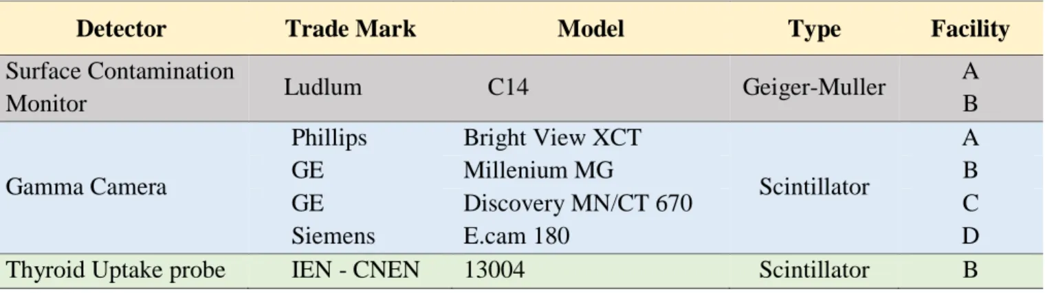
Table 1 presents the detectors which are intended to be applied in internal routine monitoring in professionals who handle 131I in the form of unsealed sources in nuclear medicine clinic.
Table 1: Detectors available evaluated in four Nuclear Medicine Clinics.
Detector Trade Mark Model Type Facility
Surface Contamination
Monitor Ludlum C14 Geiger-Muller
A B
Gamma Camera
Phillips Bright View XCT
Scintillator
A
GE Millenium MG B
GE Discovery MN/CT 670 C
Siemens E.cam 180 D
Thyroid Uptake probe IEN - CNEN 13004 Scintillator B
2.1.2 Neck-thyroid Phantom
The neck-thyroid phantom (Figure 1) used for the calibration of the detectors for the measurement of 131I in thyroid was developed at the In Vivo Monitoring Laboratory of the IRD. The
neck phantom is made of polyurethane-base tissue equivalent material. A filter paper simulating a human thyroid is spiked with a known amount of a 133Ba liquid standard source.
It is conventional practice to use sources of 133Ba as a surrogate for measuring 131I since the energy and yield of photons emitted by 133Ba are very similar to the photon emissions from 131I. Furthermore, the half-life of 133Ba is long (10.5 years) compared with 131I (8 days), so that a certified calibration standard containing 133Ba can be used for many years.
After being sealed with a plastic film, the filter paper is fixed in the proper position with an acrylic part and inserted in the neck phantom. The phantom used in this work contained originally 29771 Bq of 133Ba in 28/04/2004.
Figure 1: Neck-thyroid phantom produced in the In Vivo Monitoring Laboratory of IRD [6].
2.2 Methods
The methodology consisted basically in determining the calibration factors for the measurement of
131I in the thyroid, calculation of the minimum detectable activities and the corresponding minimum
detectable effective doses.
2.2.1 Calibration of the Detection System
Step 1 – Calculation of 131I Equivalent Activity: The 133Ba activity on phantom was corrected
between fabrication and calibration dates. The equivalent 131I activity content of the phantom is
calculated by multiplying the activity of 133Ba added to the phantom by the ratio between the sum of
133Ba and 131I photon yields in the region of interest for the measurement, using the information of
131I Eq Ac
𝐵𝑞 = A (133Ba) x
Σ ( 133Ba)
Σ ( 131I)
(1)
where 131I Eq Ac is the equivalent activity of 131I, in Bq; A (133Ba) is the activity of 133Ba present in the phantom, in Bq; Σ (133Ba) is the sum of emission intensities of 133Ba and Σ (131I) is the sum
of emission intensities of 131I.
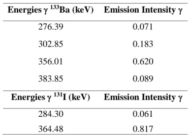
Table 2: Energy photons and emission intensity for 133Ba e 131I.
Energies 133Ba (keV) Emission Intensity
276.39 0.071
302.85 0.183
356.01 0.620
383.85 0.089
Energies 131I (keV) Emission Intensity
284.30 0.061
364.48 0.817
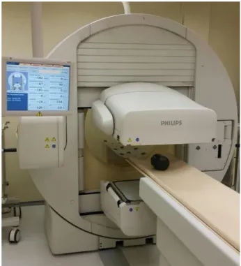
Step 2 – Determination of Measurement Setup (Figure 2): The standard geometry consisted
in positioning the phantom at 12 cm distance to the front of the Gamma-Camera (GC), 0 cm distance to Portable Surface Contamination Monitor (PSCM) probe and 0 cm distance to Thyroid Uptake probe (TUP). The count time (except for the PSCM) was determined according to detectors sensitivity for a monitoring interval of 1 and 7 days after intake, considering a weekly generic monitoring frequency, resulting in 48 monitoring periods per year.
Step 3 – Calculation of Calibration Factor (CF):
The calibration factor is given by:
𝐶𝐹𝑐𝑡𝑔/𝐵𝑞 = (𝑝ℎ𝑎𝑛𝑡𝑜𝑛 𝑐𝑜𝑛𝑡𝑠 − 𝑏𝑎𝑐𝑘𝑔𝑟𝑜𝑢𝑛𝑑 𝑐𝑜𝑢𝑛𝑡𝑠) / 131I Eq Ac (2)
The 131I equivalent activity of the phantom used in this work is in the order of 14000 Bq, depending on the date of its use for the calibration of the equipment.
Figure 2: Measurement geometry for gamma-camera.
2.2.2 Evaluation of Sensitivity
The evaluation of the sensitivity of the method for its application in routine internal monitoring is based on the calculation of the Minimum Detectable Activity (MDA), Minimum Detectable Intake (MDI) and Minimum Detectable Effective Dose (MDED).
The MDA of the method is given by equation 3 or equation 4 [7]:
MDA𝐵𝑞 = (4,65 . √N) / CF (3)
MDA𝐵𝑞 = (4,65 . σ) / CF (4)
where N is the total counts of the background of a non-exposed subject, when the calibration is made with only one measurement; σ is the standard deviation of the background, when the calibration is made in a series of background measurements; and CF is the calibration factor.
The Minimum Detectable Intake (MDI), a function of the MDA and the exposure scenario, is given by:
MDI𝐵𝑞 = MDA / m(t)𝑖𝑛ℎ 𝑜𝑟 𝑖𝑛𝑔 (5)
where MDA is the minimum detectable activity and m(t) is the retention fraction in the compartment of interest for inhalation or ingestion.
The last parameter to be calculated is the Minimum Detectable Effective Dose, which is based on the MDI, and is given by:
MDED𝐵𝑞 = MDI𝑖𝑛𝑎/𝑖𝑛𝑔 x e(g)𝑖𝑛𝑎/𝑖𝑛𝑔 (6)
where MDI is the minimum detectable intake and e(g)ina/ ing is the dose coefficient (mSv/Bq).
In order to be considered useful for internal dosimetry purposes, the technique should, at least, be able to detect an activity that would result in an annual effective dose below 1 mSv for the most likely internal exposure scenario [8]. The values of “m(t)” and “e(g)” are available in the publication 78 of the ICRP [9] and can also be generated for specific exposure scenarios and times through the software AIDE [10].
The monitoring period is determinant to evaluate the sensitivity of the proposed method to be applied on a routine internal monitoring in a specific clinic. This evaluation can be made by the following equation:
Annual Sensitivity𝑚𝑆𝑣 = MDED𝑚𝑆𝑣 x 𝑛 (7)
where MDED is the minimum detectable effective dose and n is the number of annual monitoring periods, e.g., 48 when workers are monitored once a week.
3. RESULTS AND DISCUSSION
Table 3 shows the total counts and background counts results, count times, calibration dates and its respective 131I equivalent activities present in the neck-thyroid phantom for each detector evaluated in this study.
Table 3: Calibration parameters and counts results of the detectors analyzed.
Detector Facility Count Time
(min) Counts BG Counts
Calibration Date
131I Eq Ac
(Bq)
PSCM A Instant count rate 398 70 04/17/17 13874
GC A 3 106838 26248 04/17/17 13874
PSCM B Instant count rate 408 68 06/21/17 13712
GC B 7 255825 103282 06/21/17 13712
TUP B 4 4875 755 07/04/17 13679
GC C 3 31408 4050 10/03/17 13456
GC D 3 69133 15188 03/23/18 13047
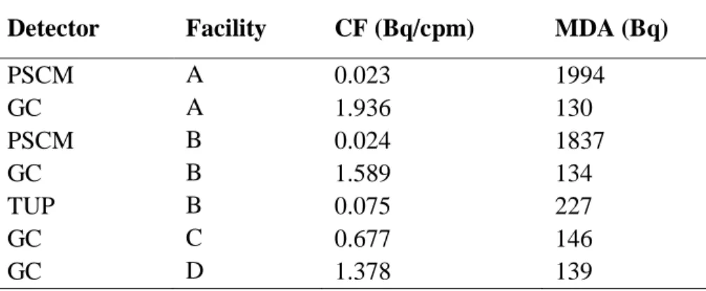
Table 4 shows the results for CF and MDA of the detectors evaluated at the different facilities using the thyroid phantom. The GCs were calibrated in a single measurement of the neck-thyroid and the background, using equation 3 to calculate the MDA, while the PSCMs and TUPs were calibrated through a series of counts of the neck-thyroid phantom and the background, using equation 4 to calculate the MDA.
Table 4: CF and MDA of the detectors analyzed.
Detector Facility CF (Bq/cpm) MDA (Bq)
PSCM A 0.023 1994 GC A 1.936 130 PSCM B 0.024 1837 GC B 1.589 134 TUP B 0.075 227 GC C 0.677 146 GC D 1.378 139
The parameter MDI depends on the exposure scenario and the time elapsed between intake and in vivo measurement. In this work it was used the retention fraction values for 1 and 7 days between 131I intake by inhalation and in vivo measurement (Table 5). Such values were obtained
through the software AIDE [10]. The MDED was calculated based on the MDI, considering the dose coefficient for inhalation associated to the corresponding intake scenario adopted in this study, as shown in Table 5.
Table 5: Retention fractions and dose coefficient for inhalation obtained with the software AIDE.
m(t) (Bq/Bq) e(g) (mSv/Bq) 1 day 7 days
1.98 x 10⁻⁵ 0.229 0.139
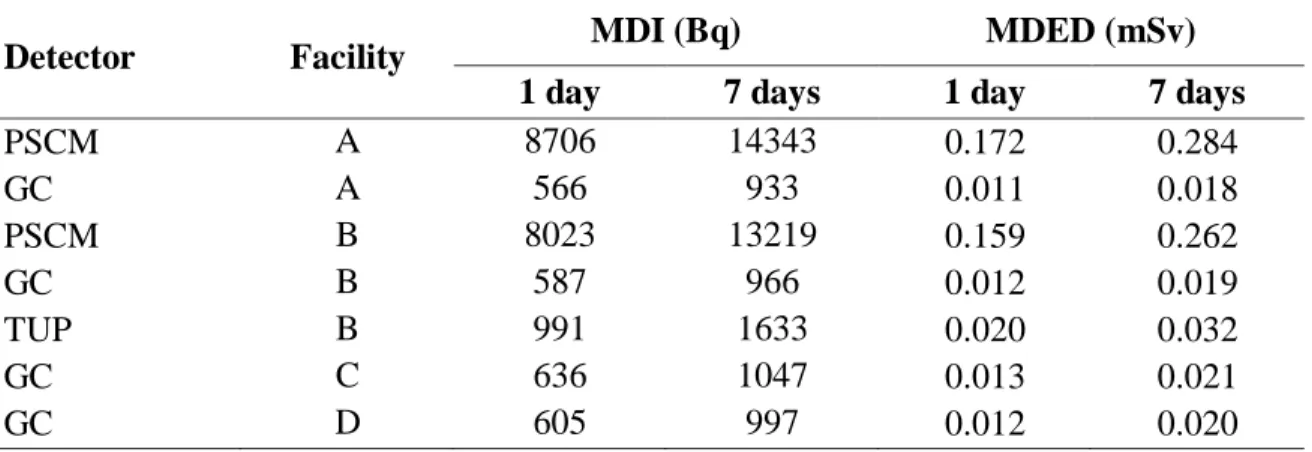
Table 6 shows the results for MDI and MDED of the detectors evaluated, considering 1 and 7 days after intake by inhalation.
Table 6: MDI and MDED of the detectors evaluated for 1 and 7 days after intake by inhalation.
Detector Facility MDI (Bq) MDED (mSv) 1 day 7 days 1 day 7 days
PSCM A 8706 14343 0.172 0.284 GC A 566 933 0.011 0.018 PSCM B 8023 13219 0.159 0.262 GC B 587 966 0.012 0.019 TUP B 991 1633 0.020 0.032 GC C 636 1047 0.013 0.021 GC D 605 997 0.012 0.020
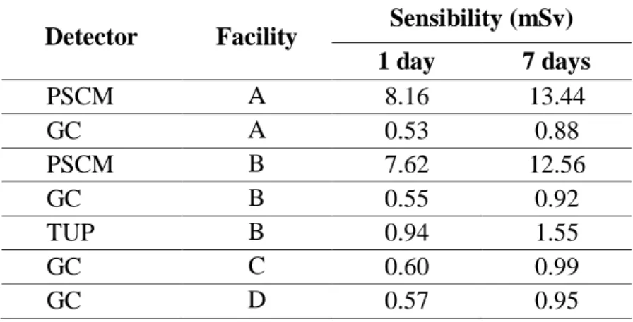
In order to evaluate the sensitivity of the detectors, it was simulated a weekly generic monitoring period, totalizing 48 annual monitoring periods. Table 7 shows the annual sensitivities obtained for a weekly monitoring period.
Table 7: Annual sensitivities (mSv) for 48 monitoring periods per year.
Detector Facility Sensibility (mSv) 1 day 7 days PSCM A 8.16 13.44 GC A 0.53 0.88 PSCM B 7.62 12.56 GC B 0.55 0.92 TUP B 0.94 1.55 GC C 0.60 0.99 GC D 0.57 0.95
The gamma-cameras available in facilities “A”, “C” and “D” are newer and more sensitive than the model in operation in facility “B”, which is approximately 20 years old. Even though, it presented enough sensitivity for occupational monitoring in a reasonable short count time.
The gamma-cameras and the thyroid uptake probe are made up of NaI scintillation detectors, which provide a high intrinsic efficiency for the detection of gamma photons emitted by 131I. However, the size of the NaI crystal used in the uptake probes area smaller in comparison with the ones used in gamma-cameras, resulting in much less sensitivity.
The surface contamination monitors evaluated in this study are Geiger-Muller detectors. Such detectors are much less sensitive than scintillator detectors. Therefore, none of then showed sufficient sensitivity for routine occupational monitoring.
The sensitive values obtained with NaI-based equipment (gamma-cameras and thyroid uptake probes), as shown in Table 4, may be increased by optimizing two basic measurement parameters; (i) reducing distance between detector and subject and (ii) increasing measurement count time.
In order to avoid disturbing the routine of the clinic, it is proposed to use the minimum count time, but keeping the necessary sensitivity of the method for its application in occupational
monitoring. The counting distance was optimized to provide a reasonable comfort to the subject. Furthermore the monitoring plan should be more effective if the measurements are schedule according to the radionuclide handling of the facility, when it is more likely to detect significant intakes by the workers.
4. CONCLUSION
Considering a generic weekly monitoring frequency, the gamma-cameras of the four facilities evaluated in this study presented enough sensitivity for use as a monitoring device up to 7 days after intake. The thyroid uptake probe showed sensitivity only for measurements performed 1 day after intake. The portable surface contamination monitors are not suitable for routine occupational monitoring. Alternatively, this device can be useful for the investigation of possible accidents involving intakes of significant amounts of 131I. It should also be highlighted that the proposed methodology is easy, simple and inexpensive, since the measurements are performed by the staff of the clinic and there is no need to purchase new equipment for the implementation of the monitoring plan.
REFERENCES
[1] IAEA - International Atomic Energy Agency. Assessment of Occupational Exposure due to
Intakes of Radionuclides. Safety Guide No. RS-G-1.2, 1999.
[2] CNEN - Comissão Nacional de Energia Nuclear. Diretrizes Básicas de Radioproteção.
Norma CNEN-NE-3.01. Rio de Janeiro: RJ, 2011.
[3] IAEA - International Atomic Energy Agency. Direct methods for measuring radionuclides
in the human body. Safety Series n. 115, 1996.
[4] CNEN - Comissão Nacional de Energia Nuclear. Instalações Autorizadas. Available at: <http://www.cnen.gov.br/instalacoes-autorizadas>. Last accessed: 06 Dez. 2017.
[5] CNEN - Comissão Nacional de Energia Nuclear. Critérios para cálculo de dose efetiva, a
[6] DANTAS, B. M.; CARDOSO, J. S.; DANTAS, A. L. A.; LUCENA, E. A.; RAMOS, M. A. P., SÁ, M. S., ALONSO, T. C.; SILVA, T. V.; OLIVEIRA, C. M.; LIMA, F. F.; OLIVEIRA, M. L.; LACERDA, I. V. B.; FAJGEL, A. Intercomparação Nacional de Medição In Vivo de
Iodo-131 na Tireoide, Projeto TC IAEA BRA 9055. Scientia Plena, 9(8), 2013.
[7] HPS - Health Physics Society. Performance Criteria for Radiobioassay, N13.30, 1996. [8] IAEA - International Atomic Energy Agency. Methods for Assessing Occupational
Radiation Doses due to Intakes of Radionuclides. Safety Report Series No. 37, Vienna,
2004.
[9] ICRP - International Commission on Radiological Protection. Individual Monitoring for
Internal Exposure of Workers. ICRP Publication 78, Vol. 27/3-4, 1998.
[10] BERTELLI, L.; MELO, D. R.; LIPSZTEIN, J.; CRUZ-SUAREZ, R. AIDE. Internal
![Figure 1: Neck-thyroid phantom produced in the In Vivo Monitoring Laboratory of IRD [6]](https://thumb-eu.123doks.com/thumbv2/123dok_br/18288798.882454/4.893.308.582.327.643/figure-neck-thyroid-phantom-produced-vivo-monitoring-laboratory.webp)