Abstract
Submitted: March 11, 2018
Modification: May 28, 2018 Accepted: July 3, 2018
Proteomic analysis of the acquired
enamel pellicle formed on human and
bovine tooth: a study using the Bauru
in situ pellicle model (BISPM)
The acquired enamel pellicle (AEP) is an organic film, bacteria-free, formed in vivo as a result of the selective adsorption of salivary proteins and glycoproteins to the solid surfaces exposed to the oral environment. Objective: This study aimed to compare the proteomic profile of AEP formed in situ on human and bovine enamel using a new intraoral device (Bauru in situ pellicle model – BISPM). Material and Methods: One hundred and eight samples of human and bovine enamel were prepared (4x4 mm). Nine subjects with good oral conditions wore a removable jaw appliance (BISPM) with 6 slabs of each substrate randomly allocated. The AEP was formed during the morning, for 120 minutes, and collected with an electrode filter paper soaked in 3% citric acid. This procedure was conducted in triplicate and the pellicle collected was processed for analysis by LC-ESI-MS/MS. The obtained mass spectrometry MS/MS spectra were searched against human protein database (SWISS–PROT). Results: The use of BISPM allowed the collection of enough proteins amount for proper analysis. A total of 51 proteins were found in the AEP collected from the substrates. Among them, 15 were common to both groups, 14 were exclusive of the bovine enamel, and 22 were exclusive of the human enamel. Proteins typically found in the AEP were identified, such as Histatin-1, Ig alpha-1, Ig alpha 2, Lysozyme C, Statherin and Submaxillary gland androgen-regulated protein 3B. Proteins not previously described in the AEP, such as metabolism, cell signaling, cell adhesion, cell division, transport, protein synthesis and degradation were also identified. Conclusion: These results demonstrate that the proteins typically found in the AEP appeared in both groups, regardless the substrate. The BISPM revealed to be a good device to be used in studies involving proteomic analysis of the AEP.
Keywords: Proteins. Saliva. Vinícius Taioqui PELÁ1
Luiza Paula Silva CASSIANO2 Talita Mendes da Silva VENTURA3 Cíntia Maria de SOUZA-E-SILVA3 Carlos Condarco GIRONDA3 Daniela RIOS3 Marília Afonso Rabelo BUZALAF3
1 Universidade Federal de São Carlos, Departmento de Genetica e Evolução, São Carlos, SP, Brasil. 2 Faculdades Integradas Aparício Carvalho, Porto Velho, RO, Brasil.
3Universidade de São Paulo, Faculdade de Odontologia de Bauru, Departmento de Ciências
Biológicas, Bauru, SP, Brasil.
Corresponding address: Marília Afonso Rabelo Buzalaf Departamento de Ciências Biológicas - Faculdade de Odontologia de Bauru - Universidade de São Paulo.
Al. Octávio Pinheiro Brisolla, 975 -Bauru-SP - 17012-901 - Brasil. Phone: + 55 14 32358346 - Fax + 55 14 32271486
Introduction
Teeth are constantly bathed by constituents
from the gingival fluid, bacterial products, and
by saliva. These constituents are rich in proteins
and glycoproteins. As a result of this exposure, a bacteria-free organic film, known as acquired enamel pellicle (AEP), is formed1. The AEP formation is quick.
Scanning electron microscopy showed that AEP can be detected even one minute after the enamel samples are exposed to the oral cavity2. Moreover, another
in vivo study using proteomic approaches detected
the presence of 89 proteins within the AEP, formed 5
minutes after dental prophylaxis3, while a recent study identified 190 proteins within the acquired pellicle,
formed in situ for 3 minutes on ceramic specimens4.
The protection of the tooth surface by the AEP is well established in the literature and has been demonstrated in several studies. The AEP acts
as a diffusion barrier or permeable membrane,
diminishing the direct contact between the acids and
the tooth surface, thus reducing the dissolution rate
of hydroxyapatite5-10. In vitro studies revealed that the first proteins to electrostatically interact with the enamel surface are proline-rich proteins (PRPs),
statherin and histatins11,12, while in vivo experiments revealed also the presence of mucins, amylase, cystatin, lysozyme and lactoferrin in the very initial
stages of pellicle formation3,13.
The comprehension of the AEP protein profile was
greatly increased with the advent of proteomic tools. Most of the AEP proteomic studies available so far were conducted in vivo3,10,13-16. While the in vivo model provides the most clinically relevant information, in
some cases it cannot be used. One of them is when
it is desirable to know the protein composition of the
acquired pellicle formed on mixed surfaces constituted by teeth and restorative materials. Another situation is when it is necessary to evaluate the protein composition of the acquired pellicle formed on dentin surfaces17, considering it is quite difficult to obtain
exposed dentin surfaces in an extent that allows
the collection of material to be analyzed. In these situations, in situ models are desirable and to the best
of our knowledge only two studies are available in the
literature so far4,17. In the study by Delecrode, et al.17 (2015), human root dentin specimens were used in a
palatal appliance. One of the main limitations of the study was the fact that only a few typical proteins of
the acquired pellicle were identified. As for the study by Delius, et al.4 (2017), the authors employed ceramic
specimens. Despite they were able to identify more
than 100 proteins, some of which are typically found
in the AEP4, the composition of the ceramic specimens is quite different of human enamel, which certainly impacts in the protein profile of the acquired pellicle.
In studies involving dental caries and erosion, bovine teeth, which are easier to obtain, are often
used as surrogate for human teeth18-20. However, there are no studies comparing the protein profile
of the AEP formed on human and bovine specimens. Considering that structural differences between these two types of substrates exist21, with bovine crystallites being thicker22 and bovine enamel presenting higher
radiographic density23 than the human counterparts, there could be differences in the protein profile of
the AEPs formed on these two types of substrates.
In addition, one of the main limitations of studies
involving proteomic analysis of the acquired pellicle is
to obtain enough material to be analyzed. Thus, it is of
great interest to develop devices to be used in in situ studies that make possible the collection of appropriate amounts of AEP to allow proper protein identification and quantification in proteomic studies. Therefore, this study aimed to compare the proteomic profile of
the acquired enamel pellicle (AEP) formed in situ on human and bovine enamel using a new device (Bauru
in situ pellicle model – BISPM), especially designed to
allow the collection of enough amount of AEP to be
analyzed using proteomic approaches.
Material and methods
Preparation of bovine and human specimens
Bovine permanent incisors and third human molar
were recently extracted, disinfected and kept in 0.1% buffered thymol solution (pH 7.0). After 30 days,
a visual inspection was conducted to evaluate the
presence of caries, stains and cracks. In these cases,
the teeth were excluded.
The selected teeth (n=108) had their crowns cut (4x4x2 mm) using a precision cutting machine (ISOMET Low Speed Saw Buehler Ltda., Lake Bluff, Illinois, USA) with two diamond discs (double-sided XL 12205 ‘high concentration’, 102x12.7x0.3 mm3; Extec
was ground flat with water-cooled silicon carbide discs (320 grade of Al2O3 paper; Buehler). Then, the
specimens were kept with wet gauze in a refrigerator
at 4°C prior to the experiment.
Ethical aspects and subjects
The local Ethics Committee approved the protocol of this research (no. 58331216.9.0000.5417; Ethics
Committee of the Bauru School of Dentistry, University of São Paulo), which followed the guidelines of good
clinical practice and conformed to the Declaration of
Helsinki. Nine young adult volunteers of both genders took part in the study after signing an informed consent document. The sample size was chosen based
on results of previous studies10,14,17. The exclusion
criteria were: smokers, presence of caries lesions, gingivitis, periodontitis, low salivary flow (unstimulated and stimulated to be greater than 0.3 and 1.0 mL/ minute, respectively) and the use of medicines that could change the salivary composition or flow-rate.
Bauru
in situ
pellicle model (BISPM)
Alginate impressions were used to make plaster models, employed to prepare silicon devices from
the mandibular arches of each volunteer. The enamel
samples were cleaned in an ultrasound (T7 Thornton, a Unique Ind. e Com. Ltda., São Paulo, SP, Brazil) for 7 minutes at 25°C. Twelve specimens (6 from each group) were placed in the recessed sites and fixed with dental wax (Asfer Indústria Química Ltda®, São Caetano do Sul, SP, Brazil) for the formation of the
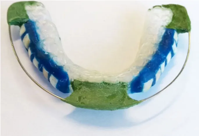
AEP in situ. A number 0.8 orthodontic wire (Morelli
Ortodontia®, Sorocaba, SP, Brazil) was suspended
above the specimens24, to avoid direct contact of the mucosa with them, thus preventing the impact
of mechanical forces on AEP formation. This wire
was fixed on the silicon devices with godiva (Kerr
Corporation®, Orange, Califórnia, USA) in the center of the apparatus and in the posterior regions (Figure 1).
In situ
experiment
The experiment was conducted during the morning to abstain from circadian effects on the composition of the pellicle16 for 3 consecutive days, aiming to obtain
enough material to be submitted to the proteomic
analysis. Firstly, the volunteers inserted the intraoral device into their mouths and for 120 minutes they were instructed not to eat or drink to allow the AEP to form on the enamel surfaces. For the AEP collection, the
intraoral device was removed from the mouth and the
samples were washed with deionized water then dried by air. An electrode filter paper 5x10 mm (Electrode Wick, Bio-Rad®, Hercules, Califórnia, USA) pre-soaked
in 3% citric acid10 was rubbed on the surface of the enamel samples with the aid of tweezers to collect the
AEP. Twelve strips were used for each participant. The
filter papers were stored separately for each group in a polypropylene microcentrifuge tube at −80°C until
the proteomic analyses.
Preparation of the AEP samples
The AEP samples were prepared for proteomic
analysis according to a recently standardized protocol15. Briefly, protein extraction was performed twice using a solution containing 6 M urea, 2 M thiourea in 50 mM
NH4HCO3 pH 7.8, and the supernatants were stored.
To increase protein recovery, the wick papers were transferred to filter tubes (Corning CostarSpin-X Plastic
Centrifuge Tube Filters®, SigmaAldrich, New York, New York, USA), centrifuged, and the supernatant was collected. The supernatants were pooled, centrifuged again and transferred to a falcon tube. Then, 50 mM
NH4HCO3 were added to dilute the urea and thiourea, and the samples were placed in Falcon Amicon tubes
(Amicon Ultra – 15 Centrifugal Filter Units – Merck
Millipore®, Tullagreen, County Cork, IE), centrifuged and concentrated to approximately 150 µL. Reduction [5 mM dithiothreitol (DTT) for 40 minutes at 37°C] and alkylation [10 mM iodoacetamide (IAA) in the absence of light for 30 minutes] were performed. Samples were then digested using 2% (w/w) trypsin (Promega®, Madison, Wisconsin, USA). Then 10 µL of 5% formic acid was placed to stop the action of trypsin. C18 Spin columns (Thermo Scientific®, Rockford, Illinois, USA) were used to desalt and purify the samples, and protein was quantified using the Bradford method (Bio-Rad®, Hercules, Califórnia, USA). The amount of
protein obtained was 14.06 and 21.81 µg for bovine and human groups, respectively. The samples were
resuspended in a solution containing 3% acetonitrile
and 0.1% formic acid to be submitted to nano
LC-ESI-MS/MS.
Accession number
Protein name Bovine
Enamel
Human Enamel
Number of matched peptides
P68032 Actin, alpha cardiac muscle 1 (b, m, n, q , u, w) Yes - 2
P68133 Actin, alpha skeletal muscle (b, m, n, q, u, w) Yes - 2
P62736 Actin, aortic smooth muscle (b, m, n, q, u, w) Yes - 2
P63267 Actin, gamma-enteric smooth muscle (b, m, n, q, u, w) Yes - 1
Q5T3N1 Annexin (Fragment) (b, l, n, p, s, u) Yes - 3 P04083 Annexin A1 (a, b, g, j, n, o, p, s, u, w) Yes - 4 P59665 Neutrophil defensin 1 (b, i, j, o, u) Yes - 5
P59666 Neutrophil defensin 3 (b, i, j, o, u) Yes - 5
Q6S8J3 POTE ankyrin domain family member E (b, m, o, u) Yes - 1
A5A3E0 POTE ankyrin domain family member F (b, m, o, u) Yes - 1
P05109 Protein S100-A8 (b, e, i, j, l, n, o, q, s, u, w) Yes - 1
Q5VXU1 Sodium/potassium-transporting ATPase subunit beta-1-interacting protein
2 (b, c, m, s, u) Yes
-1
Q9NX04 Uncharacterized protein C1orf109 Yes - 1
Q8IZS8 Voltage-dependent calcium channel subunit alpha-2/delta-3 (f, d, s, u, v) Yes - 2
I3L3I0 Actin, cytoplasmic 2 (Fragment) (b, m, r, x) - Yes 2
Q9UJY4 ADP-ribosylation factor-binding protein GGA2 (b, m, r, u) - Yes 3 G3V573 AP-5 complex subunit mu-1 (Fragment) (f, m, r, x) - Yes 1
P07814 Bifunctional glutamate/proline--tRNA ligase (b, m, n, u) - Yes 1
Q9UL45 Biogenesis of lysosome-related organelles complex 1 subunit 6 (b, m, n,
o, u) - Yes
2
P78358 Cancer/testis antigen 1 (b, m, n, u) - Yes 1
Q9NZN8 CCR4-NOT transcription complex subunit 2 (b, m, n, p, u) - Yes 1
B8ZZZ7 DNA polymerase-transactivated protein 6, isoform CRA_b (b, m, p, r, u) - Yes 1
Q8IYG6 Leucine-rich repeat-containing protein 56 (b, m, p, u) - Yes 1
Q9Y4K0 Lysyl oxidase homolog 2 (f, m, o, p, u) - Yes 1
Q9BQY4 Rhox homeobox family member 2 (b, d, e, m, p, u) - Yes 2
Q9NUQ6 SPATS2-like protein (b, c, m, n, p, u) - Yes 1
Q6SZW1 Sterile alpha and TIR motif-containing protein 1 (b, m, n, u) - Yes 2
Q5SQN1 Synaptosomal-associated protein 47 (e, m, n, u) - Yes 1
Q9BVW5 TIMELESS-interacting protein (b, m, n, p, u) - Yes 1
Q04727 Transducin-like enhancer protein 4 (b, m, p, u) - Yes 5
C9JCD5 Transmembrane 4 L6 family member 19 (b, m, o, u) - Yes 1
Q6UWW9 Transmembrane protein 207 (b, m, o, x) - Yes 1
Q5SWW7 Uncharacterized protein C10orf55 - Yes 1
Q13303 Voltage-gated potassium channel subunit beta-2 (b, c, m, n, o, u) - Yes 2
Q9P2Y4 Zinc finger protein 219 (a, c, d, m, p, u) - Yes 5
Q9NR11 Zinc finger protein 302 (a, c, d, m, p, u) - Yes 3 P60709 Actin, cytoplasmic 1 (b, m, n, q, u, w) Yes Yes 2
P63261 Actin, cytoplasmic 2 (a, d, g, j, n, q, u, w) Yes Yes 2
Q5T085 Alpha-amylase (Fragment) (a, g, o, u) Yes Yes bovine 3 / human 5 P04745 Alpha-amylase 1 (a, g, o, u) Yes Yes bovine 8 / human 10 P19961 Alpha-amylase 2B (a, g, o, u) Yes Yes bovine 8 / human 9 P01036 Cystatin-S (a, g, n, p, u) Yes Yes bonine 6 / human 6 P01037 Cystatin-SN (a, g, n, p, u) Yes Yes bovine 2 /human 3 P15515 Histatin-1 (b, i, l, o, u) Yes Yes bonine 2 / human 2 P01876 Ig alpha-1 chain C region (b, e, i, j, o, u) Yes Yes bovine 5 / human 5 P01877 Ig alpha-2 chain C region (b, e, i, j, o, u) Yes Yes bovine 7 / human 7 P61626 Lysozyme C (a, b, g, i, j, o, u, w) Yes Yes bonine 5 / human 6 P04746 Pancreatic alpha-amylase (a, g, o, u) Yes Yes bovine 6 / human 8 P02808 Statherin (b, e, i, l, o, u) Yes Yes bonine 2 / human 2 P02814 Submaxillary gland androgen-regulated protein 3B (a,g,o,u,w) Yes Yes bovine 3 / human 3 P25311 Zinc-alpha-2-glycoprotein (a, b, g, o, u, w) Yes Yes bovine 1 / human 3
Shotgun label-free quantitative proteomic
analysis
Peptides identification was performed as previously
described15 on a nanoACQUITY UPLC-Xevo QTof MS system (Waters, Manchester, New Hampshire UK). The nanoACQUITY UPLC was equipped with nanoACQUITY HSS T3, analytical reverse phase column (75 µm x 150 mm, 1.8 µm particle size, Waters).
ProteinLynx Global Server (PLGS) version 3.0 (Waters Co., Manchester, New Hampshire, UK) was used to process and search the continuum LC-MSE data. Proteins were identified with the embedded ion
accounting algorithm in the software and a search of the Homo sapiens database (reviewed only, UniProtKB/ Swiss-Prot), downloaded on June 2015 from UniProtKB (http://www.uniprot.org/). The identified proteins were classified and assigned by biological function16,25,
origin and molecular interaction (http://www.uniprot.
org/).
For label-free quantitative proteome, three MS raw files from each pooled group were analysed using the PLGS software. All the proteins identified with confidence score greater than 95% were included in
the quantitative analysis. Identical peptides from each triplicate by sample were grouped based on mass
accuracy (<10 ppm) and on time of retention tolerance <0.25 min, using the clustering software embedded in
the PLGS. Difference in expression among the groups
was expressed as p<0.05 for down-regulated proteins and 1-p>0.95 for up-regulated proteins.
Results
The use of BISPM allowed the collection of enough proteins for proper analysis. The total number of
identified proteins, considering both substrates, was 51. These proteins were classified according to their biological function, origin and molecular interaction (Figure 2). Considering these proteins, 15 were common for both groups, 14 were present only on the bovine enamel group, and 22 proteins were exclusive
of the human enamel (Figure 3).
The proteins found in both groups (Figure 2) are typical components of the AEP, such as isoforms of Cystatin, Actin, Alpha-amylase and Ig A, Histatin 1, Lysozyme C, Statherin, Submaxillary gland androgen-regulated protein 3B and Alpha-amylase. Zinc-alpha-2-glycoprotein, involved in the detection of chemical stimulus and in sensory perception of bitter taste, was also identified in both groups.
Most of the proteins found exclusively in the AEP collected from the bovine enamel are not typically described among the constituents of the AEP and are
related to cell motility (distinct isoforms of actin), immune response (Annexin A1 and distinct isoforms of neutrophil defensin), as well as binding to calcium (Protein S-100-A8 and Voltage-dependent calcium channel subunit alpha-2/delta-3) (Figure 2). As for the proteins identified uniquely in the AEP collected from human enamel, most of them are not commonly described intracellular proteins and with unknown function in the AEP (Figure 2). These proteins might
have been originated from the oral mucosa.
Accession number Protein name Ratio HE/BE p
P61626 Lysozyme C 1,1 1
P04746 Pancreatic alpha-amylase 1,18 0.99
P60709 Actin, cytoplasmic 1 0,77 0.03
P63261 Actin, cytoplasmic 2 0,76 0
P15515 Histatin-1 0,67 0
P02808 Statherin 0,73 0
Table 1- Classification and relative quantification of proteins identified in the acquired enamel pellicle collected from Human Enamel (HE)
and Bovine Enamel (BE)
Figure 3- Venn Diagram with the numbers of the exclusive
Regarding the quantitative analysis, two proteins increased in human enamel, when compared to bovine enamel (Lysozyme C and Pancreatic alpha-amylase), while two isoforms of Actin cytoplasmic, Histatin 1 and Statherin decreased (Table 1).
Discussion
The main challenge in studies involving proteomic of the AEP is to obtain enough protein to allow proper
analysis. To overcome this difficulty, we developed a new device, the BISPM that has a special design in order to optimize the collection of enough proteins from the AEP. The placement of number 0.8 orthodontics wire
suspended above the specimens to avoid direct contact of the mucosa with them24 was the main responsible
for it. In addition, the experiment was carried out in 3 consecutive days, and the samples collected from the same treatments were pooled. Furthermore, we worked with a mandibular apparatus instead of a palatal one, because more saliva is expected to bath the specimens in the first condition due to the
gravity force. These strategies were effective to allow
enough proteins in the AEP to be analysed. However,
it has been shown that the composition of the AEP changes according to its location in the dental arches15, which is a limitation of our model, despite a recent
study revealed no difference in the protective ability against initial erosion of the AEP formed in situ using palatal and mandibular intraoral device24. It is worth
mentioning that the removable apparatus developed
was well tolerated by the volunteers, without any
reported discomfort.
In this study, the total number of identified proteins, when both substrates are considered, was 51. This is quite similar to the number of proteins identified in
an experiment where the pellicle was collected from dentin specimens in situ using a mandibular device17. In the study by Delecrode, et al.17 (2015), the protocol
for collecting the acquired pellicle formed on the dentin specimens was similar to the one employed in this
study, considering that this protocol is widely used
in in vivo experiments10. Despite the similar number of proteins identified, the pattern of proteins in the study by Delecrode, et al.17 (2015) was very different
from ours. The only protein typically described in the acquired pellicle was mucin17, while in this study we were able to identify many typical proteins, such as
isoforms of Cystatin, Actin, Alpha-amylase and Ig A, Histatin 1, Lysozyme C, Statherin, Submaxillary gland androgen-regulated protein 3B and Alpha-amylase.
These differences might be explained due to the
distinct types of substrates (dentin X enamel). In a recent study, the authors collected the acquired
pellicle formed for 3 min on ceramic specimens in situ and a total of 190 proteins were identified4, among
which 58% have been described in the pellicle before4 and some of them were also identified in this study, such as Lysozyme C, IgA, Protein S-100, Cystatins,
Neutrophil defensin and Alpha amylase. The higher
number of identified proteins might be explained due
to the high total surface area of the specimens [8x3 cm2 in this study and in the one by Delecrode, et
al.17 (2015)], as well as to the protocol of collection of the acquired pellicle. As mentioned above, in this study and in the study by Delecrode, et al.17 (2015), the pellicle was collected with wick filters embedded in 3% citric acid, while in Delius, et al.4 (2017) the pellicle proteins were eluted by incubation in TRIS-HCl buffer containing SDS, followed by ultrasonication in RIPA-buffer, which cannot be done in vivo. Moreover, despite the ceramic used, Delius, et al.4 (2017)
presents protein adhesion forces close to those of
hydroxyapatite, and both substrates share common
isoelectric points26. The result of using natural teeth
certainly is much closer to the clinical condition. This study aimed to compare the protein composition of the acquired pellicle formed on bovine and human enamel. Bovine enamel has been used as surrogate for human enamel dental research. There are publications comparing both substrates
regarding chemical composition, physical properties, dental caries, dental erosion/abrasion, bonding and microleakage studies27, however, the composition of
the acquired pellicle formed on both substrates has never been compared. Bovine teeth are much easier
to obtain than human teeth and are also bigger, thus providing specimens with a higher surface area,
which is a desirable characteristic for studies involving
collection of acquired pellicle. In this study, despite
some differences found in the proteome of the acquired
pellicle formed on bovine and human enamel, most of
the proteins typically found in the AEP were present
in both substrates (Figure 2), without significant fold
change for most of them. This might be due to the similar inorganic composition of bovine and human
content in weight when compared with human enamel; and the carbonate concentrations are quite similar in both substrates27). Moreover, the crystallographic
nanoscale structure of bovine enamel is very similar to human enamel28. It should be noted that the precursor
proteins that constitute the first layers of the AEP are those with affinity to hydroxyapatite29. These results
indicate that bovine enamel can be used as a substitute for human enamel in in situ studies involving proteomic
analysis of the AEP. Moreover, the BISPM seems to
be an appropriate device to be used in such studies.
Acknowledgements
The authors thank FAPESP for the concession of a scholarship to the first author (Proc. 2014/25599-5).
References
1- Dawes C, Jenkins GN, Tonge CH. The nomenclature of the integuments of the enamel surface of teeth. Br Dent J. 1963;115;65-8. 2- Hannig M. Ultrastructural investigation of pellicle morphogenesis at two different intraoral sites during a 24-h period. Clin Oral Investig. 1999;3(2);88-95.
3- Lee YH, Zimmerman JN, Custodio W, Xiao Y, Basiri T, Hatibovic-Kofman S, et al. Proteomic evaluation of acquired enamel pellicle during in vivo formation. PloS One. 2014;8(7);e67919.
4- Delius J, Trautmann S, Médard G, Kuster B, Hannig M, Hofmann T. Label-free quantitative proteome analysis of the surface-bound salivary pellicle. Colloids Surf B Biointerfaces. 2017;152;68-76.
5- Buzalaf MA, Hannas AR, Kato MT. Saliva and dental erosion. J Appl Oral Sci. 2012;20(5);493-502.
6- Hannig M, Balz M. Protective properties of salivary pellicles from two different intraoral sites on enamel erosion. Caries Res. 2001;35(2);142-8.
7- Hannig M, Fiebiger M, Güntzer M, Dobert A, Zimehl R, Nekrashevych Y. Protective effect of the in situ formed short-term salivary pellicle. Arch Oral Biol. 2004;49(11);903-10.
8- Hara AT, Lussi A, Zero DT. Biological factors. Monogr Oral Sci. 2006;20;88-99.
9- Meurman JH, Frank RM. Scanning electron microscopic study of the effect of salivary pellicle on enamel erosion. Caries Res. 1991;25(1);1-6.
10- Siqueira WL, Zhang W, Helmerhorst EJ, Gygi SP, Oppenheim FG. Identification of protein components in in vivo human acquired enamel pellicle using LC-ESI-MS/MS. J Proteome Res. 2007;6(6);2152-60. 11- Jensen JL, Lamkin MS, Oppenheim FG. Adsorption of human salivary proteins to hydroxyapatite: a comparison between whole saliva and glandular salivary secretions. J Dent Res. 1992;71(9);1569-76.
12- Lamkin MS, Arancillo AA, Oppenheim FG. Temporal and compositional characteristics of salivary protein adsorption to hydroxyapatite. J Dent Res. 1996;75(2);803-8.
13- Delecrode TR, Siqueira WL, Zaidan FC, Bellini MR, Moffa EB, Mussi MC, et al. Identification of acid-resistant proteins in acquired enamel pellicle. J Dent. 2015;43(12);1470-5.
14- Souza e Silva CM, Silva Ventura TM, Pau L, Silva C, Lima Leite A, Buzalaf MA. Effect of gels containing chlorhexidine or epigallocatechin-3-gallate on the protein composition of the acquired enamel pellicle. Arch Oral Biol. 2017;82;92-8.
15- Ventura TM, Cassiano LP, Souza e Silva CM, Taira EA, Leite AL, Rios D, et al. The proteomic profile of the acquired enamel pellicle according to its location in the dental arches. Arch Oral Biol. 2017;79;20-9. 16- Zimmerman JN, Custodio W, Hatibovic-Kofman S, Lee YH, Xiao Y, Siqueira WL. Proteome and peptidome of human acquired enamel pellicle on deciduous teeth. Int J Mol Sci. 2013;14(1);920-34. 17- Delecrode TR, Siqueira WL, Zaidan FC, Bellini MR, Leite AL, Xiao Y, et al. Exposure to acids changes the proteomic of acquired dentine pellicle. J Dent. 2015;43(5);583-8.
18- Buzalaf MA, Hannas AR, Magalhães AC, Rios D, Honório HM, Delbem AC. pH-cycling models for in vitro evaluation of the efficacy of fluoridated dentifrices for caries control: strengths and limitations. J Oral Sci. 2010;18(4);316-34.
19- Soares FZ, Follak A, Rosa LS, Montagner AF, Lenzi TL, Rocha RO. Bovine tooth is a substitute for human tooth on bond strength studies:
a systematic review and meta-analysis of in vitro studies. Dent Mater.
2016;32(11);1385-93.
20- West NX, Davies M, Amaechi BT. In vitro and in situ erosion models
for evaluating tooth substance loss. Caries Res. 2011;45;43-52. 21- Featherstone JD, Mellberg JR. Relative rates of progress of artificial carious lesions in bovine, ovine and human enamel. Caries Res. 1981;15(1);109-14.
22- Arends J, Jongebloed WL. Crystallites dimensions of enamel. J Biol Buccale. 1978;6(3);161-71.
23- Tanaka JL, Medici Filho E, Salgado JA, Salgado MA, Moraes LC, Moraes ME, et al. Comparative analysis of human and bovine teeth: radiographic density. Braz Oral Res. 2008;22(4);346-51.
24- Mendonça FL, Jordão MC, Ionta FQ, Buzalaf MA, Honório HM, Wang
L, et al. In situ effect of enamel salivary exposure time and type of
intraoral appliance before an erosive challenge. Clin Oral Investig. 2017;21(8);2465-71.
25- Rison SC, Hodgman TC, Thornton JM. Comparison of functional annotation schemes for genomes. Funct Integr Genomics. 2000;1(1);56-69.
26- Müller C, Lüders A, Hoth-Hannig W, Hannig M, Ziegler C. Initial bioadhesoin on dental materials as a function of contact time, pH, surface wettability, and isoelectric point. Langmuir. 2010;26(6);4136-41.
27- Yassen GH, Platt JA, Hara AT. Bovine teeth as substitute for human teeth in dental research: a review of literature. J Oral Sci. 2011;53(3);273-82.
28- Teruel JD, Alcolea A, Hernández A, Ruiz AJ. Comparison of chemical composition of enamel and dentine in human, bovine, porcine and ovine teeth. Arch Oral Biol. 2015;60(5):768-75.