Brazilian Journal of Physics, vol. 35, no. 3B, September, 2005 761
Analysis and Discussion of Trace Elements in Teeth of Different Animal Species
F. O. Falla-Sotelo, M. A. Rizzutto, M. H. Tabacniks, N. Added, M. D. L. Barbosa, Instituto de F´ısica, Universidade de S˜ao Paulo, S˜ao Paulo, SP, Brazil
R. A. Markarian, A. Quinelato, M. Mori, and M. Youssef Faculdade de Odontologia, Universidade de S ˜ao Paulo, S˜ao Paulo, SP, Brazil
Received on 15 June, 2005
Human, bovine and swine teeth were analyzed by Proton Induced X-ray Emission (PIXE). The aim of this work was to determine the concentration of trace elements in enamel and dentine of different animal species. PIXE analysis was carried out at the Laboratrio de Anlise de Materiais por Feixes Inicos da USP (LAMFI) using a 2.4 MeV proton beam to probe the samples. Healthy teeth from So Paulo region were analyzed. Thirteen elements were measured and quantified in the samples: P, S, Cl, K, Ca, Cr, Mn, Fe, Ni, Cu, Zn, Sr and Ba. The measured ratio of Ca:P in dentine and enamel teeth is the same expected for hydroxyapatite: 2.13, for all three types. Trace element concentrations were found to be very similar between the three species, except for S, Cl, Fe, Cu and Sr. Ni and Cu concentrations were found to be close to 1 ppm, which is also close to the detection limits of the SP-PIXE system.
I. INTRODUCTION
Trace elements play an important and complex role in the human and animal metabolism. The trace elements in te-eth have been examined for a number of reasons, for exam-ple there are some studies of dental health where trace ele-ment concentrations have been correlated with the presence of dental caries [1–3]. Some elements such as Al, Fe and Sr are caries inhibitory and Cu, Mn and Cd are caries promo-ting [2], however the combinations of Mn and Cd may have inhibitory role, while Al and Sr can promote caries. The mi-neral tissue of the tooth consists of hydroxyapatite crystals Ca10(PO4)6(OH)2 with incorporated trace elements, which can provide information of the habitat environment or dietary habits. The tooth is a bio-indicator of great interest because it contains information on deposited elements in the tooth ma-terial. Mammalian teeth contain three tissues such as enamel, dentine and cementum. In order to find compatible human teeth for substitution in the dentistry laboratory practice and chemical tests, a comparison of the trace elements in the ena-mel and dentine teeth was made, between the human and ani-mal teeth (swine and bovine). This work was performed with Particle Induced X-ray Emission (PIXE) spectroscopic tech-nique [4]. The trace elements concentrations in enamel and dentine of human, swine and bovine teeth were obtained with the thick target PIXE (ttPIXE) analysis calculated for a given matrix as hydroxyapatite throught the CLARA program [5].
II. EXPERIMENTAL PROCEDURE
Healthy teeth were collected from So Paulo region. Bo-vine incisive, swine molars and human molars (control group), were divided in three groups of ten teeth. All teeth were weighed and sterilized using one autoclave cycle and finally stored in dry air at 4oC in individual containers until analysis. To obtain a flat surface with the exposed dentine, the molar’s crown was sliced and the bovine incisive had their buccal
fa-ces cut with a diamond disk, to avoid metal contamination of the cut surface. The PIXE measurement of the samples was performed at the LAMFI Laboratory. Samples were irradiated in vacuum with a 2.4 MeV proton beam with 4 mm diame-ter. The sliced teeth were mounted on a multiposition target tower at the center of the PIXE chamber [6]. In the reaction chamber the target was positioned at 135owith respect to the proton beam, the target tower allows vertical translation. Ty-pical beam currents used were about a few nA to reduce dead time and pile-up. The acquisition time was 1200 seconds for each sample. The X-rays were observed with a Si(Li) detec-tor with a resolution of about 145 eV at MnKα. The X-ray spectra were optimized for detection of elements above P, and a ”funny filter” made of Mylar (300µmthick with a 10 % hole and a plain Mylar 50µm thick) was placed between the tar-get and detector to reduce the count-rate of Ca-Kαpeak. The PIXE spectra were analyzed by AXIL-X-Ray analysis soft-ware [7]. The program evaluates the integral of thick target correction factor, using Newton-Cotes algorithm [8]. Total X-ray ionization cross sections are calculated using Johansson and Johansson [9] polynomial fit. X-ray intensity ratios were taken from Scofield [10] and Perujo [11] while the fluores-cence yields are from Bambyneck [12]. Stopping powers for protons up to 10 MeV, are calculated using the fitted curve given by Ziegler et al. [13]. Mass absorption coefficients are calculated using the XCOM software developed by Ber-ger and Hubbell [14].
III. RESULTS AND DISCUSSION
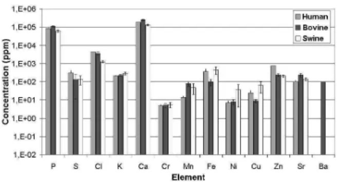
The enamel and dentine mean trace element concentration of the three teeth species are summarized in figure 1 and 2. The dentine and enamel comparison between the three spe-cies shows no statistical differences among the major concen-tration elements (Ca and P).
762 F. O. Falla-Sotelo et al.
FIG. 1: Mean trace element concentration (in ppm) in enamel of human, swine and bovine teeth. Lines on top of each bar indicate sample mean standard deviation.
FIG. 2: Mean trace element concentration (in ppm) in dentine of human, swine and bovine teeth. Lines on top of each bar indicate sample mean standard deviation.
found to be close to 1 ppm, which is also close to the detection limits of the SP-PIXE system. The bovine dentine and enamel shows clear evidence of Ba concentration. Concentrations of Cl, Mn, Fe, Cu and Zn are higher in enamel compared with dentine. The measured ratio of Ca:P = 2.17 (22) and 2.13(38) in human, 2.32(19) and 2.08(18) in bovine and 2.21(19) and 1.92(8) in swine in enamel and dentine respectively, is similar to the expected data for hydroxyapatite: 2.13.
IV. CONCLUSION
Trace elements in dentine and enamel for human, bovine and swine teeth were compared for the first time. Results from this study shows no statistical differences among the major concentration elements (Ca and P). Thirteen elements were measured and quantified in the samples: P, S, Cl, K, Ca, Cr, Mn, Fe, Ni, Cu, Zn, Sr and Ba. This study demonstrates that Ba exist only in bovine teeth. The measured ratio Ca:P in ena-mel and dentine respectively, is similar to the expected data for hydroxyapatite.
Acknowledgments
M.A.R and M.H.T. authors wish to grateful to FAPESP for financial support. F.O.F.S, M.H.T. and N.A. acknowledge CNPq for financial support.
[1] M. A. Chaudhri, and T. Ainsworth, Nucl Instr. Meth.181, 333 (1981).
[2] H. J. Annegarn, A. Jodaikin, P. E. Cleaton-Jones, J. P. F. Sells-chop, and C. C. P. Madiba, Nucl Instr. Meth.181, 323 (1981). [3] M. E. Curzon, and D. C. Crocker, Arch. Oral Biol. 23, 647
(1978).
[4] S. A. E. Johansson and J. L. Campbell,PIXE: A novel technique for elemental analysis(John Wiley&Sons, Inc., 1988). [5] J. Aburaya, M. H. Tabacniks, M. D. L. Barbosa, N. Added,
M. A. Rizzutto, and M. D. L. Barbosa, Submitted to Nucl. Instr. and Meth. (2005).
[6] M. H. Tabacniks, Physics Institute Report - IF/USP1469, 50 (2000).
[7] P. Van Espen, H. Nullens, and F. Adams, Nucl. Instr. and Meth.
145, 579 (1977).
[8] A. Hadding, Anorg. Allgerm. Chem122, 195 (1922).
[9] S. A. E. Johansson and T. B. Johansson, Nucl. Instr. and Meth.
137, 476 (1976).
[10] J. H. Scofield, Phys. Ver.A9, 1041 (1974).
[11] A. Perujo, J. A. Maxwell, W. J. Teesdale, and J. L Campbell, J.Phys.B20, 4973 (1987).
[12] W. Bambyneck, private comunication of material presented verbally at the International Conference on X-ray and Inner Shell Process in Atoms, Molecules and Solids, University of Leipzig, (1984).
[13] J. F. Ziegler, J. P. Biersack, and U. Littmark,The Stopping and Range of Ions in SolidsNucl. Instr. Meth.1, (Pergamon Press, (1985).