RESUMO.- [Indicadores hematológicos e bioquímicos de maturidade em potros e relação com suas características placentárias.] A saúde do neonato está diretamente relacionada às condições gestacionais e eficiência placentária. Os objetivos deste estudo foram: (1) avaliar parâmetros hematológicos e
bioquímicos de potros nascidos de éguas com placentite ao nascimento e com 24h de vida e (2) verificar se a patologia placentária exerceu influência no grau de maturidade através da resposta hemato-bioquímica destes neonatos. De acordo com os resultados histopatológicos placentários (controle e placentite) e grau de maturidade neonatal (maturo e imaturo), os potros foram divididos em três grupos: grupo controle (n=22); e potros nascidos de éguas com placentite classificados como (2) maturos (n=26) e (3) imaturos (n=10). Foi avaliado hematócrito e concentrações sanguíneas de fibrinogênio, proteína plasmática total, leucócitos totais, lactato, glicose, creatinina, uréia, albumina, bilirrubinas, triglicerídeos, colesterol, cálcio, fósforo, magnésio, aspartato aminotransferase (AST), creatina quinase (CK), fosfatase ABSTRACT.- Feijó L.S., Curcio B.R., Pazinato F.M., Almeida B.A., Moraes B.S.S., Borba L.A. &
Feijó J.O. & Nogueira C.E.W. 2018. [Hematological and biochemical indicators of maturity in foals and their relation to the placental features.] Pesquisa Veterinária Brasileira 38(6):1232-1238. Departamento de Clínica Veterinária, Universidade Federal de Pelotas, Campus Universitário, Capão do Leão, RS 96160-000, Brazil. E-mail: [email protected]
Newborn’s health is directly related to gestational conditions and placental efficiency. The aims of this study were: (1) To evaluate hematological and biochemical parameters of foals born from mares with placentitis at birth and at 24h of age, (2) to verify if placental pathology had any influence on neonatal maturity degree through hematological and biochemical response of those foals. According to placental findings (control and placentitis) and neonatal maturity degree (mature and immature), foals were divided into three groups: (1) Control group (n=22), foals born from mares with placentitis and classified as (2) Mature (n=26), and (3) Immature (n=10). The hematocrit and plasma concentration of fibrinogen, total plasma protein, white blood cells count, lactate, glucose, creatinine, urea, albumin, bilirubin, triglyceride, cholesterol, calcium, phosphorus, magnesium, aspartate aminotransferase (AST), creatine kinase (CK), alkaline phosphatase (ALP), and gamma-glutamyltransferase (GGT) were measured. Placental features were significantly different between neonatal maturity degree (P=0.001). Mares that had acute placentitis foaled more immature neonates (n=8/10; 80%). Concentrations of fibrinogen (P=0.003), creatinine (P=0.021), total cholesterol (P=0.014), AST (P=0.001), GGT (P=0.002), total (P=0.001) and unconjugated bilirubin (P=0.010) were higher at birth in the Immature group, whereas albumin levels were lower (P=0.002). Foals born from mares with placentitis presented hyperlactatemia at 24h of age (P=0.002). Acute placentitis had an influence on the neonatal maturity, allowing an accelerated but incomplete fetal maturation. The monitoring of lactate, fibrinogen, creatinine, bilirubin, cholesterol, albumin, AST, and GGT levels, associated with clinical, physical, and behavior evaluation may contribute as indicators of neonatal maturity.
INDEX TERMS: Immaturity, hematology, biochemistry, placentitis, maturation, neonatal, foals, Equids.
PVB-5503 MF
Hematological and biochemical indicators of maturity
in foals and their relation to the placental features
1Lorena S. Feijó2, Bruna R. Curcio2*, Fernanda M. Pazinato2, Bruno A. Almeida3,
Bruna S.S. Moraes2, Luciana A. Borba2, Josiane O. Feijó2 and Carlos E.W. Nogueira2
1 Received on June 21, 2017.
Accepted for publication on October 31, 2017.
2 Departamento de Clínica Veterinária, Universidade Federal de Pelotas (UFPel), Campus Universitário, Capão do Leão, RS 96160-000, Brazil. *Corresponding author: [email protected]
3 Instituto de Ciências Biológicas, Universidade Federal do Rio Grande (FURG), Campus Carreiros, Rio Grande, RS 96203-900, Brazil. E-mails: [email protected], [email protected], [email protected], [email protected], [email protected], [email protected]
ors o the
os om suas
alcalina (FA) e gama glutamiltranferase (GGT). As características placentárias foram significativamente diferentes entre os graus de maturidade neonatal (P=0.001). Éguas com placentite aguda produziram mais potros imaturos (n=8/10; 80%). No nascimento, os potros imaturos apresentaram maiores concentrações de fibrinogênio (P=0,003), creatinina (P=0,021), colesterol total (P=0,0014), AST (P=0,001), GGT (P=0,002), bilirrubina indireta (P=0,010) e total (P=0,001) e menor concentração de albumina (P=0,002). Os potros nascidos de éguas com placentite apresentaram hiperlactatemia com 24h de vida (P=0,002). A placentite aguda exerceu influência na maturidade neonatal, permitindo uma maturação fetal acelerada, porém, incompleta. Mensurações dos níveis sanguíneos de lactato, fibrinogênio, creatinina, colesterol total, AST, GGT, bilirrubinas e albumina, associado à avaliação clínica, física e comportamental, podem contribuir como indicadores de maturidade neonatal.
TERMOS DE INDEXAÇÃO: Imaturidade, hematologia, bioquímica, placentite, maturação, neonatal, potros, Equidae.
INTRODUCTION
Insufficiency in placental structure and function may cause disorders in fetal growth and maturity (Whitwell 1980), and might lead to fetal death, abortion or stillbirth. Conditions that affect uteroplacental unit may decrease fetal nutrients and oxygen supply (Cottrill et al. 1991). Thus, placentitis is the most common cause of premature birth and neonatal mortality (Giles et al. 1993).
Maturation of the fetal hypothalamic-pituitary-adrenal axis (HPA) plays a central role triggering the maturity of organic systems in preparation to extrauterine life (Rossdale et al. 1997). In equines, fetal maturation is considered delayed in comparison with other species because it starts 4-5 days before delivery and continues in the first days of postnatal life (Rossdale et al. 1984). However, in situations of chronic stress, such as placental lesions and hypoxia episodes, early maturation of fetal HPA axis has been reported (Rossdale et al. 1991).
Therefore, newborn’s health is directly related to gestational conditions and placental efficiency (Hay Junior 1995). Hematological and biochemical perinatal evaluation may be useful for early identification of foals at risk for developing clinical and metabolic disorders (Axon 2011, Pirrone et al. 2014). Parameters in hematology and biochemistry were previously described in neonatal foals (Bauer et al. 1984, Harvey et al. 1984, Axon & Palmer 2008). However, clinical studies about placental evaluation and analysis of indicators focused in neonatal maturity have not been completely elucidated to date, even though placentitis is frequently diagnosed in mares. Moreover, reference values for some parameters immediately after delivery in foals are lacking. Thus, the aims of this study were: (1) to evaluate hematological and biochemical parameters of foals born from mares with placentitis at birth and 24h of age and (2) to verify if placental pathology had any influence on neonatal maturity degree through hematological and biochemical response of those foals.
MATERIALS AND METHODS
A longitudinal case-control study was conducted in a population of Thoroughbred mares and their foals from a farm at Aceguá, Rio Grande do Sul, Brazil, during three breeding seasons (2011 to 2013).
All procedures were approved by Ethical Committee on Animal Experimentation of the Veterinary College, UFPel (protocol 4750).
Animals. All mares were maintained in a semi-extensive system
and assessed daily for the presence of mammary gland development and vulvar discharge. Transrectal ultrasonography of the caudal placental pole was performed monthly to measure the combined thickness of the uterus and placenta (CTUP) and to assess for signs of chorioallantois separation from the endometrium (Renaudin et al. 1997). Mares with clinical signs of placentitis such as increased CTUP values, placental separation, vulvar discharge and premature mammary gland development were treated with trimethoprim sulfamethoxazole (25mg/kg, IV, q 12h; Trissulfin® - Ouro Fino Saúde
Animal, Cravinhos, SP) and flunixin meglumine (1.1 mg/kg, IV, q 24h;
Banamine® - Schering Plough Saúde Animal, São Paulo, SP) for 10 days
and altrenogest (0.088mg/kg, PO, q 24h; Regumate® - Merck Animal
Health Corporate headquarters, Summit, NJ, USA) until delivery. Thirty days prior to the delivery date, mares were conducted to paddocks near the maternity barn where they would stay until the moment of delivery. After chorioallantois rupture, mares were brought into the stable for assisted foaling.
Case selection criteria. The following data were recorded for
each mare at the time of foaling: age, gestational length (days) and type of parturition. Foal’s blood was withdrawn from the jugular vein 10 minutes and 24h after birth. Blood samples were placed into plastic vials containing clotting activator for serum biochemistry, ethylenediaminetetraacetic acid (EDTA) for hematology and sodium fluoride for glucose and lactate concentrations. Blood culture was also performed in all foals at birth. Neonatal maturity was defined through physical characteristics and behavior assessment. Immaturity was set for foals that presented low birth weight, small body size, short and shiny hair coat, prominent rounded head, droopy ears and lips and/or lack muscle tone (Rossdale et al. 1984). Low birth weight was considered in animals that weighted less than 50.9 kg as established for Thoroughbred foals (Whitwell & Jeffcott 1975). The following parameters were used for neonatal behavior assessment: suckle reflex (<20 min), standing (1-2h) and suckling (within 2 h) (Koterba 1990).
Immediately after foaling, placentas were evaluated and classified according to the gross and histologic findings. Placentas that presented no gross or histologic lesions were classified as Control (n=22). Placentitis (n=36) was set to placentas that showed gross lesions as discoloration and avillous areas on chorionic surface, allantochorion thickening and presence of inflammatory exudate near the cervical star and uterine body. Moreover, in histologic evaluation, placentitis were categorized as acute or chronic as described by Hong et al. (1993). Acute placentitis were identified by inflammatory cellular infiltrates with prevalence of neutrophils, microcotyledons necrosis, and suppurative exudate on the chorionic surface. Chronic placentitis were associated with a severe tissue reaction characterized by infiltration of mononuclear inflammatory cells in the intervillous space, necrosis of chorionic villi, chorionic stroma, villous stroma, and adenomatous hyperplasia of the allantoic epithelium.
Exclusion criteria. Foals in Control group were excluded from
this study if: (1) they were not foaled spontaneously and intervention was needed; (2) they did not show normal behavior or mentation; (3) they took longer time to present the parameters previously mentioned in behavior assessment; (4) they presented any signs of clinical disorder in the first 72h of life.
Groups. All births were assisted, and the mares and their foals
into three groups: Control group with 22 healthy delivered foals, after normal pregnancy and parturition, normal physical characteristics and behavior; Mature group with 26 foals born from mares with placentitis and normal parturition or dystocia, normal physical characteristics and behavior; Immature group with 10 foals born from mares with placentitis and normal parturition or dystocia, presenting physical characteristics associated with immaturity and abnormal behavior.
Hematological and biochemical analysis. The hematocrit
(Hct) and white blood cells (WBC) count were determined using an automated hematology analyzer (CC-530, CELM CIA, São Caetano do Sul, Brazil). Total plasma protein and fibrinogen concentration were measured by refractometry technique. Serum samples for biochemistry analysis were centrifuged at 2.220 g for 10 minutes
and then immediately frozen at -80°C until processment. Specimens containing clots or hemolysis were excluded. In each sample, the concentrations of lactate, glucose, creatinine, urea, albumin, bilirubin (conjugated, unconjugated and total), triglyceride, total cholesterol, calcium, phosphorus, magnesium, aspartate aminotransferase (AST), creatine kinase (CK), alkaline phosphatase (ALP), and gamma-glutamyltransferase (GGT) were measured by spectrophotometry using commercial kits (Labtest Diagnóstica SA, Lagoa Santa, Brazil). The coefficients of variation were below than 10% in all assays.
Statistical analysis. All analyses were carried out using the
commercial software Statistix 10.0 (Analytical Software, Tallahasse, FL, USA). All variables were submitted to the Shapiro-Wilk normality test. Continuous data (gestational length and mare’s age) were assessed by ANOVA One-Way. Comparisons between neonatal groups and values at birth and 24h of age were analyzed by ANOVA repeated measures and posthoc LSD test. Fisher’s exact test was used to analyze categorical data (fetal membranes pathological features, placentitis treatment and neonatal maturity degree). Statistical significant was set at p<0.05. Continuous results are expressed as mean ± standard error of the means, whereas categorical results were represented as percentage and proportions to facilitate interpretation.
RESULTS
The gestational length was shorter (P=0.01) in Immature group (329±3.3 days) than in Control (344±2.2 days) and Mature group (342±2.1 days). No difference was found in mare’s age (Control group: 8.6±0.8; Mature group: 9.6±0.7; Immature group: 10.2±1.2 years). Twelve mares (n=12/36; 33%) with placental lesions confirmed by histologic evaluation were treated for placentitis and their foals were allocated in Mature (n=8/26; 31%) and Immature group (n=4/10; 40%).
However, treatment management had no influence on neonatal maturity of foals born from mares with placentitis. In addition, placental pathological features presented by those mares treated for placentitis were not different between acute (n=6/19; 31%) and chronic lesions (n=6/17; 35%).
On the other hand, fetal membranes histological features were significantly different (P=0.001) between neonatal maturity groups. Mares that had acute placentitis foaled more immature neonates (n=8/10; 80%) than mares with chronic placentitis (n=2/10; 20%) and control group (n=0/10; 0%).
Only one case of dystocia was observed in Immature group (n=1/10; 10%). Other five foals (n=5/10; 50%) were delivered at premature placental separation and they were classified as affected by perinatal asphyxia syndrome (PAS) based on history and clinical signs, especially because of the neurologic dysfunction. The clinical signs included loss or absence of suckling reflex, dysphagia, seizures, hyperactivity, and weakness. Other causes of neurologic diseases as meningitis and trauma were excluded. Blood culture was negative in all foals. In addition, all foals were considered viable during this study.
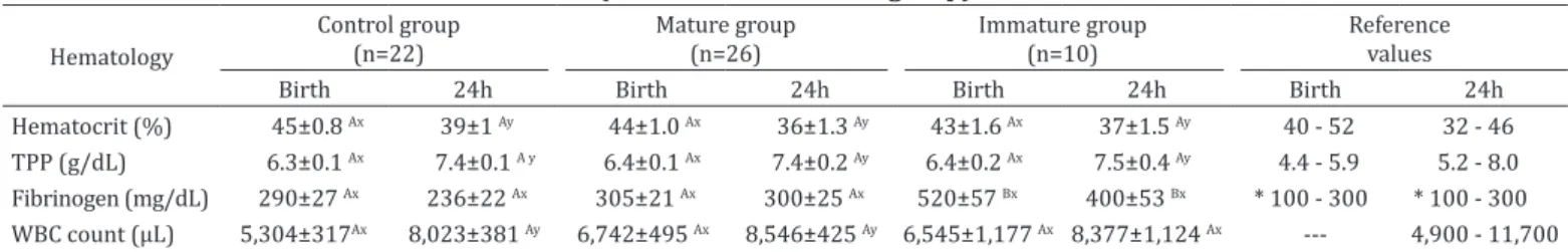
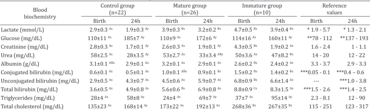
Hematological parameters evaluated at birth and at 24 hours after delivery are shown in Table 1. Plasma fibrinogen concentration was higher at birth (P=0.003) and 24h of age (P=0.014) in Immature group, whereas the number of WBC increased significantly in Control (P=0.001) and Mature group (P=0.006) at 24h of age. Neonatal biochemical parameters and statistical comparisons are illustrated in Table 2 and 3. Foals born from mares with placentitis presented plasma lactate concentration significantly higher than Control group at birth (P=0.001) and at 24h of age (P=0.002). There was no difference found in urea levels at birth between groups. However, foals from Control (P=0.001) and Mature group (P=0.002) showed a decreased in this parameter at 24h of age.
Foals born from mares with infected placentas showed elevated concentrations of total bilirubin at birth (P=0.001). Concentrations of creatinine (P=0.021), total cholesterol (P=0.014), AST (P=0.001), GGT (P=0.002), and unconjugated bilirubin (P=0.010) were higher at birth in Immature group. Serum concentration of albumin was significantly lower (P=0.002) in Immature group compared with foals from Mature group. There was no difference found in the others variables between groups.
Table 1. Hematological parameters at birth and at 24h of age of the Control group and foals born from mares with placentitis (Mature and Immature group)
Hematology Control group(n=22)
Mature group (n=26)
Immature group (n=10)
Reference values
Birth 24h Birth 24h Birth 24h Birth 24h
Hematocrit (%) 45±0.8 Ax 39±1 Ay 44±1.0 Ax 36±1.3 Ay 43±1.6 Ax 37±1.5 Ay 40 - 52 32 - 46
TPP (g/dL) 6.3±0.1 Ax 7.4±0.1 A y 6.4±0.1 Ax 7.4±0.2 Ay 6.4±0.2 Ax 7.5±0.4 Ay 4.4 - 5.9 5.2 - 8.0
Fibrinogen (mg/dL) 290±27 Ax 236±22 Ax 305±21 Ax 300±25 Ax 520±57 Bx 400±53 Bx * 100 - 300 * 100 - 300
WBC count (µL) 5,304±317Ax 8,023±381 Ay 6,742±495 Ax 8,546±425 Ay 6,545±1,177 Ax 8,377±1,124 Ax --- 4,900 - 11,700
Data are expressed as medians ± SEM (standard error of the means). TPP = total plasma protein, WBC = white blood cells; AB Different capital letters in rows indicate significant differences (p<0.05) between groups with LSD’s test, xy Different small letters in rows indicate significant differences (p<0.05)
DISCUSSION
This present study showed that abnormal pregnancy carried with placental pathology has its effects on fetal maturational process. In horses, the maturational changes begin in late gestation and continue into the first few days of neonatal life (Rossdale et al. 1984). This indicates the importance of the final maturational process. Alsothis implies that adrenocortical function may not be fully mature at birth even in full-term foals (Rossdale et al. 1997). Acute placentitis was the most common placental dysfunction that resulted in neonatal immaturity. In addition, mares with acute placentitis had shorter gestational length than other mares, including those diagnosed with chronic placentitis. Rossdale (1993) reported that consequences of disturbances in the uterine environment probably depend on the severity and timing of the insult. Mature group had gestational length similar with the Control mares and chronic placentitis was the most common placental lesion found in this group. Chronic placentitis is characterized as placental insufficiency (Giles et al. 1993) and this result suggests that longer gestational length provided a fetal metabolic adaptation
to this uterine environment which allowed an improvement on the maturation process of the fetus. Furthermore, mares of Control and Mature group had pregnancies longer than 340 days, which is reported for Thoroughbred breed (Rossdale & Short 1967).
On the other hand, shorter gestational length presented by mares with acute placentitis probably is the major result to explain the accelerated but incomplete maturation showed by the Immature group. Challis et al. (2001) described that premature maturation of the HPA axis often causes an inappropriate developmental stage for some body systems, causing asynchrony of organ maturation and significant postnatal problems. Previous clinical study performed by Rossdale et al. (1991) reported that maturation of fetal HPA axis may be initiated precociously in situations of chronic stress, such as placental lesions and hypoxia episodes. Although, neonatal maturity was not significantly different between newborns foaled by mares with placentitis treated or non-treated in the present study. Therapy must be instituted early in mares with placentitis and aggressive treatment has improved foal viability in experimental models (LeBlanc 2010). However,
Table 2. Biochemistry metabolites and small inorganic molecules at birth and 24h of age of the Control group and foals born from mares with placentitis (Mature and Immature group)
Blood biochemistry
Control group (n=22)
Mature group (n=26)
Immature group (n=10)
Reference values
Birth 24h Birth 24h Birth 24h Birth 24h
Lactate (mmol/L) 2.9±0.3 Ax 1.9±0.3 Ay 3.9±0.3 Bx 3.2±0.2 Bx 4.7±0.5 Bx 3.9±0.4 Bx * 1.9 - 5.7 * 1.3 - 2.1
Glucose (mg/dL) 110±11 Ax 185±7 Ay 110±9 Ax 172±6 Ay 114±16 Ax 160±11 Ay **78 - 112 **137 - 193
Creatinine (mg/dL) 2.8±0.3 Ax 1.7±0.1 Ay 2.6±0.3 Ax 1.9±0.1 Ay 4.3±0.5 Bx 1.9±0.2 Ax 1.6 - 2.4 1 - 1.1
Urea (mg/dL) 58±2.5 Ax 28±3.5 Ay 53±2.7 Ax 33±3.4 ABy 50±3.6 Ax 47±8.2 Bx 14 - 20 12 - 22
Albumin (g/dL) 3.1±0.1 ABx 2.9±0.1 Ax 3.2±0.1 Ax 2.9±0.1 Ax 2.6±0.2 Bx 2.4±0.2 Ax 3.3 - 3.7 2.9 - 3.3
Conjugated bilirubin (mg/dL) 0.6±0.1 Ax 0.5±0.1 Ax 1.0±0.1 ABx 0.9±0.1 Bx 1.5±0.2 Bx 1.4±0.2 Bx ***0.05 - 0.1 ***0.4 – 0.6
Unconjugated bilirubin (mg/dL) 2.9±0.5 Ax 4.3±0.7 Ax 4.5±0.6 Ax 5.9±0.7 Ax 6.8±0.9 Bx 6.6±1.4 Ax --- ***1.0 - 3.8
Total bilirubin (mg/dL) 3.6±0.5 Ax 4.9±0.8 Ax 5.6±0.6 Bx 6.9±0.8 Ax 8.8±0.9 Cx 8.3±1.5 Ax ***1.5 - 2.6 ***1.4 - 2.5
Triglycerides (mg/dL) 28±4 Ax 58±8 Ay 24±4 Ax 69±7 Ay 37±7 Ax 95±14 Ay 2.3 - 8.1 12 - 90
Total cholesterol (mg/dL) 135±23 Ax 168±14 Ay 173±22 Ax 192±13 Ax 268±36 Bx 267±35 Bx 115 - 251 123 - 317
Data are expressed as medians ± SEM (standard error of the means). AB Different capital letters in rows indicate significant differences (p<0.05) between groups with LSD’s test, xy Different small letters in rows indicate significant differences (p<0.05) between time points with LSD’s test; Reference
values = Aoki & Ishii 2012; * Castagnetti et al. 2010, ** Kitchen & Rossdale 1975, *** Barton & Leroy 2007 (---: indicates there is no reference value described for this analysis in this neonatal age).
Table 3. Serum electrolyte and enzymes at birth and 24h of age of the Control group and foals born from mares with placentitis (Mature and Immature group)
Blood biochemistry
Control group (n=22)
Mature group (n=26)
Immature group (n=10)
Reference values
Birth 24h Birth 24h Birth 24h Birth 24h
Calcium (mg/dL) 12.9±0.3 Ax 12.5±0.3 Ax 12.8±0.2 Ax 12.3±0.2 Ax 13.6±0.4 Ax 12.0±0.5 Ay 12.02 - 12.07 11.0 - 12.2
Phosphorus (mg/dL) 5.9±0.3 Ax 7.1±0.4 Ay 5.5±0.2 Ax 5.9±0.3 Ax 5.8±0.4 Ax 6.6±0.6 Ax --- * 3.8 -7.4
Magnesium (mg/dL) 2.9±0.1 Ax 3.9±0.2 Ay 2.7±0.1 Ax 3.4±0.2 Ay 2.9±0.2 Ax 3.3±0.3 Ax **0 - 4.28 * 0.6 - 4.2
AST (U/L) 96±10 Ax 196±17 Ay 95±9 Ax 203±11 Ay 139±15 Bx 185±30 Ax 49 - 111 113 - 275
CK (U/L) 138±15 Ax 235±26 Ay 194±18 Ax 308±29 Ay 167±33 Ax 212±49 Ax 126 - 452 134 - 312
ALP (U/L) 977±41 Ax 858±50 Ax 933±40 Ax 838±62 Ax 975±144 Ax 957±191 Ax --- * 861 - 2,671
GGT (U/L) 19±2 Ax 29±2 Ay 14±2 Ax 25±2 Ay 31±4 Bx 24±5 Ax 15 - 27 16 - 52
Data are expressed as medians ± SEM (standard error of the means). AST = Aspartate aminotransferase, CK = Creatine kinase, ALP = Alkaline phosphatase, GGT = Gamma-glutamyltransferase; AB Different capital letters in rows indicate significant differences (p<0.05) between groups with LSD’s test, xy Different
results of experimentally induced placentitis may be different from spontaneous infections and treatment goals may not be accomplished. Clinical studies of placental spontaneous infections and treatment management in these cases require further investigations.
The results of the present study have shown that foals born from mares with placentitis may present some differences in hematological and biochemical evaluation. In the current study, Hct values are similar to previous studies performed in hot-blooded neonatal foals, such as Thoroughbred and Quarter Horse (Harvey et al. 1984, Harvey 1990). The decrease of Hct and increase of TPP levels observed in all groups might be related to absorption of colostral proteins, which enhance plasma volume through osmotic effects (Harvey 1990). Foals of the Immature group showed elevated levels of fibrinogen and this has been reported in foals from placentitis (Lins et al. 2012, Feijó et al. 2014). Hyperfibrinogenemia in foals less than 2 days of age is an indicator of in utero sepsis and inflammation, since this indicates an inflammatory response at least 24 to 48 hours (Morresey 2005, Axon & Palmer 2008). On the other hand, there was no difference found in fibrinogen concentrations in the Mature group; this finding suggests an appropriate fetal adaptation to placental dysfunction. There was no difference found in WBC count among the groups; however, an increase was observed at 24h of age in all groups, although no statistical difference was found in the Immature group. Harvey (1990) described that immature foals tend to show leukopenia and maintain low values in total leukocyte count due to inability of the adrenal gland to secret corticosteroid hormones. Therefore, in our study, the number of WBC did not provide evidence for the organic immaturity in neonatal foals.
Foals born from mares with infected placentas had significantly higher plasma lactate concentrations at birth and 24h of age than Control group. Hyperlactatemia at birth is considered normal due to cortisol and catecholamines release or to physiologic hypoxia during the birth process, not representing clinical importance (Rossdale et al. 1991, Castagnetti et al. 2010). Nevertheless, plasma lactate in equine neonates is age-dependent and healthy foals should be able to reduce blood levels during the first hours after delivery, reaching 2.1mmol/L at 24h of age (Castagnetti et al. 2010). Therefore, the maintenance of hyperlactatemia observed in Mature and Immature group may represent inadequate tissue perfusion and failure in clearance due to hepatic immaturity. Furthermore, this confirms the importance of the plasma lactate in clinical diagnostics, particularly in foals that experienced any degree of placental lesion.
There was no difference found in blood glucose concentration among the groups. Vaala (1999) reported that pre-suckle hypoglycemia in the newborn foal is associated to placental dysfunction and PAS. Asphyxia leads to the rapid metabolism of glucose by the brain and other tissues for energy (Hollis et al. 2008). Although PAS was diagnosed in five immature foals, hypoglycemia was not identified in this group.
Creatinine concentrations are variable during the initial 24h of life and clinical studies performed in the immediate postpartum (presuckle) period have been reported values as 1.1-2.3mg/dL (Aoki and Ishii 2012) and 2.3-3.0mg/dL (Pirrone et al. 2014). Therefore, the hypercreatininemia detected in the Immature group at birth (>3mg/dL) demonstrated
its importance in neonatal evaluation. Vaala (1999) and Morresey (2005) documented that hypercreatininemia in first hours of life is an indicator of placental dysfunction and fetal stress, whereas placenta is the mainly responsible for the elimination of fetal excretions. On the other hand, normal levels found in Mature group suggest that serum creatinine may be a biomarker of placental dysfunction only in acute placentitis cases, when the adjustment of fetoplacental unit has not occurred yet.
In neonatal foals, serum urea concentrations at birth are within the adult reference range and generally this drops during the following days (Bauer et al., 1984). In this current study, elevated levels of urea in all groups may be considered a singular result of the local population since similar results have been described in a clinical study performed in the same area (Lins et al. 2012). Nevertheless, Immature group showed that urea concentrations remain increased at 24h of age which may be attributed to hepatic immaturity rather than to impaired renal function. Moreover, no other evidences were found to support primary renal dysfunction. In the present study, the lowest concentrations of serum albumin were showed by the immature foals, although no statistical difference was found between control and Immature group. This could be attributed to liver disorder since hypoalbubinemia in newborn foals may be associated with hepatic immaturity, as described by Stoneham (2006).
Mature and Control group showed increased concentrations of serum total bilirubin at birth and at 24h of age (Table 3). However, newborn foals frequently have elevated total bilirubin and mild icterus. Hyperbilirubinemia appears to be a transient finding in the first week of life and this has been well documented in equine neonatology (Bauer 1990, Hawthorne 1990, Palmer 2006, Axon & Palmer 2008). Koterba (1990) reported normal neonatal serum bilirubin levels at 24h of age range 1.39-5.48mg/dL. Nevertheless, some clinical studies have been reported lower reference values (Bauer et al. 1989, Barton & Leroy 2007). However, when hyperbilirubinemia reaches levels >7.5 mg/dL, as showed by Immature group, this could be attributed to intrauterine distress (Palmer 2006). Newborn foals that experienced placental dysfunction can demonstrate hyperbilirubinemia due to reduction of bilirubin excretion through placenta, whereas fetal liver capacity to excrete bilirubin is minimal (Bauer et al. 1984). In addition, increased levels of AST and GGT associated to hyperbilirubinemia in the Immature group at birth provide evidence of hepatic immaturity, as described by Feary (2011).
(2008) described that abnormal electrolyte concentrations in the newborn foal reflect placental dysfunction, and a recent study (Mariella et al. 2016) documented that foals affected by PAS showed hypermagnesemia at admission when compared with healthy, septic and premature foals. However, there was no difference found in serum concentrations of electrolytes between groups, although foals suffered from placental dysfunction in both Mature and Immature group.
CONCLUSIONS
Our findings suggest that acute placentitis had an influence on neonatal maturity, allowing an accelerated but incomplete fetal maturation.
The Immature group demonstrated abnormal adaptative processes to extrauterine life with some organic disorders caused by hepatic and adrenocortical immaturity.
The Mature group had gestational length similar with the Control mares, despite the placental insufficiency, since chronic placentitis was the most common placenta lesion found in this group.
This result suggests that a fetal adaptation in this pathologic uterine condition has occurred which provided an adequate neonatal maturation.
The condition may have allowed their foals to present a hematological and biochemical panel similar to foals born from healthy pregnancies. However, those newborn foals still should be considered at risk for developing clinical disorders.
Therefore, during the first 24h of life, the assessment of lactate, fibrinogen, creatinine, bilirubin, cholesterol, albumin, AST, and GGT levels, associated with clinical, physical, and behavior evaluation may contribute as indicators of neonatal maturity.
These parameters evaluation may be aided in the screening of foals that suffered from placental dysfunction.
Acknowledgements.- We thank the Brazilian sources FAPERGS, CAPES, and
CNPq for providing scholarships to graduate students. Also, we thank the members of the farm who provided the animals for research.
REFERENCES
Aoki T. & Ishii M. 2012. Hematological and biochemical profiles in peripartum mares and neonatal foals (heavy draft horse). J. Equine Vet. Sci. 32(3):170-176. http://dx.doi.org/10.1016/j.jevs.2011.08.015.
Axon J.E. 2011. Critical Care: assessment, p.167-176. In: McKinnon A.O., Squires E.L., Vaala W.E. & Varner D.D. (Eds), Equine Reports. 2nd ed. Wiley-Blackwell, Oxford, UK.
Axon J.E. & Palmer J.E. 2008. Clinical pathology of the foal. Vet. Clin. N. Am., Equine Pract. 24(2):357-385. http://dx.doi.org/10.1016/j.cveq.2008.03.005. PMid:18652960.
Bauer J.E. 1990. Normal blood chemistry, p.602-614. In: Koterba A.M., Drummond W.H. & Kosch P.C. (Eds), Equine Clinical Neonatology, Lea and Febiger, Philadelphia.
Bauer J.E., Asquith, R.L. & Kivipelto J. 1989. Serum biochemical indicators of liver function in neonatal foals. Am. J. Vet. Res. 50(12):2037-2041. Barton M.H. & LeRoy B.E. 2007. Serum bile acids concentrations in healthy and
clinically ill neonatal foals. J. Vet. Intern. Med. 21(3):508-513. PMid:17552459. Bauer J.E., Harvey J.W., Asquith R.L., McNulty P.K. & Kivipelto J. 1984. Clinical chemistry reference values of foals during the first year of life. Equine Vet. J. 16(4):361-363. http://dx.doi.org/10.1111/j.2042-3306.1984.tb01944.x. PMid:6479134.
Berryhill E.H., Magdesian K.G., Kass P.H. & Edman J.E. 2017. Triglyceride concentrations in neonatal foals: serial measurement and effect of age and illness. Vet. J. 227:23-29. http://dx.doi.org/10.1016/j.tvjl.2017.08.002. PMid:29031326.
Brewer B.D. 1990. Neonatal infection, p.295-317. In: Koterba, A.M., Drummond, W.H. & Kosch P.C. (Eds), Equine Clinical Neonatology. Lea and Febiger, Philadelphia.
Castagnetti C., Pirrone A., Mariella J. & Mari G. 2010. Venous blood lactate evaluation in equine neonatal intensive care. Theriogenology 73(3):343-357. http://dx.doi.org/10.1016/j.theriogenology.2009.09.018. PMid:19962183. Challis J.R., Sloboda D., Matthews S.G., Holloway A., Alfaidy N., Patel F.A., Whittle W., Fraser M., Moss T.J. & Newnham J. 2001. The fetal placental hypothalamic-pituitary-adrenal (HPA) axis, parturition and post natal health. Mol. Cell. Endocrinol. 20(1-2):135-144. http://dx.doi.org/10.1016/ S0303-7207(01)00624-4. PMid:11738803.
Cottrill C.M., Jeffers-Lo J., Ousey J.C., McGladdery A.J., Ricketts S.W., Silver M. & Rossdale P.D. 1991. The placenta as a determinant of fetal well-being in normal and abnormal equine pregnancies. J. Reprod. Fert. 44:591-601. PMid:1795302.
Feary D.J. 2011. Critical care-monitoring, p.177-188. In: McKinnon A.O., Squires E.L., Vaala W.E. & Varner D.D. (Eds), Equine Reproduction. 2nd ed. Wiley-Blackwell, Oxford, UK.
Feijó L.S., Curcio B.R., Haetinger C., Pazinato F.M., Kasinger S., Santos R.S.D., Ladeira S.R.L. & Nogueira C.E.W. 2014. Maturidade de potros nascidos de éguas com placentite. Arq. Bras. Med. Vet. Zootec. 66(6):1662-1670. http://dx.doi.org/10.1590/1678-7635.
Giles R.C., Donahue J.M., Hong C.G., Tuttle P.A., Petrites-Murphy M.B., Poonacha K.B., Roberts A.W., Tramontin R.R., Smith B. & Swerczek T.W. 1993. Causes of abortion, stillbirth, and perinatal death in horses: 3527 cases (1986-1991). J. Am. Vet. Med. Assoc. 203(8):1170-1175. PMid:8244867. Harvey J.W. 1990. Normal hematologic values, p.561-570. In: Koterba A.M.,
Drummond W.H. & Kosch P.C. (Eds), Equine Clinical Neonatology. Lea and Febiger, Philadelphia.
Harvey J.W., Asquith R.L., McNulty P.K., Kivipelto J. & Bauer J.E. 1984. Haematology of foals up to one year old. Equine Vet. J. 16(4):347-353. http://dx.doi.org/10.1111/j.2042-3306.1984.tb01940.x. PMid:6479131. Hawthorne T.B. 1990. Neonatal hyperbilirubinemia, p.589- 601. In: Koterba A.M., Drummond W.H. & Kosch P.C. (Eds), Equine Clinical Neonatology. Lea and Febiger, Philadelphia.
Hay Junior W.W. 1995. Current topic: metabolic interrelationships of placenta and fetus. Placenta 16(1):19-30. http://dx.doi.org/10.1016/0143-4004(95)90078-0. PMid:7716125.
Hollis A.R., Furr M.O., Magdesian K.G., Axon J.E., Ludlow V., Boston R.C. & Corley K.T. 2008. Blood glucose concentrations in critically ill neonatal foals. J. Vet. Intern. Med. 22(5):1223-1227. http://dx.doi.org/10.1111/j.1939-1676.2008.0174.x. PMid:18691362.
Hong C.B., Donahue J.M., Giles Junior R.C., Petrites-Murphy M.B., Poonacha K.B., Roberts A.W., Smith B.J., Tramontin R.R., Tuttle P.A. & Swerczek T.W. 1993. Etiology and pathology of equine placentites. J. Vet. Diagn. Invest. 5(1):55-63. http://dx.doi.org/10.1177/104063879300500113. Kitchen H. & Rossdale P.D. 1975. Metabolic profiles of newborn foals. J.
Reprod. Fert. 23(23):705-707. PMid:1060869.
Koterba A.M. 1990. Physical examination, p71-85. In: Koterba A.M., Drummond W.H. & Kosch P.C. (Eds), Equine Clinical Neonatology. Lea and Febiger, Philadelphia.
LeBlanc M. 2010. Ascending placentitis in the mare: an update. Reprod. Dom. Anim. 45(Suppl. 2):28-34. http://dx.doi.org/10.1111/j.1439-0531.2010.01633.x. PMid:20591062.
Lins L.A., Finger I.S., Fernandes C.G., Curcio B.R., Corrêa M.N. & Nogueira C.E.W. 2012. Resposta clínica e metabólica de potros neonatos em relação aos achados histopatológicos da placenta na égua. Arq. Bras. Med. Vet. Zootec. 64(6):1436-1441. http://dx.doi.org/10.1590/S0102-09352012000600005.
Mariella J., Isani G., Andreani G., Freccero F., Carpenè E. & Castagnetti C. 2016. Total plasma magnesium in healthy and critically ill foals. Theriogenology 85(2):180-185. http://dx.doi.org/10.1016/j.theriogenology.2015.09.011. PMid:26498390.
Morresey P.R. 2005. Prenatal and perinatal indicators of neonatal viability. Clin. Tech. Equine Pract. 4(3):238-249. http://dx.doi.org/10.1053/j. ctep.2005.07.005.
Palmer J. 2006. Recognition and resuscitation of the critically ill foal, p.121-131. In: Paradis M.R. (Ed.), Equine Neonatal Medicine: a case-based approach. Elsevier Saunders, Philadelphia.
Pirrone A., Antonelli A., Mariella J. & Castagnetti C. 2014. Gross placental morphology and foal serum biochemistry as predictors of foal health. Theriogenology 81(9):1293-1299. http://dx.doi.org/10.1016/j. theriogenology.2014.02.011. PMid:24661433.
Renaudin C.D., Troedsson M.H.T., Gillis C.L., King V.L. & Bodena A. 1997. Ultrasonographic evaluation of the equine placenta by trans rectal and transabdominal approach in the normal pregnant mare. Theriogenology 47(2):559-573. http://dx.doi.org/10.1016/S0093-691X(97)00014-9. PMid:16728008.
Rossdale P.D. 1993 Clinical view of disturbances in equine foetal maturation. Equine Vet J. 14:3-7
Rossdale P.D. & Short R.V. 1967. The time of foaling of thoroughbred mares. J. Reprod. Fert. 13(2):341-343. http://dx.doi.org/10.1530/jrf.0.0130341. PMid:6022637.
Rossdale P.D., Ousey J.C. & Chavatte P. 1997. Readiness for birth: an endocrinological duet between fetal foal and mare. Equine Vet. J. 24(24):96-99. PMid:9355809.
Rossdale P.D., Ousey J., Silver M. & Fowden A. 1984. Studies on equine prematurity 6: guidelines for assessment of foal maturity. Equine Vet. J. 16(4):300-302. http://dx.doi.org/10.1111/j.2042-3306.1984.tb01931.x. PMid:6090120.
Rossdale P.D., Ousey J.C., Cottrill C.M., Chavatte P., Allen W.R. & McGladdery A.J. 1991. Effects of placental pathology on maternal plasma progestagen and mammary secretion calcium concentrations and on neonatal adrenocortical function in the horse. J. Reprod. Fert. 44:579-590. PMid:1665519. Stoneham S.J. 2006. Assessing the newborn foal, p.31-38. In: Paradis M.R.
(Ed.), Equine Neonatal Medicine: a case-based approach. Elsevier Saunders, Philadelphia.
Vaala W.E. 1999. Peripartum asphyxia syndrome in foals. Proc. Am. Assoc. Equine Pract. 45:247-253.